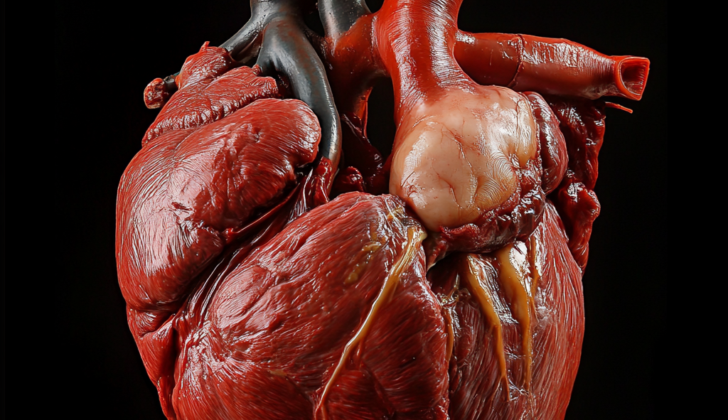
What is Cardiac Rhabdomyoma (Heart Muscle Tumor)?
Cardiac rhabdomyoma is a rare, non-cancerous tumor that originates from striated muscles, which are muscles controlled by our will such as skeletal muscles. This type of tumor often occurs in the head and neck. There are two types — those that occur in the heart or cardiac, and those that happen elsewhere or extracardiac. The extracardiac type is further divided into adult, fetal, and germ cell tumors.
Cardiac rhabdomyoma, in particular, is the most common heart tumor in children, usually being detected before their first birthday. These tumors are considered hamartomas, which are non-cancerous, disorganized overgrowths of cells in an area of the body where these cells are normally found. Most cardiac rhabdomyomas are linked with a condition called tuberous sclerosis (TS) and are found in the heart muscle of the ventricles or atria, the intersection of the large veins and atria, or the outer layer of the heart.
Typically, individuals with cardiac rhabdomyomas have multiple tumors which tend to shrink on their own; thus, surgical removal is not recommended unless the patient is showing symptoms. Symptoms usually occur when the tumor blocks the flow of blood, possibly leading to congestive heart failure. Additionally, abnormal heart rhythms or arrhythmias can occur and may range from a slow heartbeat due to problems with the sinus or atrioventricular node (a part of the heart that ensures its contractions are in sync) to accelerated heartbeats in the atria or ventricles.
What Causes Cardiac Rhabdomyoma (Heart Muscle Tumor)?
Rhabdomyoma, a certain type of muscle tumor, might be caused by changes in genes during muscle development. Currently, no other causes are known. It’s also interesting to note that between 80% and 90% of rhabdomyomas found in the heart are linked to a condition called tuberous sclerosis.
Risk Factors and Frequency for Cardiac Rhabdomyoma (Heart Muscle Tumor)
Primary cardiac tumors, specifically cardiac rhabdomyomas, are quite rare, especially in children. They make up about 45% of all cardiac tumors found in children. Both boys and girls can get this type of tumor, and it doesn’t favor any particular race.
Signs and Symptoms of Cardiac Rhabdomyoma (Heart Muscle Tumor)
Cardiac rhabdomyoma is a type of heart tumor that can be detected in unborn babies using ultrasound scans. The way it presents can vary extensively. It can cause problems like heart blocks, a condition called hydrops fetalis, or fluid surrounding the heart. In severe cases, it can cause the baby to pass away in the womb. After birth, they often don’t show any symptoms. If there are symptoms, they usually indicate heart failure and trouble in the heart’s blood flow due to the tumor. Shortness of breath is one of the common symptoms.
These tumors are diagnosed more often in unborn babies than in newborns due to the higher sensitivity of fetal heart ultrasound scans. Having cardiac rhabdomyoma is also closely associated with a genetical condition known as tuberous sclerosis. Some physical signs of tuberous sclerosis such as light-colored spots on the skin, rough patches of skin or benign growths on the skin can also be part of the patient’s condition.
Testing for Cardiac Rhabdomyoma (Heart Muscle Tumor)
If you are diagnosed with cardiac rhabdomyoma, which often relates to tuberous sclerosis, your doctor will necessarily perform an electrocardiogram (EKG). The EKG is a type of test that checks your heart’s electrical activity and is non-invasive and painless. Even if you don’t have heart-related symptoms, you’ll need an EKG. After that, regular EKGs will be taken every 2 to 5 years for monitoring.
Your doctor will also likely use a diagnostic tool called an echocardiography (ECHO). This is an ultrasound test that uses sound waves to build detailed pictures of your heart. On these images, a cardiac rhabdomyoma appears as multiple bright and rounded clusters in the ventricular myocardium, which is the middle part of your heart’s walls. These clusters can even stick out into the inner space of the heart, and they look more solid and brighter than the normal heart tissue.
However, these images might be misinterpreted as a different type of heart tumor, like an atrial myxoma, especially if the location of the rhabdomyoma is unusual or if there is only one large tumor. In such cases, your doctor may also perform a cardiac magnetic resonance imaging (MRI). This is another type of imaging test that helps doctors see the heart in more detail. An MRI can better outline the tumor, which can be particularly useful if surgical removal is being considered. This type of imaging also gives a more up-to-date estimate of how well the heart is pumping blood.
Treatment Options for Cardiac Rhabdomyoma (Heart Muscle Tumor)
Cardiac rhabdomyomas are heart tumors that usually don’t cause any symptoms and tend to shrink on their own. However, in rare cases, they can affect the heart’s normal functioning and lead to congestive heart failure, a condition where the heart can’t pump blood as well as it should. In such cases, certain medications may be prescribed.
Drugs called angiotensin-converting (ACE) enzyme inhibitors, digitalis, and diuretics can all be used to help manage heart function in these patients. In very ill newborns with heart instability, a drug called Prostaglandin E can be used. Antiarrhythmic drugs can also be given when the heart’s usual electrical signals that coordinate heartbeats are not working properly.
If medications aren’t enough and the heart is still not functioning well due to a large tumor, surgery may be recommended. This could entail removing some or all of the tumor, but the preference is usually to only remove part of it. This is because completely removing it could risk damaging the heart muscle or other important structures in the heart.
In extremely rare and severe cases, when the tumor is so large that it has taken over the normal heart tissue, a heart transplant might be considered. However, this is not a common occurrence.
What else can Cardiac Rhabdomyoma (Heart Muscle Tumor) be?
When diagnosing certain heart conditions, the doctor might consider these possible causes:
- Cardiac fibroma (a heart tumor)
- Atrial myxoma (a noncancerous heart tumor)
- Hemangioma (a birthmark that looks like a rubbery bump)
- Teratoma (a tumor typically found in the tailbone, ovaries, or testicles)
- Thrombus (a blood clot)
- Inflammatory myofibroblastic tumor (a rare tumor that can occur anywhere in the body)
What to expect with Cardiac Rhabdomyoma (Heart Muscle Tumor)
Patients who undergo surgery to remove a type of tumor called a rhabdomyoma generally have a fair to good outlook. The highest risk is associated with cardiac rhabdomyomas – those that occur in the heart. They can continue to grow and block an important pathway in the heart, leading to issues with the blood flow. They can also cause rapid heart rhythms or disrupt the normal electrical impulses that control the heart’s rhythm.
An important sign of a rare genetic disease known as tuberous sclerosis is the presence of fetal cardiac rhabdomyoma. This implies that the baby, while still in the womb, might have tuberous sclerosis, therefore other organs like the kidney or brain should be checked for similar tumors. Often, a heart rhabdomyoma is the first symptom of tuberous sclerosis, which can later lead to neurological problems.
Possible Complications When Diagnosed with Cardiac Rhabdomyoma (Heart Muscle Tumor)
The problems you can encounter with cardiac rhabdomyoma include:
- Infections
- Changed heartbeat patterns, which could be too slow (bradycardia) or too fast (tachycardia)
- Heart failure, which is the heart’s inability to pump blood effectively
- Issues with blood circulation (hemodynamic compromise)
Recovery from Cardiac Rhabdomyoma (Heart Muscle Tumor)
After surgery, standard aftercare like regular bandage changes and removing stitches, is required. Pain relievers such as acetaminophen, codeine, and oxycodone can assist in managing pain after surgery.
Preventing Cardiac Rhabdomyoma (Heart Muscle Tumor)
The patient needs to understand how their condition progresses over time. Regular check-ups are advised in order to monitor any changes or onset of symptoms. This helps in managing the condition more effectively.


