What is Cutaneous Vascular Malignancies, Angiosarcoma and Kaposi Sarcoma?
In the past, it has been tough to accurately identify and diagnose blood vessel abnormalities in the head and neck without using a tissue sample or biopsy. This usually depended on the doctor’s experience and their ability to observe how the abnormality progressed over time. However, progress was made when these abnormalities were split into two groups: vascular tumors and vascular malformations.
With recent advances in medical imaging technology and the processing of tissue samples, doctors have been able to gain a better understanding of these abnormalities. Moreover, additional subcategories have been defined. Vascular tumors include various types such as hemangiomas, tufted angiomas, pyogenic granulomas, and hemangiopericytomas. Vascular malformations are further divided into groups like lymphatic, venous, mixed, arteriovenous, or capillary.
The type of treatment and the patient’s prognosis depend on correctly identifying these blood vessel abnormalities. Because of this, a team of healthcare professionals is often required to manage these cases correctly. This article will discuss various harmful blood vessel abnormalities that commonly occur on the skin of the face and neck, specifically Kaposi sarcoma and angiosarcoma.
What Causes Cutaneous Vascular Malignancies, Angiosarcoma and Kaposi Sarcoma?
Kaposi sarcoma is a type of skin cancer that grows in the cells lining the lymph or blood vessels and is believed to occur due to a combination of genetics and viral influences. In particular, the human herpesvirus 8 (HHV-8) has been strongly linked to the development of Kaposi sarcoma, but a person’s overall health also contributes to the risk.
Another form of skin cancer, cutaneous angiosarcoma, is primarily associated with radiation treatment for other types of cancer. It usually occurs 5 to 10 years after the radiation therapy. Also, prolonged swelling of the limbs (chronic lymphedema), which could be caused by factors like surgical removal of lymph nodes, inherited conditions like Milroy disease, or chronic infections, can increase the risk. This is often referred to as Stewart-Treves syndrome.
Sometimes, skin injuries are also found to be a minor association because injuries often draw attention to the aggressive growth. Other risk factors that have been suggested include exposure to arsenic and certain plastics (polyvinyl chloride), previous infections from herpes zoster (shingles), and the presence of abnormal blood vessel formations.
However, it’s important to note that many skin angiosarcomas develop without any link to pre-existing conditions or injuries.
Risk Factors and Frequency for Cutaneous Vascular Malignancies, Angiosarcoma and Kaposi Sarcoma
Kaposi sarcoma is a type of cancer that can be broken down into four distinct categories:
- The classic type primarily occurs in Mediterranean males from 50 to 70 years of age.
- The endemic type is usually found in children from southern and eastern Africa, and it accounts for about half of all soft tissue tumors in children. This type can be very aggressive.
- The immunosuppressed type is typically seen in people with weakened immune systems due to medication or organ transplants. Mediterranean males are particularly susceptible to this type.
- The AIDS-related type is the most common and is especially prevalent among men who have sex with men who have HIV. It’s the most common cancer found in people with AIDS.
On the other hand, angiosarcomas are quite rare. They are cancers consisting of cells from either blood or lymph vessels and can invade nearby structures. These cancers are very uncommon and only make up between 1% and 2% of all sarcomas in the head and neck area. The most familiar manifestation of an angiosarcoma is when it affects the skin, known as cutaneous angiosarcoma.
- It mostly appears on the face and scalp, particularly in older white males between the ages of 60 and 80.
- This type of cancer often arises in contexts of chronic lymphedema (when the body’s tissues swell up due to damage to the lymphatic system) or after radiation therapy.
- In instances not related to these factors, the causes are usually unknown.
Signs and Symptoms of Cutaneous Vascular Malignancies, Angiosarcoma and Kaposi Sarcoma
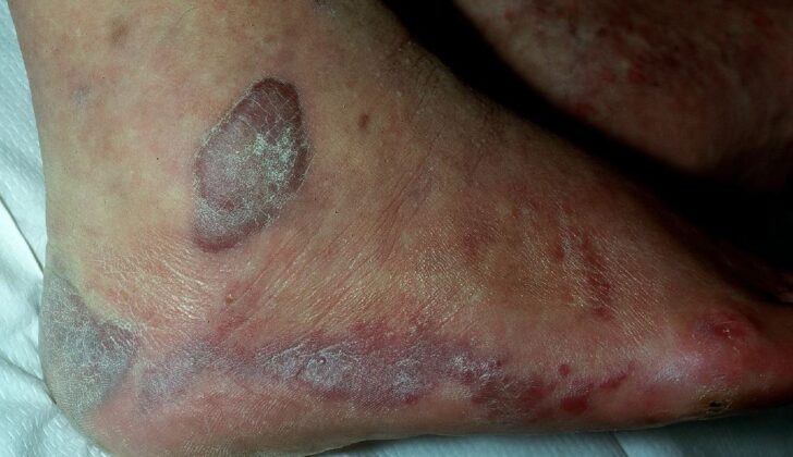
Kaposi sarcoma and cutaneous angiosarcoma are two conditions that are difficult to diagnose due to their varied appearances. Kaposi sarcoma can present itself in different ways such as spots, patches, raised bumps, scaly areas, and nodules, which are usually purple/red in color. This condition frequently shows up as purple patches on the lower legs. However, the spots can also appear on the face and neck, particularly around the nose, eyelids, ears, and in the mouth (especially on the hard roof part). Depending on the individual, these lesions can either grow quickly or slowly. It’s also important to review patient’s overall health because symptoms like unexplained fever, sweating at night, and weight loss can indicate the presence of cancer. Kaposi sarcoma associated with AIDS often appears on the head and neck, while the type common in certain geographic regions may involve neck lymph nodes and the mucosal lining of the mouth and nose.
Cutaneous angiosarcoma, on the other hand, can often appear harmless at first. That’s why it’s often called “the great mimicker.” These skin cancers may appear as violet nodules and scaling areas, bruise-like spots, and might look like patches of bruises and spots. Some patients have skin lesions that look like blood clots underneath the skin, rosacea, skin infection, and swelling caused by fluid build-up.
Testing for Cutaneous Vascular Malignancies, Angiosarcoma and Kaposi Sarcoma
A punch biopsy, which is similar to using a hole puncher on the skin, is often used to examine different skin abnormalities such as vascular lesions. This type of biopsy can help confirm whether someone has Kaposi sarcoma or cutaneous angiosarcoma, which are types of skin cancer. But, this test can sometimes miss the presence of these cancers. Therefore, multiple biopsies might be necessary.
If a skin abnormality appeared after radiation treatment, the biopsy sample should be taken from the area away from the edges to avoid simply identifying changes caused by radiation. Blood tests might also be needed, particularly in individuals with HIV. When Kaposi sarcoma is present, the CD4 count, a type of blood cell crucial for the immune system, is usually below 200 cells/mL.
In addition to biopsies and blood tests, advanced imaging methods such as positive emission topography (PET) MRI and CT scans with contrast dyes, can be used to see if larger skin abnormalities have spread into nearby structures. Angiosarcomas, another type of cancer, can show up as irregular masses on these scans.
Treatment Options for Cutaneous Vascular Malignancies, Angiosarcoma and Kaposi Sarcoma
The treatment for Kaposi sarcoma varies depending on the type. If you have Classic Kaposi sarcoma and the affected area is small and hasn’t spread, you may undergo surgery to remove it. However, if multiple areas are affected, radiation treatment is usually used. If the condition keeps coming back or gets worse, you might receive a combination of treatments such as radiation, chemotherapy, and surgery. If Kaposi sarcoma arises as a result of diseases that weaken the immune system, the first step might be to remove or change the medicines that caused it. If this doesn’t help, the next step would be radiation and chemotherapy. For people with Kaposi sarcoma related to AIDS, treatments can include radiation, freezing off the lesions (cryosurgery), injecting a drug named vincristine directly into the lesions, or taking HIV drugs in combination with the hormone interferon-alpha. In fact, if the correct HIV drugs are used, the sarcoma can sometimes vanish by itself.
Cutaneous angiosarcoma, a rare skin cancer, is typically treated with surgery and radiation. Research indicates that the results are better if both treatments are used than if only one is selected. However, surgery might not always be the best option, depending on the location, size, and visibility of the tumor. On average, patients receive high-energy radiation of a specific dose, either as a supplement to surgery or as a standalone treatment. It can be hard for doctors to guarantee that they’ve removed all of the cancer during surgery because it’s sometimes difficult to see the extent of the disease. Sometimes patients may also receive initial chemotherapy – though experiences vary across hospitals. Some hospitals didn’t see a benefit to including chemotherapy; others saw the five-year survival rate rise by over half. Nevertheless, no clear treatment guidelines exist yet about using chemotherapy due to the lack of comprehensive studies for cutaneous angiosarcoma.
What else can Cutaneous Vascular Malignancies, Angiosarcoma and Kaposi Sarcoma be?
When examining potentially dangerous blood vessel-related skin conditions on the face and neck, doctors consider conditions such as Kaposi sarcoma and cutaneous angiosarcoma. Along with these, they also consider several other conditions. These include:
- Skin T-cell lymphomas (cancers that affect a type of white blood cell)
- Bacillary angiomatosis (a rare bacterial infection)
- Pyogenic granuloma (a noncancerous skin growth)
- Malignant melanoma (a dangerous type of skin cancer)
- Squamous cell carcinoma (a common type of skin cancer)
- Basal cell carcinoma (the most common type of skin cancer)
- Merkel cell carcinoma (a rare type of skin cancer)
- Progressive pigmented purpura (a group of disorders characterized by spots and bruising on the skin)
Doctors need to carefully consider these possibilities and conduct necessary tests to provide a correct diagnosis.
Surgical Treatment of Cutaneous Vascular Malignancies, Angiosarcoma and Kaposi Sarcoma
Surgery is not usually the go-to choice for treating Kaposi sarcoma. This is because it often shows up in multiple places in the body, which means you need treatment that works throughout your system.
On the other hand, a combination of substantial surgery and radiation is the best treatment for a skin cancer called cutaneous angiosarcoma – it’s the only treatment that could possibly get rid of the cancer.[27] Doctors aim to remove the cancer with a 1-cm border of healthy tissue around it as much as possible. However, this can be tough, especially when the cancer has spread to critical structures in the head and neck. After the surgeon removes the cancer, they usually patch up the area with a graft or flap.
An interesting fact is that one institution found that when they tried to remove the cancer with a 1-cm border of healthy tissue, they were able to do so in 77% of their patients with cutaneous angiosarcoma in the head and neck.[28]
What to expect with Cutaneous Vascular Malignancies, Angiosarcoma and Kaposi Sarcoma
The outlook for patients with Kaposi sarcoma varies based on the type of the disease. For AIDS-associated Kaposi sarcoma, the outlook has considerably improved with the advent of antiretroviral therapy (ART). Patients with early-stage disease (T0) have an approximately 92% chance of living for at least five more years, and those with more advanced disease (T1) have an 83% five-year survival rate.
Classic Kaposi sarcoma generally has a good outlook due to its slow progression. In a study that tracked patients for nearly five years, it was found that only 2% died due to the widespread disease, while 24% died from other types of cancer, and 22% died from other medical conditions.
On the other hand, the outlook for patients with cutaneous angiosarcomas, a type of skin cancer, is often dire. These tumors spread very quickly, and it’s common that the cancer has already spread to other parts of the body at the time of diagnosis. Common sites for spread include the lungs, liver, lymph nodes, spleen, and brain. On average, patients live for about 4 months after the cancer spreads, and have a 10% to 35% chance of living for five more years. A study in 2011 found a 5- and 10-year survival rate of approximately 33.6% and 13.8%, respectively. Predictors of a poorer outcome include a primary tumor larger than 5 cm, the presence of additional skin lesions, and the scalp being the location of the primary tumor.
Possible Complications When Diagnosed with Cutaneous Vascular Malignancies, Angiosarcoma and Kaposi Sarcoma
Complications that arise from all types of skin-related blood vessel cancers in the head and neck area mainly involve structure invasion. This means the tumors can grow on different parts including eyelids, nostrils and the inside of the mouth. This can disrupt the normal functioning of these structures.
When it comes to angiosarcomas, more complications are observed due to the frequent spread of the cancer to vital organs via the bloodstream. The lungs are often affected. The most severe possible complication from this type of cancer is death.
Recovery from Cutaneous Vascular Malignancies, Angiosarcoma and Kaposi Sarcoma
If a skin related blood vessel tumor needs to be surgically removed, it’s important that the patient continues to meet with the surgeon who carried out the procedure for regular check-ups. Stitches on the face are usually left in place for 3 to 5 days, whereas those on the scalp are usually left in for 7 to 10 days. To take care of the wound, it’s advisable to clean it with soap and water and keep it moist using petroleum jelly or antibiotic cream.
Preventing Cutaneous Vascular Malignancies, Angiosarcoma and Kaposi Sarcoma
If someone notices a new lump or spot on their head or neck, especially if they have HIV, they should get it checked by a doctor. This is important because it allows any serious conditions to be identified and treated promptly. Additionally, those people living with HIV who don’t regularly take their prescribed medicines need to learn about Kaposi Sarcoma. This education can help them understand the risks and take better care of their health.


