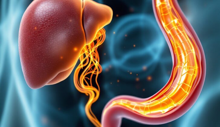
What is Hepatobiliary Tract Cancer?
Cholangiocarcinoma was initially a term used to describe a type of cancer that occurs in the bile ducts inside the liver. Today, however, the term has broadened to include cancer that appears in the bile ducts both inside and outside the liver, and in areas near the liver like the hepatobiliary tract. Additionally, since the gallbladder and a part of the small intestine called the ampulla of Vater are part of the biliary tract system, cancers found in these places are often considered cholangiocarcinoma in research and published reports.
Cholangiocarcinoma is an uncommon and oftentimes deadly form of cancer. This is mainly because it is usually found at a late stage where it is locally advanced – which means it has spread to nearby tissues or lymph nodes – and cannot be removed by surgery.
What Causes Hepatobiliary Tract Cancer?
Chronic inflammation, or long-term swelling and irritation in the body, can possibly lead to a type of cancer called biliary tract cancer. The most significant risk factor for developing a subtype of this cancer, cholangiocarcinoma, is having a specific inflammatory bowel disease known as primary sclerosing cholangitis (PSC). This condition is particularly associated with another inflammatory bowel disease, ulcerative colitis.
Additionally, some other known risk factors for cholangiocarcinoma are choledochal cysts (abnormal fluid-filled sacs in the bile ducts), hepatolithiasis (stones in the liver), and liver fluke (a type of parasite).
Intrahepatic cholangiocarcinoma, a specific type of cholangiocarcinoma that occurs within the liver, can also be associated with other long-term liver conditions. These include cirrhosis caused by hepatitis C virus (HCV), hepatitis B virus (HBV), metabolic syndrome (a combination of health problems like high blood pressure and high blood sugar), and fatty liver disease that occurs both with and without the presence of alcohol.
Thorotrast, a substance that was previously used in medical imaging, was discontinued as it was linked with a high risk of cholangiocarcinoma due to its cancer-causing potential.
A connection has also been made between cholangiocarcinoma and certain genetic conditions, like Lynch syndrome and multiple biliary papillomatosis, which causes many noncancerous tumors to form in the bile ducts.
Risk Factors and Frequency for Hepatobiliary Tract Cancer
Cholangiocarcinoma, a type of gastrointestinal cancer, makes up 3% of all such cancers. According to US Cancer Statistics, it’s estimated that there will be 52,450 new cases and 32,750 expected deaths. These cases include liver, intrahepatic bile duct, gallbladder, and other biliary cancers. All together, these make up 6% of all new cancer cases, making cholangiocarcinoma the fifth deadliest type of cancer.
There are three main types of cholangiocarcinoma, classified by where they are found in the body. Each type accounts for a different percentage of total cases:
- Intrahepatic makes up 5% to 10% of cases
- Perihilar makes up 50% to 60% of cases
- Distal biliary tract cancers make up 20% to 30% of cases
The chance of having cholangiocarcinoma increases as people get older, except for those related to a condition called primary sclerosing cholangitis, which affects younger patients more. It is also slightly more common in males, except for gallbladder cancer, which is more common in females.
Signs and Symptoms of Hepatobiliary Tract Cancer
Bile duct obstruction is often a sign of a type of cancer called extrahepatic cholangiocarcinoma. The symptoms can include yellowing of the skin or eyes (jaundice), pain in the upper right side of your abdomen, stools that are pale in color, dark-colored urine, and itching skin. Less specific symptoms of this advanced cancer can include fever, night sweats, weight loss, and a general feeling of discomfort or illness. It’s also possible for a patient to have a rare condition called acute cholangitis.
Another type of cancer called intrahepatic cholangiocarcinoma may not bring about noticeable symptoms initially. Often, this type is discovered accidentally during tests for liver disease or through abnormal laboratory findings.
In a physical check-up, a doctor might notice signs like jaundice, pain in the upper right side of the abdomen, an enlarged liver, or a condition called Courvoisier sign, which is a hard, non-sensitive lump in the gallbladder area. This sign is more likely if the bile duct obstruction has been caused by a slowly worsening cancer rather than a non-cancerous, sporadic obstruction.
- Jaundice or yellow skin and eyes
- Pain in the upper right side of the abdomen
- Pale colored stools
- Dark-colored urine
- Itchy skin
- Fever and night sweats
- Weight loss and general discomfort
- Enlarged liver
- Hard, non-sensitive lump in the gallbladder area (Courvoisier sign)
Testing for Hepatobiliary Tract Cancer
If you experience symptoms like yellowing skin (which doctors refer to as jaundice) or pain in the upper right section of your abdomen, your doctor will likely order some tests to investigate. These tests include complete blood count (measures different types of cells in your blood), basic chemistry panel (measures different minerals and proteins in your blood), and liver function tests. These tests might show that there is a blockage in your bile, an important substance your liver makes to help digest food. This blockage can cause damage to your liver.
To get a better look at your liver and any possible blockages, your doctor will likely order an ultrasound or a CT scan, which are types of imaging that can provide detailed pictures of your body’s internal structures. However, these imaging techniques have some limitations and might not clearly distinguish between harmless and harmful lesions (abnormal areas) within the liver. In such cases, a form of imaging called dynamic magnetic resonance (MR) and MR cholangiopancreatography can provide more accurate information, including distinguishing a form of liver cancer called intrahepatic cholangiocarcinoma from other types of liver cancer.
Sometimes, it can be challenging to get a small sample of the abnormal tissue for more detailed examination and diagnosis. For blockages that are outside of the liver (extrahepatic obstruction), doctors prefer using techniques such as endoscopic ultrasound or endoscopic retrograde cholangiopancreatography as these can provide a direct view of the bile duct, allow for sampling of the tissue, and possibly even place a stent (tube) to open up the blockage.
To distinguish suspicious looking areas within the liver, doctors frequently collect certain tumor markers from your blood. These include the carcinoembryonic antigen, alpha-fetoprotein, and carbohydrate antigen 19-9. If these markers are found in larger amounts in your blood, they can support the diagnosis of certain types of liver cancer.
In order to understand the extent of the disease, doctors might do a CT scan of the chest, abdomen, and pelvis, if not already done. If there is no evidence of the disease having spread to other parts of the body and the doctor is considering surgery as a possible treatment, a scan called positron emission tomography (PET)/CT might be ordered to look for hidden areas of disease that can prevent a major surgery.
Staging, or determining the extent of the disease, in cholangiocarcinoma, a form of bile duct cancer, is separated based on the location of the cancer. The 5-year survival rate is generally higher for those patients who do not have multiple tumors, blood vessel invasion, or spread of the disease to lymph nodes on the surgical specimen evaluation.
Treatment Options for Hepatobiliary Tract Cancer
Treatments for cholangiocarcinoma, or bile duct cancer, aren’t entirely standard and often depend on the individual case. In very specific situations, patients might get pre-surgical treatments, like chemotherapy or radiation-therapy to shrink the tumor. However, these are uncommon and best utilized in a clinical trial setting, an experiment for testing a new treatment’s effectiveness.
The only treatment known to cure this cancer is surgery, and the kind of operation depends on the cancer’s location. If it’s near the tail end of the bile duct, a Whipple procedure might be carried out, which involves removing the bile duct’s part, the gallbladder, and parts of the pancreas and the small intestine. When the cancer is inside the liver, the surgeon might have to remove part of the organ, which is known as a hepatectomy. The surgical procedures for cancer at the junction of the bile duct and the liver differ according to the Bismuth-Corlette classification, which categorizes the tumors based on their location and spread. Broadly, the interventions range from removal of the bile ducts to removal of part of the liver along with adjacent lymph nodes.
Post-surgery, patients should receive additional therapy 8 to 12 weeks after their operation. This adjuvant therapy — extra treatment to lower the risk of the cancer returning — is particularly significant when tests show cancer cells in the blood vessels, lymph nodes, or at the edge of the area from where the tumor was removed. The specific form of adjuvant therapy — chemotherapy, radiation, or a combination of both — can vary according to the patient’s condition and is often decided in the context of a clinical trial.
For different types of cholangiocarcinoma, the National Comprehensive Cancer Network (NCCN) consensus-based guidelines recommend various treatment plans. However, these recommendations have to be considered against vital factors including the exact location of the cancer, its spread, and whether the cancer was completely removed during surgery.
No single therapy can cure patients whose cholangiocarcinoma cannot be surgically removed or has spread beyond the bile ducts. For these advanced cases, the focus is typically on improving the quality of life or prolonging survival time. Participating in clinical trials to try new treatments is always a relevant option. Patients who are generally in good health might also be recommended a combination of chemotherapy drugs.
After treatment, the NCCN suggests regular follow-up tests. These may include imaging scans every 6 months for the first 2 years and then yearly until the 5-year mark. Doctors might also monitor patients’ liver function tests and specific tumor marker levels (CEA and CA19-9) at regular intervals for 5 years.
What else can Hepatobiliary Tract Cancer be?
When dealing with certain medical issues, doctors may have to consider several possible conditions. These could include things like:
- Primary sclerosing cholangitis
- Secondary sclerosing cholangitis
- Recurrent pyogenic cholangitis
- AIDS-related cholangiopathy
- Autoimmune pancreatitis-cholangitis syndrome
- Hepatobiliary inflammatory pseudotumor
- Mirizzi Syndrome
- Xanthogranulomatous cholecystitis or cholangitis
- Biliary sarcoidosis
- Chemotherapy-induced biliary sclerosis
- Intrabiliary metastasis
Each of these conditions have specific symptoms and characteristics, and the doctor will have to consider each one in order to make the right diagnosis.
Surgical Treatment of Hepatobiliary Tract Cancer
Surgery is the only treatment that can completely cure certain patients. However, the type of surgery performed depends on where the problem area is located in the body. If the issue is found near the end of the pancreas, a procedure known as a pancreaticoduodenectomy, or the Whipple procedure, is typically used. If the problem is inside the liver, a surgery called hepatectomy, which is the removal of part or all of the liver, might be needed. The removal of lymph nodes, a procedure known as lymphadenectomy, is rarely done. For issues located at the junction of the bile duct and the liver, also known as hilar lesions, the type of surgery performed can vary based on the Bismuth-Corlette anatomical classification, which is a system doctors use to describe the extent and location of the problem.
What to expect with Hepatobiliary Tract Cancer
The likelihood of survival after being diagnosed with a tumor highly depends on two factors: the located area of the tumor, as well as the extent or stage of the tumor.
If the tumor is within the liver’s bile duct (intrahepatic), the survival rate over five years varies:
- For localized or early stage (stage I) tumors – the survival rate is 15%
- When the tumor starts to spread regionally (stage II and III), the survival rate drops to 6%
- If the tumor has traveled to distant parts of the body (stage IV), the survival rate further declines to 2%
In contrast, if the tumor lies in the bile duct outside the liver (extrahepatic), the five-year survival outlook is relatively more positive:
- For localized or early stage (stage I) tumors – the survival rate is 30%
- When the tumor starts to spread regionally (stage II and III), the survival rate stands at 24%
- Just like intrahepatic tumors, if the tumor has spread to distant parts of the body (stage IV), the survival rate is expected to be about 2%
This statistics show that the early detection of these types of tumors can greatly improve the chances of survival.
Possible Complications When Diagnosed with Hepatobiliary Tract Cancer
Cancer can cause a blockage in the flow of bile, leading to various infections. More severe complications like cholangitis, a condition where the bile duct is infected, or sepsis and septic shock, a wide-spreading and severe infection, can even lead to death.
Complications may also occur due to the treatments for cancer, such as surgery, chemotherapy, and radiotherapy.
Common Side Effects:
- Infections due to bile blockage
- Cholangitis
- Sepsis and septic shock
- Potential death
- Complications from cancer treatments like surgery, chemotherapy, and radiotherapy
Preventing Hepatobiliary Tract Cancer
Biliary cancers, which affect the part of the body that creates and moves bile, are often linked with a condition called cirrhosis. Cirrhosis involves damage and scarring of the liver, often caused by factors like excessive alcohol intake, hepatitis B or C infections, among others. As a way to prevent cirrhosis, it’s recommended to avoid excessive alcohol consumption, injecting drugs, and to maintain safe sexual practices.
If you’re diagnosed with biliary cancer, you should know that an array of specialized experts forms your healthcare team to manage and monitor your case. It’s very crucial for improving your health outcome and survival rate that you maintain a regular schedule of check-ups, chemotherapy, and radiotherapy sessions.


