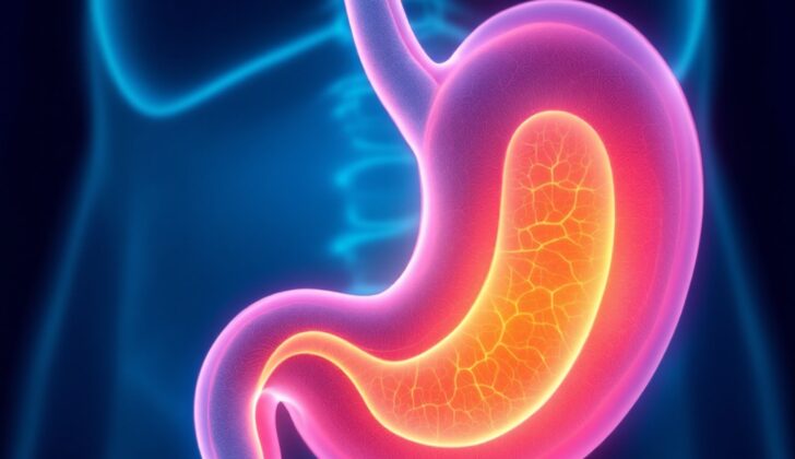
What is Linitis Plastica?
The term “Linitis plastica” (LP) has been used in medicine since 1779 – introduced by Lieudaut – to describe a hard and deadly type of stomach tissue. Later on, in 1859, Dr. William Brinton further explained this as a harmless condition of the stomach that brings peculiar changes, such as a noticeably thick stomach wall and beneath-the-surface swelling made up mostly of connective and muscle tissues. Microscopically, the condition showed bands that looked like linen, hence the name ‘linitis’. However, because the tissue showed few malignant (cancerous) cells, there was a lot of debate about whether it was a harmless or malignant disorder. The issue was eventually resolved in 1953 by Dr. Arthur Stout, who confirmed LP as cancerous. He described that it creates a lot of scar-like tissue that resemble fibres.
It is important to note that LP is a variant of stomach cancer. Although sometimes referred to as Borrmann type 4, especially in the Asian population, this term isn’t entirely accurate. Other terms that are sometimes used include Schirrous carcinoma, Lauren carcinoma, or signet cell carcinoma. These different terms can be confusing, but they all refer to the same underlying condition.
What Causes Linitis Plastica?
The exact cause of a condition called linitis plastica isn’t fully understood yet, and scientists are still studying it. When it comes to stomach cancer, linitis plastica is typically more common among American and Asian populations, particularly in Japan, Korea, and China. This higher occurrence might be connected to their typical eating habits, like eating a diet low in fiber, or to specific aspects of their genetic background.
It’s important to note that linitis plastica isn’t related to infections caused by a bacterium called H.pylori, or to a condition called chronic active gastritis, which is long-term inflammation of the stomach lining. However, certain genes, like the CDH1 gene and the HER2 gene, do play a significant role in this condition.
Risk Factors and Frequency for Linitis Plastica
Linitis plastica is a type of stomach cancer that represents 7 to 10% of all primary stomach cancer cases. When looking at all types of stomach cancers, it accounts for between 3 to 19% of cases. This form of cancer is more common in younger individuals and females.
Signs and Symptoms of Linitis Plastica
Linitis plastica is a disease that often does not show any symptoms until it has progressed significantly. The stomach typically doesn’t pick up on the problem until the capacity of the stomach is severely reduced. When symptoms do show, they often include indigestion, vomiting linked to backflow of food from the esophagus, and difficulty swallowing due to thickening of the stomach wall, compromising its ability to stretch.
- Indigestion
- Vomiting linked to backflow of food from the esophagus
- Difficulty swallowing due to thickening of the stomach wall
- Stomach pain
- Feeling a lump in the upper central region of the abdomen
- Unintentional weight loss
Testing for Linitis Plastica
Diagnosing a condition called linitis plastica can be challenging. This disease affects a deeper layer of the stomach wall (the submucosa), leaving the surface layer (mucosa) relatively untouched. This means that during a type of diagnostic procedure called an upper gastrointestinal endoscopy, where small samples of tissue (biopsies) are taken, those samples might not show signs of the disease, leading to inconclusive results.
That’s where a diagnostic tool called an endoscopic ultrasound (EUS) becomes very important. The EUS can show if the deeper layer of the stomach wall has thickened or is infiltrated by the disease. It also provides information about the size and structure of nearby lymph nodes, which are part of your immune system, and if the disease has spread to other organs.
While this method is helpful, it is not 100% accurate. The EUS can accurately diagnose the extent (or “T stage”) of linitis plastica between 64% to 92% of the time and can accurately identify if the disease has spread to lymph nodes (or “N stage”) between 50% to 90% of the time.
Doctors can use various types of biopsies during these diagnostic procedures. In a regular upper endoscopy, brush cytology (gently brushing the surface to collect cells) and superficial biopsy (taking a small sample of the surface layer of tissue) may be used. During an EUS, a fine-needle biopsy (taking a small sample of tissue using a thin needle) may be performed, especially in the case of enlarged lymph nodes.
Another important tool for diagnosing linitis plastica is a high-resolution computed tomography (CT) scan with contrast dye. This is the best initial test for the condition because it can detect thickening in the deeper layers of the stomach wall, as well as signs that the disease has spread to the lining of the abdomen (peritoneum), liver, and lymph nodes. Taking this scan removes any doubts and helps to rule out other possible diseases. The scan can also provide early detection of linitis plastica, especially since up to 30% of biopsies may show negative results in the early stages of the disease due to the unaffected superficial layer.
If a CT scan cannot be performed for whatever reason, an MRI (Magnetic Resonance Imaging) scan can be used instead. This is about as effective as a CT scan in diagnosing linitis plastica.
Another way to diagnose linitis plastica, especially when it has spread to the lining of the abdomen, is peritoneal lavage. This is a procedure where fluid is injected and then collected from the abdomen using a tube inserted during a small procedure called a laparoscopy.
There are also certain proteins called CEA and CA19-9 that may be higher in people with linitis plastica and could help in predicting the course of the disease. However, these proteins are not very reliable for diagnosing linitis plastica through a blood test, although they may be helpful if tested in fluid collected from peritoneal lavage.
Treatment Options for Linitis Plastica
Cancer specialists often see palliative systemic therapy as the primary strategy. They think that, in most cases, the disease has likely spread by the time it’s detected and that surgery only provides relief for about 20% of patients. However, there are no other alternatives, and surgery is still viewed as the only potential cure for the disease, as it can help to extend survival.
For some specific cases of localized linitis plastica—a rare type of stomach cancer—a combination of surgery to remove the stomach (gastrectomy) and systemic therapy, such as radiation or chemotherapy, may lead to promising outcomes. Patients treated this way tend to live longer.
However, completely removing the stomach (total gastrectomy) is seen as the only potential cure, but not everyone can go through this procedure. This is because often, the disease is at an advanced stage when first detected. Also, because it’s a major surgery, there’s a high risk of complications and even death, which can impact how long patients live after the procedure. Plus, patients may suffer from poor nutrition after the surgery, mainly in the form of anemia, which makes recovery more challenging.
Treatment methods can vary between western and eastern countries. In western countries, physicians typically recommend removing the entire stomach and nearby lymph nodes (a process known as a D2 lymph node dissection), along with chemotherapy either before or after the surgery. However, in eastern countries, the standard treatment is chemotherapy after total gastrectomy. In Japan, a specific combination of chemotherapy drugs is usually prescribed after surgery.
Another approach to treatment is personalized medicine, where therapy is tailored to each patient, based on the specific characteristics of their disease and its progression. This could involve a combination of different chemotherapy drugs or sequential treatment courses. This strategy has shown promising results in certain advanced cases.
What else can Linitis Plastica be?
When trying to diagnose linitis plastica, a rare type of stomach cancer, doctors should also consider other conditions that might cause similar symptoms. These include:
- Atrophic gastritis, which can cause indigestion and poor nutrition due to damage and loss of the stomach lining.
- Hypertrophic H.pylori chronic gastritis, which can cause the stomach lining to thicken in a way that may look like linitis plastica. However, the stomach will still be able to stretch as usual.
- Corrosive gastritis, which will often come with damage to the esophagus and a history of consuming a harmful substance.
- Different forms of widespread inflammation like lymphoma, GIST, non-Hodgkins lymphoma, adenocarcinoma, Bormann type 3 and 4. Differentiating these conditions will need examination of the affected tissue.
- “Watermelon stomach,” or Gastric Antral Vascular Ectasia, which can be linked with high blood pressure in the veins of the liver and a loss of stomach flexibility due to blood build up. Symptoms of this condition may also include liver failure.
In addition, a history of partial gastrectomy, a surgery that removes part of the stomach, can also lead to loss of stomach volume and flexibility.
Surgical Treatment of Linitis Plastica
Patients who undergo surgery for the condition live much longer, around 16.7 months, on average, compared to patients who only receive medical treatment, who on average live around 3.6 months. These survival rates are similar to rates reported in patients with a kind of cancer which is not LP adenocarcinoma.
During surgery, the doctor makes sure to remove all of the tumor by leaving a clear, cancer-free margin around it. The surgeon also performs a real-time check known as a frozen section examination to make sure the edges are indeed free from cancer. This helps cure the condition and lessens the chance of the disease coming back.
What to expect with Linitis Plastica
Linitis plastica is a condition that often comes with a serious outlook, mainly because it’s usually discovered at an advanced stage, when it has already spread to other parts of the body. This late detection happens because there are typically no clear signs or symptoms in the early stages of the disease. This is because the lining of the stomach (gastric mucosa) isn’t often affected, so patients don’t usually feel sick until the disease is quite advanced.
If surgical treatment is chosen, the average survival time varies from 5 to 17 months. The chance of surviving for five years after the diagnosis is quite low, ranging from 3 to 10%.
Possible Complications When Diagnosed with Linitis Plastica
Complications of this condition arise from a hardened, “leathery” stomach, which leads to difficulties in food digestion and absorption. This can result in poor nutrition and severe wasting of the body. As the disease worsens, a surgical bypass might be necessary to relieve the lack of movements in the rigid stomach walls and to address any blockages. The disease could spread to different organs, such as the liver or the peritoneum (the lining of the abdomen), leading to fluid in the abdomen (ascites), liver cell failure, or blockage of bile ducts.
Common Complications:
- Difficulty in food digestion and absorption
- Poor nutrition
- Severe body wasting
- Need for surgical bypass to relieve stomach blockages
- Spread of disease to other organs like the liver or peritoneum
- Fluid build-up in the abdomen (ascites)
- Liver cell failure
- Blockage of bile ducts (biliary obstruction)
Preventing Linitis Plastica
It is very helpful for patients with linitis plastica, a type of advanced stomach cancer, to understand their condition, as it usually diagnosed at a late stage when a cure is not possible. Understanding the condition and the various treatment options is extremely important for the patient. One of these options may include personalized medicine, which is a treatment plan tailored to the individual patient’s needs and is often used in advanced cases.
It’s also important for the patient to understand their nutritional needs. Depending on the patient’s condition, they might need to eat and drink regularly, or they might need to receive nutrients in a different way, such as through an intravenous (IV) drip. In some situations, surgery may be necessary. All these elements should be discussed with the patient so they can play an active role in their care and to ensure the best possible outcome for them.
Genetic testing could also be beneficial because it can help understand the risk of the disease for the patient’s family members. If there is a high risk, preventative surgery might be considered. Therefore, the patient needs to be well-informed and understanding in order to discuss these possibilities with their family.


