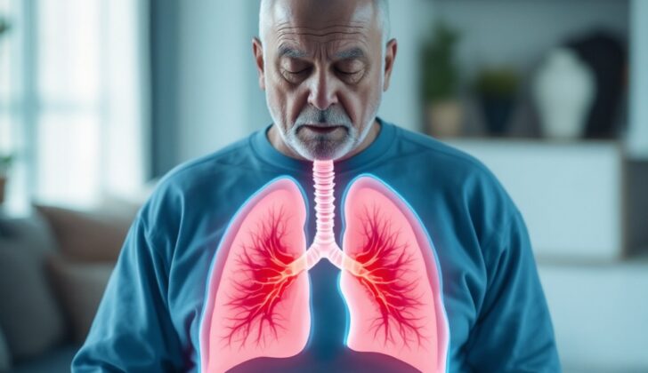
What is Lung Metastasis?
Metastasis refers to the spreading of tumor or cancer cells from the original site where they developed to nearby areas and also to far-off parts of the body. It’s a significant factor that contributes to illness and death, as its occurrence often signals that the original tumor is highly aggressive.
The process of metastasis, when it occurs in the lungs, involves multiple steps. The tumor cells first separate from the original tumor site, then invade blood or lymph vessels. From there, the cells force their way into their new location, where they set up a supportive environment for their growth and the distribution of blood. Certain types of cancer commonly move to the lung tissues, such as breast, lung, colon, uterine, and head/neck cancers.
Some cancers also spread to the internal bronchial tree of the lungs, including colon, kidney, lung cancers, and lymphomas. Others, such as bone and testicular cancers, can also end up in the lungs. There are even some rare forms of cancer that can do the same, including adrenal, thyroid, choriocarcinoma, and hypernephroma cancers.
There are instances when the original tumor site cannot be identified; these are referred to as cancers of unknown primary (CUP). Most CUPs are a type of cancer called adenocarcinomas. Other types, like squamous cell and undifferentiated carcinomas, are reported less frequently.
What Causes Lung Metastasis?
Tumors have different characteristics such as their makeup, genes, and the ways they develop, that determine where in the body they will spread. Tumors can move to the lungs through the blood or lymph system, or by directly invading the area.
1. Spread through the blood: This happens when tumors are in parts of the body where the veins carry blood back to the lungs. This includes tumors in the head and neck, thyroid gland, adrenal glands, kidneys, testicles, skin (melanoma), and bone (osteosarcoma).
2. Spread through the lymph system: This can happen in two ways. Tumors can move forward through the thin muscle separating the chest and abdomen (diaphragm) or the surface of the lungs (pleural), or backward from lymph nodes in the chest region. Examples include cancers of the lung, stomach, breast, pancreas, uterus, rectum, and prostate.
3. Direct spread to the lung coverings: This happens when tumors spread through the blood and reach the lung coverings (pleura), through the lymph system, or from already established liver tumors. This includes cancers of the lung, breast, pancreas, and stomach.
More than one lung bumpy growth, or nodules, found along with the original tumor is usually a sign of lung cancer that has spread from another area. However, single nodules found when primary tumors are present could be a sign of either lung cancer that has spread (specifically for melanoma, a type of skin cancer, and sarcoma, a type of tissue cancer) or new separate lung cancer.
Risk Factors and Frequency for Lung Metastasis
The lungs are usually the second place cancers from other parts of the body are likely to spread, with 20% to 54% of all cancers eventually spreading to the lungs. People with cancer that has spread to the lungs generally have different treatment options and typically have a worse outlook than those with the original, or primary, cancer. This spread, known as distant metastasis, plays a key role in determining the stage of the cancer. For instance, the 5-year survival rate for breast cancer drops from 96% to just 21% if it spreads distantly. Patients with colorectal cancer that has spread to the lungs or liver have a survival rate of less than 10% after 5 years, compared to 91% for those without such spread. This underlines the challenges of current treatment options when cancer has spread. Roughly 1,500 people die everyday from cancers that have spread.
A study of 228 patients with lung nodules showed that the average age was around 62 years old, and roughly 53.5% were male, 46.5% female. The cancer had originally started in the following areas:
- Colorectal: 25.8%
- Head and neck: 19.4%
- Urologic (kidney, ureter, prostate, testes): 14.7%
- Gastrointestinal non-colorectal cancer: 10.9%
- Breast cancer: 10.5%
- Melanoma: 6.5%
- Gynecologic cancer (ovarian, endometrial, cervical): 6.1%
- Other primary sites (sarcoma, thyroid, squamous cell): 6.1%
About a quarter of patients had nodules in other parts of the lungs as well. Close to half of the patients had one nodule, while a slight majority had multiple nodules. The nodules were mostly between 20 and 30 mm (50%), 10 to 20 mm (28.5%), or less than 10 mm (21.5%). In about 88.5% of the cases, the nodules were hollow (cavitary) or had lost their structure (necrotic).
After testing a tissue sample (biopsy), it was found that 64% of the patients had cancer that had spread, while around 26.3% had a new, separate lung cancer. Only about 9.6% had no cancer. It was also found that having multiple lung nodules or cavitary nodules were associated with a higher possibility of cancers turning up in other parts of the body. Hence, it cannot be assumed that lung nodules are cancerous without a biopsy. In one case, a single patient had both a new lung cancer and breast cancer that had spread to the lung.
Signs and Symptoms of Lung Metastasis
Patients with lung metastasis, or the spread of cancer to the lungs, may have previously been diagnosed with a primary tumor somewhere else in their body, or they may discover their condition when the lung metastasis is found. This can occur as a single cancer spread (solitary metastasis), or there can be several (multiple metastases). Some individuals may not exhibit any symptoms (asymptomatic), while others will experience a range of symptoms.
For some people, the presence of lung metastasis is surprising, as they may not even realize they have it until lung nodules are discovered during medical examinations for unrelated issues. Other individuals may begin to experience general health symptoms, such as fatigue, nausea, loss of appetite, and weight loss.
Specific symptoms related to lung metastasis can include:
- Pain or fluid in the lung lining (pleurisy/pleural effusion)
- A cough, which may or may not produce mucus
- Difficulty breathing (dyspnea)
- Coughing up blood (hemoptysis)
- Metastasis to the scalp
- Disturbances in the balance of fluids, salts, and minerals in the body (electrolyte disturbances)
- Pancoast tumor, a type of lung cancer located in the upper part of the lung
- Superior vena cava syndrome, a condition in which the large vein carrying blood from the head, arms, and upper body back to the heart is compressed or obstructed.
In a survey about common symptoms associated with metastatic cancers, vomiting was reported in 40 cases (25%), low back pain in 38 cases (24%), loss of appetite in 32 cases (20%), and shoulder pain in 27 cases (17%).
Upon examination, the doctor may find that the patient’s lung sounds are normal, but there could also be signs of a problem. For example, there may be a wheezing sound if there is a bronchogenic mass, crackling noises if fluid or post-obstructive pneumonia is filling the alveoli (air sacs in the lungs), or decreased breath sounds if there is pleural effusion (fluid around the lungs) or atelectasis (collapsed lung).
Further physical indicators of lung metastasis can include:
- Abnormal enlarging and rounding of the fingertips (digital clubbing)
- Weight loss
- Enlarged lymph nodes (lymphadenopathy)
- Signs of Pancoast tumor (including Horner syndrome, a condition that affects the eyes and face)
- Signs of Superior vena cava syndrome
Testing for Lung Metastasis
If your doctor suspects that your cancer may have spread to other parts of your body, they might carry out specific blood tests to look for signs of this. These tests can pick up on things like anemia (low levels of blood cells), hypercalcemia (high levels of calcium in your blood) and SIADH (a condition that affects your body’s water balance). However, these tests alone can’t provide a definitive diagnosis of metastatic disease (cancer that has spread).
To get a clearer picture, your doctor may also order a chest X-ray, particularly if you’re showing symptoms or if you already have a known primary tumor (the original site of the cancer). While X-rays are cost-effective and easy to perform, they do have limitations. For example, they might miss small areas of cancer spread or a dispersed pattern of tiny cancer spots. Some studies suggest that using X-rays with high kilovolts radiation can help to detect nodules as small as 5 to 10 mm.
Another imaging option is a computed tomography (CT) scan of the chest, which can pick up more detail than a standard X-ray. Techniques called helical or multi-plane projection or maximum intensity projection can make the CT scan even more sensitive. Up to 97% of nodules, including very tiny ones as small as 3 mm, can be detected using this method. However, the usefulness of CT scans can be hampered by false negatives, which can occur due to inconsistent breathing during the scan.
Positron Emission Tomography (PET) with a substance called fluorodeoxyglucose (FDG) is another method used to find areas of cancer spread in the body. Combining this with a CT scan (PET-CT) can help to pinpoint the exact location of the cancer.
Magnetic resonance imaging (MRI) has not shown to be better than a CT scan for detecting lung metastases (cancer spread to the lungs). However, it is useful for showing if the tumor has spread into the great vessels (main blood vessels), heart chambers, chest wall, and spine. It can also help to rule out cancer spread to the liver. A procedure where a flexible tube is inserted into your airways (flexibltracheobronchoscopy with endobronchial ultrasound) is often part of the pre-surgery tests. This allows doctors to examine the lining of the airways and confirm the type of cancer present. It also helps to check the state of the lymph nodes (small glands that are part of the immune system) around the airway and in the mediastinum (the area between the lungs).
Certain image features on a CT scan can suggest different types of tumors. For example, the way cancer spots are spread throughout the lungs can indicate that the original cancer was a thyroid cancer. On the other hand, large single cancer spots could suggest certain other types of cancers. The appearance and location of metastasis seen in imaging studies can sometimes help to determine the path that the tumor cells took to get there.
For example, the spread of cancer cells through the blood typically results in cancer spots appearing on the base and outer parts of the lungs. On a chest X-ray, signs of cancer spread through the lymph system might appear as changes to the pattern of the lung markings or thickened lines (interlobular septa). High-resolution CT scans can be particularly good at picking up these patterns.
Cancer spread to the pleura (lining of the lungs) can show up as nodules or plaque-like formations on X-rays and CT scans. Almost half of all cancer patients with this kind of spread will have a buildup of fluid in the pleural space, which is known as a malignant pleural effusion. This is most common in people with lung, breast, ovary, or lymphoma primary tumors.
Treatment Options for Lung Metastasis
When it comes to treating lung tumors that have spread from another part of the body, different options are available. These options include surgery, chemotherapy, and radiation. However, it’s important to note that there are no definitive studies proving which of these treatments might increase survival rates. There are also no proven protocols specifying whether surgery, radiation, or chemotherapy (or some combination of these) is the best way to manage tumors that have spread to the lungs.
Chemotherapy
Chemotherapy is a type of drug treatment that helps kill cancer cells, but it usually can’t fully cure lung tumors, except for a few types of cancer. For instance, a chemotherapy drug named cisplatin is often very effective in treating testicular cancer; it has a high success rate and can result in long-term recovery. Chemotherapy also plays a crucial part in treating bone cancers known as osteogenic sarcomas.
Before surgery, doctors often use chemotherapy to reduce the size of the tumor and control the spread of the disease in the body – this is known as neoadjuvant chemotherapy. After the surgery, further chemotherapy (known as adjuvant chemotherapy) may be used to kill any remaining cancer cells. A study of people who had chemotherapy before and after surgery showed that slightly over half were free of the disease two years later.
Immunotherapy
Immunotherapy is a newer type of treatment that stimulates the body’s immune system to fight cancer. Some types of cancer, like skin melanoma and kidney cancer, respond well to this treatment. Several studies have shown that patients with melanoma who had surgery followed by vaccine immunotherapy had better survival rates than those who didn’t have surgery. Immunotherapy often has fewer side effects than traditional chemotherapy and may provide long-term benefits.
Certain chemicals naturally produced by the body, such as tumor necrosis factor (TNF)-alpha, interferon (IFN)-gamma, and interleukin (IL)-2, have been shown to help treat various tumors, but they can lead to severe side effects, which require reduction in the dosage or cessation of treatment.
Radiation
Radiation therapy uses high-energy beams to kill cancer cells. However, this treatment doesn’t significantly improve survival rates in people with tumors that have spread to the lungs, except in patients with lymphomas. One key obstacle is that the high doses of radiation needed to control the tumor may damage the surrounding healthy lung tissue. To mitigate this problem, doctors sometimes use intratumoral radiation, inserting radioisotopes directly into the tumor. This method can be beneficial when other treatments are not suitable.
The main role of radiation therapy in treating lung metastases is to relieve pain and discomfort from metastases that invade the chest wall. When combined with other treatments, radiation therapy can improve outcomes and decrease the chances of recurrent cancer. Certain types of cancer, like Ewing’s sarcoma, can respond well to a combination of traditional chemotherapy and whole-lung radiation.
What else can Lung Metastasis be?
When a doctor is trying to diagnose a condition related to lung metastasis, they consider a number of possibilities:
- A tumor that started in the lung
- Pneumonia
- Fungal infection/mycetoma
- Wide-spread tuberculosis in the lungs (known as miliary)
- Hamartoma (a benign tumor-like growth)
- Adenomatous hyperplasia (a benign enlargement of lymph nodes)
- Amyloidosis (a build-up of abnormal proteins)
- Solitary fibrous tumor (a rare kind of tumor)
- Melanoma (a new cancer that starts in the skin cells)
- Anthracosis (a lung disease caused by inhaling coal dust)
- Scar tissue from previous injury or surgery
By carefully examining these possibilities, doctors can properly diagnose what’s causing a patient’s illness.
Surgical Treatment of Lung Metastasis
In the field of cancer treatment, a medical procedure called surgery can be useful if cancer has spread only to the lungs. However, since it’s difficult to predict a person’s survival without performing the operation, and no comprehensive study has been done to test the usefulness of surgery for this situation, the decision to opt for surgery must be made individually for each patient.
To decide if a patient should have surgery to remove lung growths, doctors consider things like:
- Whether the operation is technically possible
- Whether the patient’s general health allows for surgery
- If the original cancer is under control
- If the patient has no cancer spread to other parts of the body outside the lungs.
Those who are likely to have successful outcomes after the surgical removal of lung growths tend to share some features. These include:
- It’s been a long time since the original tumor was treated and the spread to the lungs was discovered.
- No cancer cells are found in the lymph nodes in the chest – these nodes are small, bean-shaped structures that help with the body’s immune response.
- A small number of growths exist in the lungs.
The most common type of surgery done is one that spares as much lung tissue as possible, but occasionally, sections of the lung, like the lobe or segment, may need to be removed. If this isn’t possible due to multiple growths or one that is in the central part of the lung, a laser or procedure to remove the entire lung may be needed. The role of minimally invasive surgery, known as VATS, in achieving the same treatment results as traditional “open” surgery is still being studied.
In one research, cancer growths that weren’t detected before surgery were discovered in 20% of patients through feeling the lung tissue during the operation. Hence, less invasive surgical procedures are not generally suggested for curative intent as they do not allow touching of the lungs. The 5-year survival rates, meaning the percentage of people who live at least five years after the surgery, largely vary depending on the type of original cancer they have. For example, for kidney cancer, it is between 35.5% and 47%, for colorectal cancer – between 39.1% and 67.8%, for soft-tissue cancer – between 29% and 52%, for bone cancer – between 38% and 49.7%, and for non-seminomatous germ cell tumors (cancers that develop from reproductive cells) – between 79% and 94%.
Patients who have the best outlook are those who had all of the cancer (known as R0) removed and went more than three years without any sign of disease after their original tumor was treated. In one study that showed a significantly better five-year survival after complete removal (36%) compared to incomplete removal (13%), it revealed how surgery could potentially improve survival, even though the study didn’t include a group of patients who didn’t have surgery for comparison. Furthermore, if a person has only one new cancer growth in the lungs, a further investigation is needed to decide if additional surgery is necessary. The chances are better if more time has passed between the first surgery for lung metastases and the appearance of the new growth in the lungs. When re-surgery was necessary due to new growth, survival times showed differences depending on the number of re-surgeries performed: more than 60 months after one re-surgery, 34.7 months after two re-surgeries, and 45.6 months after three or more re-surgeries. For patients who weren’t able to undergo surgery, the average survival was 8 months.
When it comes to specific types of cancer, the following considerations are made:
- Colorectal cancer: Only 1% to 2% of patients have surgery to remove growths in the lungs. Stage IV cancers usually survive for 24 months, and with surgery, a 68% can make it to 5 years. If liver growths are also present, the 5-year survival goes down to 42% after both liver and lung growths are removed.
- Kidney cancer (renal cell carcinoma): Chemotherapy is crucial, but curative surgery can still be considered, especially if there is no spread to the chest lymph nodes; the presence of these nodes can reduce survival after surgery from 64-92 months to 26-29 months.
- Breast cancer: The average survival time for patients with lung metastasis is 21 months, and 15.5% of patients lived for more than 3 years. Lung metastasis is uncommon, and studies show survival rate between 40% and 50% using all available systemic treatment options; compared to those who didn’t get surgery, the survival rate was 32%. Single new growths during treatment should be removed surgically, especially if no metastases are present in other parts of the body outside the chest.
- Head and neck cancers: Survivals of 20% to 59% have been reported after surgery. Since lung cancer often co-occurs with head/neck cancer, it’s difficult to distinguish between spread from the original tumor and a new lung cancer, even after taking a small sample of the tissue for examination.
- Melanoma: Even though 70% of melanomas spread to other areas, only 10% involve the lungs. After surgery, 5-year survival varies from 21% to 35%.
- Non-seminomatous germ cell tumors: Surgery is needed to remove all residual tumors after chemotherapy, and having normal levels of biomarkers (substances that indicate cancer is present) after treatment doesn’t mean the remaining tumor should not be removed. Surgery is also needed if the disease persists or returns after chemotherapy treatment or if the response to treatment was partial.
- Soft tissue sarcoma: These are usually found as growths in the lungs during the course of the disease. As they only slightly respond to chemotherapy, surgical removal is preferred. The 5-year survival after surgery ranges from 29% to 52%.
- Osteosarcoma (bone cancer): Despite using a combination of chemotherapy, surgery, and radiotherapy, the 5-year survival varies from 40% to 20%. The outlook is poor when bone cancer initially spreads. When the disease is found at the same time as the treatment, complete removal after chemo and surgical removal of the original tumor should be the goal.
- Hepatocellular carcinoma (liver cancer): In one case, successful treatment of multiple lung metastases after liver resection for liver cancer has been reported with combined chemotherapy.
- Chondrosarcoma (cancer of cartilage cells): Metastasectomy and radiofrequency ablation (a treatment method that uses heat) affect the prognosis in patients who have lung metastasis. Three and 5-year survivals were 51.5% and 45.7%.
One study that followed 42 patients for 6 to 98 months after metastasectomy for different types of cancers reported 3-year and 5-year overall survival rates of 45.7% and 34.6%, respectively; these values were higher compared to survival rates after surgery for stage IIIA non-small cell lung cancer (24.9% to 33%). While the presence of lymph node spread was found to be a significant factor for prognosis, the 5-year survival was 46.9% and 25.0%, respectively. Thus, doctors always check the lymph nodes during lung metastasectomy to predict outcomes better. A more successful outcome is likely if patients receive surgical removal of lymph nodes around the lungs or in the chest during the removal of lung metastases. The study also reported similar survival rates with or without further treatments after surgery – 31.4% and 36.6%, respectively. It suggested that the effectiveness of surgical removal was roughly the same, whether or not the patient had one or more metastatic lung tumors. Patients who died after surgery from multiple metastases also had metastases to other organs, like bone, liver, and brain, emphasizing the need for a detailed examination of other organs before surgery.
What to expect with Lung Metastasis
The outlook of lung cancer spreading (lung metastasis) varies widely and depends on the type of tumor, specific molecular markers in the body, how far the disease has spread (extent), and the treatments used.
For instance, in the case of colorectal cancer, without treatment, the average survival time (median survival) is 8 months. The odds of surviving for a year (1-year survival) is 30%.
In the case of liver cancer (hepatocellular carcinoma), amongst people who couldn’t undergo surgery (unresectable groups), the median survival time was around 7.5 months. The survival rates after the first, third, fifth and the tenth were noted to be 34.1%, 8.1%, 3.5%, and 2.1% respectively.
For renal cell (or kidney) cancer, the median survival time is between 8 to 12 months. The five-year survival rate is low and varies between 2 to 3%.
Chondrosarcoma, which is a type of bone cancer, has a survival rate of 51.5% and 45.7% after 3 and 5 years respectively post development of lung metastasis.
For breast cancer patients with lung metastases, the median survival was found to be 21 months. If the metastases were only in the lungs, survival increased to 25 months. In another exploration, breast cancer patients with lung metastases treated with systemic chemotherapy observed a median survival of around 22.5 months. However, if they underwent surgery to remove the cancer from the lung (pulmonary metastasectomy), the median survival increased to approximately 35 to 75.6 months, and the five-year survival rate was between 38% and 54%. Factors such as age, race, certain cancer subtypes, and grade were found to increase the risk, while certain other subtypes, having insurance, and being married were associated with a better prognosis.
As for melanoma, a type of skin cancer, the mean survival rate for advanced cases is only 6 to 8 months with a five-year survival rate of about 5%. The most common site for metastasis is the lung, occurring in 40% of cases. Having complete surgery to remove the cancer is beneficial and can result in a 5-year survival rate as high as 39%, compared with a survival rate of 3% to 5% for those receiving systemic therapy.
For non-seminomatous germ cell tumors, the median survival time after post-chemotherapy surgical removal could potentially stretch to as long as 23.4 years. The types of tumors included in the study included teratoma, persistent NSGCT and degenerative non-germ cell cancer.
Finally, regarding ovarian cancer, a study that involved 357 patients found that cancer has spread to the chest in 169 patients (44.5%), and only 5.6% were alive after 5 years compared with 49% of patients with no evidence of chest involvement. Another study involving 255 patients with ovarian epithelial carcinoma noted that 38% had distant metastasis with a median survival of 6 months since the diagnosis of the spread. Lung metastases (parenchymal lung metastases) were identified in 7.1% of patients, with a median survival of 8 months.
Possible Complications When Diagnosed with Lung Metastasis
Chemotherapy often brings along some uncomfortable and acute side effects for cancer patients. Common complaints from patients, no matter the type of cancer — breast, colorectal, or lung, include symptoms like chest pain, constipation, diarrhea, difficulty breathing, fatigue, mouth sores, pain, skin rashes, vomiting, and anemia.
Key Chemotherapy Side Effects:
- Chest pain
- Constipation
- Diarrhea
- Difficulty breathing
- Fatigue
- Mouth ulcers and pain
- Skin rash
- Vomiting
- Anemia
Notably, mouth ulcers and intestinal damage (mucositis) could lead to weight loss, anemia, fatigue, and even sepsis. Some chemotherapy drugs may lead to nerve damage in limbs (chemotherapy-induced peripheral neuropathy), while others may cause liver damage, bone marrow toxicity, muscle loss, and changes in muscle function.
After thoracic surgery, around 9.3% of patients may face complications. Complications may include infection, lung collapse (atelectasis), irregular heartbeat (arrhythmia), stroke, heart attack (myocardial infarction), air leak from lungs for more than 3 days, and kidney failure. The calculation was based on the past data of 776 surgeries, where unfortunately, a few (0.2%) even lost their lives due to respiratory failure and stroke within 30 days after surgery.
Postsurgical Complications:
- Infection (in 19 cases)
- Lung collapse (in 29 cases)
- Irregular heartbeat (in 18 cases)
- Stroke (in 2 cases)
- Heart attack (in 3 cases)
- Air leak from lungs for more than 3 days (in 28 cases)
- Kidney failure
Radiation therapy, like chemotherapy, also has its side effects, including radiation pneumonitis (lung inflammation due to radiation) and the risk of new tumors post the radiation treatment.
Radiation Side Effects:
- Radiation pneumonitis
- Post-radiation tumors
Preventing Lung Metastasis
It’s very important for patients to understand the need to follow through with scheduled check-ups and treatments. By sticking to this schedule, they can ensure the best possible results for their health.


