What is Lymphangitic Carcinomatosis?
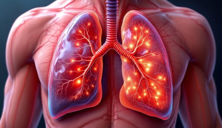
Lymphangitis carcinomatosis is a condition in which cancer spreads and causes inflammation in the lymph vessels, which are small tubes that carry a clear liquid called lymph throughout your body. This usually happens when cancer spreads from the primary location where it started. Often this occurs in the lymph vessels found in the area between the lungs (known as pulmonary lymphangitic carcinomatosis). Though less common, this cancer spread and inflammation can also occur in the lymph vessels of the skin, the duodenum (a part of the small intestine), and the kidneys.
In this summary, we are focusing on pulmonary lymphangitic carcinomatosis, which is the most common type of lymphangitic carcinomatosis. In medical terms, you may hear it referred to as pulmonary tumor embolism. These two terms are generally used interchangeably, even though they describe slightly different conditions—pulmonary lymphangitic carcinomatosis refers to when the cancer is primarily in the area between the lungs and there are no cancer cells in the lung arteries or capillaries (the smallest blood vessels), while a pulmonary tumor embolism refers to when there are cancer cells in the lung vessels.
This type of cancer spread was first described by Dr. Gabriel Andral in 1829 in a case of uterine cancer. More detailed understanding of the condition was achieved later in 1874. Usually, pulmonary lymphangitic carcinomatosis is indicative of a late-stage cancer with a short expected lifespan, however, recent reports suggest increased survival rates. The types of cancer that most commonly lead to lymphangitic carcinomatosis include breast, lung, stomach, prostate, pancreas, colon, cervix and uterine cancers.
What Causes Lymphangitic Carcinomatosis?
Only a small portion, about 6-8%, of lung metastases, which is when cancer spreads to the lung from another part of the body, is caused by a condition known as lymphangitic carcinomatosis. This condition is a type of lung cancer that affects the lymph vessels, which are thin tubes that branch out to all parts of the body to fight infection. Around 80% of these cases are caused by a group of cancers known as adenocarcinomas, which commonly occur in organs that produce fluids such as mucus, like the breast, lung, stomach, colon, or prostate, to name a few.
There have been unique cases where liver cancer showed up as lymphangitic carcinomatosis in the lungs just eight weeks after a liver transplant and kidney cancer doing the same barely two weeks after the removal of the main kidney tumor.
Even though this condition is uncommon in children, there have been reports of it in cases of kidney cancer, a type of bone cancer called chondrosarcoma, skin, colon, and cancers where the primary location is unknown.
Risk Factors and Frequency for Lymphangitic Carcinomatosis
Pulmonary lymphangitic carcinomatosis is a condition that is difficult to diagnose and measure. This is because its symptoms are often subtle, and doctors usually only confirm its presence after a patient’s death. Despite this, we know it only makes up around 6 to 8% of lung metastasis cases.
This condition typically appears in people between 40 to 49 years of age, with an average age of around 49. It equally affects both genders. The most common cause is cancer originating in the breast, occurring in 17.3% of cases. Lung and stomach cancer each accounts for 10.8% cases. This information is supported by two separate studies conducted by Bruce et al. and Ikezoe et al.
- Pulmonary lymphangitic carcinomatosis is difficult to diagnose due to subtle symptoms.
- It represents only 6 to 8% of lung metastasis cases.
- It typically occurs in individuals between 40 to 49 years of age.
- The average age at diagnosis is around 49.
- There is no gender preference; it occurs equally in men and women.
- The most common cause is breast cancer, accounting for 17.3% of cases.
- Lung and stomach cancers each cause about 10.8% of cases.
Signs and Symptoms of Lymphangitic Carcinomatosis
Pulmonary lymphangitic carcinomatosis is a medical condition where cancer spreads and blocks the lungs’ lymph vessels. Its most common symptom is a worsening shortness of breath that gradually develops over days or weeks. It’s present in about 60% of patients. While this condition usually occurs in patients already diagnosed with cancer, sometimes it can be the first sign of a hidden cancer. If the cancer blocks the lymph vessels near the surface of the lungs, it can cause chest pain. Coughing, blood in the spit, weight loss, and tiredness are less common symptoms. Some patients don’t have any symptoms, and the diagnosis is made incidentally while monitoring an existing cancer with imaging scans.
During a physical exam, the patient might have a low-grade fever, rapid breathing, and a fast heart rate. When the doctor checks the lungs, typically, they sound clear. Sometimes there can be additional sounds. Pulmonary lymphangitic carcinomatosis can present similarly to pulmonary tumor embolism, making diagnosis difficult. Signs of heart strain caused by high pressure in the lungs and right heart failure, such as a loud second lung sound, a forceful heart beat, a raised pressure in the neck veins, and bluish skin, are less likely to be seen in pulmonary lymphangitic carcinomatosis compared to pulmonary tumor embolism.
Testing for Lymphangitic Carcinomatosis
If your doctor thinks you might have a condition called pulmonary lymphangitic carcinomatosis, they’ll rely on several things to make a diagnosis. They’ll consider your overall health, whether you’ve had other medical problems in the past, and any symptoms you’re experiencing. To confirm their suspicions, they’ll usually carry out certain imaging tests, and perhaps also take a sample of your cells for lab testing. This condition can sometimes be confused with similar conditions, like pulmonary tumor embolism, so your doctor will want to rule these out.
However, using imaging tests to identify this disease can be tricky. Pulmonary lymphangitic carcinomatosis and conditions similar to it are often hard to detect in the early stages. Despite this, doctors find imaging helpful to identify what may be causing symptoms like shortness of breath.
In some cases, your doctor may use an X-ray to view your lungs. While an X-ray might not always show the disease, especially in the early stages, they can provide a clearer picture as the disease progresses.
Another scanning method that might be used is a CT scan. This process maps out the shape and substance of your lungs to form a more detailed picture. The scan will help your doctor identify if there is any thickening of the tissue within your lungs, and consequently, any patterns that might indicate the presence of the disease.
V/Q scans, another method of imaging, are not as effective in diagnosing this condition as they often appear normal. However, they can sometimes help in predicting how the disease might progress, which aids doctors in deciding on the most suitable treatment option.
F-18 FDG PET/CT is a specific type of scan that has shown high accuracy in detecting the disease. It works by injecting a small amount of a radioactive drug into your body, which then helps to highlight cancerous cells in the scan images.
Doctors also carry out laboratory tests to aid in the diagnosis. These tests often show non-specific results, which are supportive rather than conclusive elements in the identification of the disease. You may undergo a few different tests including a white blood cell count, a D-dimer test, arterial blood gas analysis, and pulmonary function tests.
For an accurate diagnosis, a sample of the infected tissue may be taken through a process called a biopsy. This, however, may not be possible in many situations. If your symptoms get worse rapidly and you’re finding it difficult to breathe, this process can be risky and might be skipped. In such cases, a diagnosis will generally be made based on your history, symptoms, and the results of your imaging tests.
Your doctors may also use a bronchoscopy, which involves inserting a tube through your mouth or nose to examine your lungs and air passages. A small sample of lung tissue or fluid can be taken during this procedure for further testing to help confirm the diagnosis. A procedure to remove a lung sample for testing will only be done if you are well enough.
In some instances, a diagnosis may be assumed based on your prior health history, current symptoms, and the outcomes of your imaging tests. This often applies to people who have a history of advanced cancer and are not in an ideal condition to undergo invasive diagnostic procedures. In such cases, doctors, patients, and their families often agree on a treatment plan based on these assumptions rather than opting for invasive diagnostic processes. It would be essential to discuss and decide on the best approach with your healthcare provider in such instances.
Treatment Options for Lymphangitic Carcinomatosis
Once a diagnosis of pulmonary lymphangitic carcinomatosis, a type of lung cancer that spreads along your lungs’ lymph vessels, is confirmed, the outlook remains quite poor. This condition may be the first symptom that signals the existence of another cancer in the body. If this is the case and the primary cancer has been found, doctors will consider options such as surgery, chemotherapy or radiation therapy to treat the underlying cancer.
Only cancers that react to anti-cancer drugs will get better with chemotherapy. For example, these reactions have been observed in cases of Wilms tumor (a kidney cancer in children) and trophoblastic tumors (a condition where cancerous cells grow in the womb). There have also been a few cases where significant improvements have been observed in cancer patients after being treated with hormone therapy, chemotherapy, tyrosine kinase inhibitors(a type of targeted cancer therapy like apatinib), and certain monoclonal antibodies(a type of targeted cancer treatment including bevacizumab and cetuximab). A specific case reported that intravenous eribulin, a type of chemotherapy, led to a rapid control of symptoms and partial disappearance of pulmonary lymphangitic carcinomatosis in a patient suffering from breast cancer that had spread to other parts of the body (metastatic breast cancer).
Additionally, supportive care is crucial to ease symptoms and maintain patient comfort while definitive therapy is attempted. This involves measures such as providing supplemental oxygen, using non-invasive or mechanical ventilation, or administering medication that support the heart’s function. For patients with advanced cancer who are experiencing poor quality of life and showing signs of pulmonary lymphangitic carcinomatosis, another approach focuses more on making them comfortable with diagnostics that do not require invasive procedures. These patients are often treated with oxygen supplementation and antibiotics, with the aim of easing symptoms rather than curing the disease.
Although there’s no scientific evidence to support the efficacy, some doctors prescribe intravenous steroids to relieve symptoms. If a patient experiences difficulty breathing (dyspnea) and anxiety, medications like opiates and benzodiazepines might be used carefully to alleviate these symptoms. If heart failure is detected, diuretics and pulmonary vasodilators are administered. If a patient’s cancer progresses and doesn’t respond to treatment, efforts will switch to palliative measures (aiming to alleviate symptoms instead of curing the disease) to ensure the patient’s comfort.
What else can Lymphangitic Carcinomatosis be?
When diagnosing pulmonary lymphangitic carcinomatosis, it can often be confused with pulmonary tumor embolism, which shows similar symptoms, especially those related to pulmonary hypertension. However, both conditions are serious, indicating advanced stages of cancer, and are managed in somewhat similar ways. Pulmonary lymphangitic sarcomatosis is another rare condition that closely resembles these two in terms of symptoms and imaging features.
This disease can also be mistaken for a number of others due to overlap of symptoms. These include:
- Pulmonary embolism (blockage in one of the pulmonary arteries)
- Obstructive airway disease
- Commonly acquired pneumonia, especially cases that are viral, unusual, or caused by fungi
- ARDS (Acute Respiratory Distress Syndrome)
- Pulmonary hypertension
- Heart failure
In addition, the listed interstitial (relating to small spaces or gaps in the tissue) lung diseases should be considered too:
- Acute idiopathic interstitial pneumonitis
- Cryptogenic organizing pneumonia
- Severe eosinophilic pneumonia
- Sarcoidosis
- Hypersensitivity pneumonitis
- Alveolar hemorrhage syndrome
- Interstitial lung diseases associated with connective tissue disorder
- Drug-induced lung disease
- Radiation pneumonitis
- Lymphomas
- Vasculitis
A rare form of non-Langerhans histiocytosis known as Erdheim-Chester disease, presenting with respiratory symptoms and very similar radiology, should also be considered.
What to expect with Lymphangitic Carcinomatosis
Pulmonary lymphangitic carcinomatosis is usually diagnosed in late-stage cancer patients, with a typical life expectancy of about six months. However, some reports have shown that survival up to 3 years or more is possible with the current treatments available.
There’s been some research done which observed that around half of patients passed away three months following the onset of respiratory issues, while almost 72% of patients passed away within seven months. A recent review has shown an increase in survival of patients from the year 2000 to 2018, comparing to a shorter survival rate from 1970 to 1999. Notably, hospital admission has been associated with a shorter survival timeframe.
Despite these odds, increased survival has been documented in lymphangitic carcinomatosis with particular treatments, like hormonal therapy (for example, prostate cancer patients), chemotherapy (for instances in breast, ovary, cervix, and stomach cancer), tyrosine kinase inhibitors (a drug named apatinib), and monoclonal antibodies (for example, colon cancer patients).
Possible Complications When Diagnosed with Lymphangitic Carcinomatosis
In severe cases of advanced pulmonary lymphangitic carcinomatosis, a lung cancer that affects the lymph vessels in the lungs, patients might experience serious oxygen deficiency and breathing failure. This situation might require the use of a breathing tube and a machine to support their breathing. The severe oxygen deficiency happens because the disease has widely spread in the lungs, which impedes the normal gas exchange process.
Complications might also occur due to the original cancer and the side effects of cancer-fighting drugs. There is a high risk of severe blood clot in the lung worsening the already compromised breathing function, especially in patients who are mainly inactive due to constant breathlessness, associated with higher risks of forming blood clots in deep veins.
Moreover, an episode of lower lung infection could potentially worsen breathlessness and the underlying breathing failure. Although extensive organ support measures, such as ventilators or other life-supporting devices, might not be beneficial in most cases, some patients, for example those with chronic obstructive pulmonary disease (COPD), might benefit if their breathing crisis is caused by an infection that can be treated with antibiotics. Negative consequences such as lung damage and infections can arise if invasive ventilatory support, such as a ventilator, is used.
If the cause of the breathing failure (for example, pulmonary lymphangitic carcinomatosis or interstitial pneumonia – inflammation of the lung tissues) is unknown at admission, especially for patients without any previously known cancers, complications might result from attempts at obtaining a definite diagnosis, which might involve taking a sample of lung tissue either through bronchoscopy (a procedure that looks inside the lung’s airways) or through open surgery.
Complications of advanced pulmonary lymphangitic carcinomatosis:
- Oxygen deficiency
- Breathing failure
- Blood clots in lungs
- Lower lung infection
- Complications from the use of life-supporting devices
- Potential complications from diagnostic procedures
Preventing Lymphangitic Carcinomatosis
Keeping open and clear communication between the healthcare team and the patient and their family is essential, especially during difficult times. Lymphangitic carcinomatosis, a severe condition often linked to advanced types of cancer, is known to reduce life expectancy significantly. In order to make informed decisions, complications, expected survival time, and the potential limitations of advanced life support need to be thoroughly explained to the patient and their family. This allows everyone involved to develop a clear plan of action together. If the patient is stable enough, procedures like a transbronchial lung biopsy or surgical lung biopsy could be considered to confirm the diagnosis.
However, many patients with advanced cancer may not be fit enough for a diagnostic procedure. These patients are often treated with care that aims to relieve symptoms, also known as palliative care, based on the presumed diagnosis of pulmonary lymphangitic carcinomatosis. While treating, factors like a possible lower respiratory infection, which could cause a sudden severe breathing problem, should always be considered. In some instances, treating these reversible factors might be appropriate, and this would be decided on a case-by-case basis.
In some rare cases, a patient might show signs of pulmonary lymphangitic carcinomatosis even without a known history of an underlying cancer. In such situations, an extensive evaluation is needed to confirm a diagnosis of lung disease, and search for a previously unknown primary cancer. Good communication skills on the part of the doctors and healthcare team are again essential to help the patient and their family agree to a solid plan of action.


