What is Lymphomatoid Papulosis?
Lymphomatoid papulosis is a type of skin condition which falls under the category of non-aggressive T-cell lymphoma. This means it’s a condition involving a certain type of white blood cell (T-cell) that doesn’t typically behave as aggressively as some other types of cancer. This condition is marked by skin changes which appear as recurrent (repeating), spontaneously healing bumps and nodules, some of which may become necrotic (dead tissue). These skin changes often spread to other parts of the body and resemble a type of cancer that’s positive for a marker called CD30 when examined under a microscope.
Lymphomatoid papulosis is relatively rare, making up about 12% of skin lymphomas – cancers that develop from lymphocytes (a type of white blood cell). This condition is grouped with a condition called primary cutaneous anaplastic large cell lymphoma because they both involve cells that express the CD30 marker.
One important thing to note for those with lymphomatoid papulosis is that they have an increased risk of developing other conditions that affect the blood and lymph system. This includes conditions such as mycosis fungoides (a type of skin lymphoma), erythrodermic T-cell lymphoma (a serious form of skin lymphoma), Hodgkin disease (a cancer of the lymphatic system), or large-cell CD30+ lymphoma (a type of lymphoma where large cancerous cells express the CD30 marker).
What Causes Lymphomatoid Papulosis?
The exact cause behind lymphomatoid papulosis, a skin condition, is yet to be determined. There were suggestions that it could stem from a viral infection, such as the Epstein-Barr virus or other types of herpesviruses. However, research has consistently found no connection.
We don’t fully understand why the skin lesions associated with this condition sometimes get better on their own. It’s suspected that interactions between certain proteins called CD30 and its partner, CD30L, may play a role. These proteins might trigger a process called apoptosis, which is when cells effectively ‘self-destruct’, and this could help the skin lesions to recede. However, this is still just a theory, and more research is needed to confirm it.
Risk Factors and Frequency for Lymphomatoid Papulosis
Lymphomatoid papulosis is a type of skin lymphoma that makes up about 12% of all skin lymphomas. It can happen to people of any age, but generally starts earlier than a similar disease known as CD30+ anaplastic large-cell lymphomas. It’s not very common in children. Most people are diagnosed with this condition between the ages of 35 and 45. There are around five times as many women diagnosed with this condition as men.
Signs and Symptoms of Lymphomatoid Papulosis
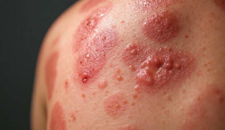
The typical signs of this condition include a red-brown rash of raised spots that might bleed or develop into sores. These spots usually heal within 2 to 12 weeks, leaving behind lighter or darker patches of skin, or pitted scars. It’s common to see spots of different ages at the same time. This condition tends to come and go, with periods of no symptoms in between outbreaks. There might be sores in the mouth, but this is rare. The number of spots and where they appear can vary from person to person and between outbreaks. Spots can appear anywhere, but they tend to show up on the body and arms. Usually, the rash doesn’t cause any discomfort.
- Red-brown rash of raised spots
- Spots may bleed or turn into sores
- Spots heal in 2 to 12 weeks, leaving discolored or pitted scars
- Spots of different ages may be present at the same time
- Condition comes and goes, with symptom-free periods
- Sores in the mouth may occur, but it’s rare
- Number and location of spots can vary
- Spots most commonly appear on the body and arms
- Rash is usually not uncomfortable
Testing for Lymphomatoid Papulosis
If your doctor suspects you have a particular skin condition called lymphomatoid papulosis, they need to do a skin biopsy. This is a small procedure where they take a tiny piece of your skin for examination under a microscope. This test can confirm the diagnosis and rule out other skin diseases that have similar symptoms. When they check the skin sample, they can figure out the type of lymphomatoid papulosis – A, B, or C.
After a diagnosis of lymphomatoid papulosis, no additional tests are necessary. Sometimes, doctors might take a blood sample looking for abnormal cells, but that’s not helpful in this case. But, they usually do a complete blood count and test for a protein called lactate dehydrogenase as part of the initial assessment.
At the same time, it’s also important for your doctor to check your skin carefully. That’s because lymphomatoid papulosis is often associated with another skin condition called cutaneous lymphoma. If you have any signs of this condition, your doctor will notice it during the skin examination. They usually don’t recommend imaging tests (like CT scans or MRIs) unless there’s a reason to suspect a more serious condition known as secondary lymphoma.
Treatment Options for Lymphomatoid Papulosis
Since Lymphomatoid papulosis is a rare and complex disease, there’s no single, standardized way to treat it. Current treatment options can reduce or control the symptoms but are typically not entirely effective. For patients with a small number of non-scarring lesions, no treatment might be necessary. However, those with more serious or unpleasant symptoms might benefit from methotrexate, a drug administered either orally or through injection. Methotrexate, which is used regularly, is the preferred systemic (affecting the whole body) treatment for lymphomatoid papulosis, irrespective of the disease’s specific type.
Treatment with methotrexate usually starts with low doses and gradually increases until the disease is under control. Once the disease is under control, the dosage is gradually reduced until the lowest effective dose is found, or the treatment can be halted entirely. Unfortunately, complete and sustainable remission of the disease is seen in less than a third of patients undergoing methotrexate treatment.
Another intervention that can be offered is phototherapy, which involves using ultraviolet light to treat the skin. Psoralen combined with Ultraviolet A (PUVA) therapy has been used for over three decades to treat lymphomatoid papulosis. It often helps lesions to disappear after roughly 15 sessions, but the disease tends to come back frequently. Other treatment options may include local chemotherapies, bexarotene, interferon, or excision (removal) surgery or radiotherapy for larger skin tumors that have developed as part of lymphomatoid papulosis and don’t go away on their own after 4 to 12 weeks.
Treating lymphomatoid papulosis in children is particularly challenging due to the potential risks of the phototherapy at this age and the reservations about using methotrexate in pediatrics. Most doctors tend to either withhold treatment or use potent topical corticosteroids on the initial inflammatory stages of papules (small, red, raised spots). However, if there’s a risk of the lesions leaving scars, general treatment may be required, and Ultraviolet B (UVB) phototherapy could become the preferred choice – even though its results in children are not always consistent.
All patients with lymphomatoid papulosis require long-term follow-up due to the potential risk of the disease developing into systemic lymphoma, a type of cancer that affects the entire body.
What else can Lymphomatoid Papulosis be?
Lymphomatoid papulosis is a chronic disease that is characterized by repeated flare-ups and healing, often leaving scars each time. Because these signs are very typical of the disease, it’s not frequently mistaken for other conditions. However, in cases where there are fewer skin lesions, it could be misdiagnosed as:
- Insect bites
- Prurigo (itchy rash)
- Lichenoid pityriasis (skin disease causing patches)
- Folliculitis (inflamed hair follicles)
- Scabies (itchy skin condition caused by microscopic mites)
On a microscopic level, it can be challenging to distinguish between lymphomatoid papulosis subtype B and a type of cancer called mycosis fungoides, or lymphomatoid papulosis subtype C and a type of lymphoma. So, doctors usually rely on what they observe during a physical exam to make the diagnosis.
It’s important to tell apart mycosis fungoides associated with lymphomatoid papulosis, which usually has a good prognosis, from a more dangerous form of the disease called transformed mycosis fungoides. This distinction is based on the medical history of the patient and the expertise of the doctor.
What to expect with Lymphomatoid Papulosis
Patients with lymphomatoid papulosis, a skin disease, have a chance of developing another blood-related disorder. These disorders include mycosis fungoides (a type of skin lymphoma), erythrodermic T-cell lymphoma (a rare and aggressive type of non-Hodgkin lymphoma), Hodgkin disease (a form of cancer that starts in white blood cells), or large-cell CD30+ lymphoma (a type of non-Hodgkin lymphoma). The blood disorder can occur before, after, or at the same time as lymphomatoid papulosis.
This risk ranges from 2% to 15% after 5 years of having the disease, and it increases the longer the disease lasts. Risks are higher for older patients and those with a specific type of abnormal cell (T-cell clone) in their skin lesions.
This disease can significantly affect quality of life, primarily because it is a long-term condition and can affect visible areas of skin. Regardless of the risk of developing a secondary disease, it’s important to know that lymphomatoid papulosis itself has an excellent prognosis. Most patients nearly have a 100% chance of survival 10 years from diagnosis.
Possible Complications When Diagnosed with Lymphomatoid Papulosis
People with lymphomatoid papulosis have a higher chance of developing other types of cancers. These can include:
- Mycosis fungoides (a type of skin cancer)
- Erythrodermic T cell lymphoma (a rare type of Non-Hodgkin lymphoma)
- Hodgkin disease (a cancer of the lymphatic system)
- Large cell CD30+ lymphoma (a rare and aggressive type of lymphoma)
Preventing Lymphomatoid Papulosis
Patients need to know that they can’t pass their condition on to others. It’s important for everyone to know how to properly care for their wounds to prevent infection. Wounds or sores should be gently cleaned with soap and water twice daily. A layer of petroleum jelly, like Vaseline, should then be applied to keep the wound moist and deter infection. If the condition causes crusty sores, cover them lightly with a bandage.
Patients must also watch for certain signs and report them to their doctor. These include:
- Skin bumps or sores that don’t get smaller after three months.
- Swollen lymph nodes in the neck, underarm, or groin area.
- Fever.
These signs could mean an infection has started or could be a sign of cancer related to the condition.


