What is Malignant Atrophic Papulosis?
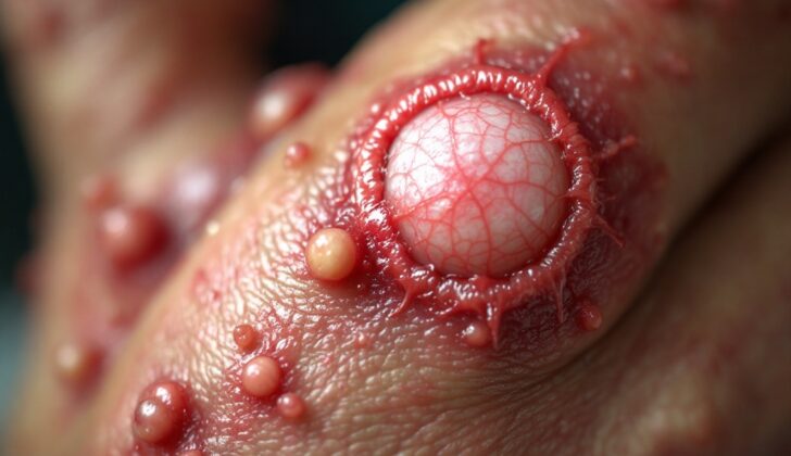
Malignant atrophic papulosis, also known as Kohlmeier-Degos disease or Degos disease, is a rare medical condition that affects the blood vessels in your skin, gut, and central nervous system (the brain and spinal cord). This condition is recognized by specific skin bumps that have a white, shrunken center surrounded by a tiny, dilated blood vessels.
Medical professionals distinguish two forms of this disease. One type is life-threatening and impacts the whole body, while the other is a less serious form that only affects the skin. Because of these differences, some doctors suggest the more general name of “atrophic papulosis” to include both forms. The disease could then be broken down into two categories: the severe, body-wide form (malignant atrophic papulosis) and a more moderate form that only affects the skin (benign atrophic papulosis).
What Causes Malignant Atrophic Papulosis?
Atrophic papulosis is a condition that was first identified in 1941 by Kohlmeier, but doctors are still not entirely sure what causes it. Some believe that it may run in families, as it has been seen to occur among multiple generations of family members. This suggests that the disease might be passed down from parent to child in an “autosomal dominant” way—meaning you only need a gene from one parent to potentially get the disease.
Beside the possibility of genetics playing a role, there are three other main ideas about what might cause this disease. The first is vasculitis, which is inflammation of the blood vessels. The second is coagulopathy, which is a condition that prevents blood from clotting properly. The third is a primary dysfunction of the endothelial cells, which are cells that line the inside of the heart and blood vessels. These three ideas aren’t seen as being exclusive of each other, and it’s possible that a combination of these factors, with certain environmental circumstances, could lead to someone developing the disease.
Risk Factors and Frequency for Malignant Atrophic Papulosis
Atrophic papulosis, a skin condition, typically begins to show symptoms between the ages of 20 to 50. However, there have been rare cases where it was reported in newborns. While it’s slightly more common in females, both males and females have the same chances of a favorable outcome, despite what was previously suggested in medical literature.
Signs and Symptoms of Malignant Atrophic Papulosis
Skin sores that come from this condition start out small, from 2.0mm to 5.0mm (about the size of a grain of salt to a grain of rice), and they can appear anywhere on the body or limbs. Over time, about 2 to 4 weeks, these sores change. The middle sinks in and the sores turn into large bumps from 0.5cm to 1.0cm wide (about the size of a small marble) with a pale, white center and a red, web-like edge. Common areas like the palms of the hands, soles of the feet, scalp, and face are usually spared of these sores.
In some cases, the disease can affect other parts of the body, not just the skin. This internal disease can come on suddenly or may not show up until many years after the skin sores have appeared. Depending on the organs that get affected, patients may experience symptoms like bleeding from the gut, belly pain, tingling sensations, changes in vision, and breathing or heart problems.
Testing for Malignant Atrophic Papulosis
If you’re suspected of having atrophic papulosis, which is a skin condition, your doctor will look for specific skin signs during a physical examination. This can then be confirmed through something called histopathology, which is the study of changes in tissues caused by disease. Unlike some conditions, there are no specific changes that occur in your blood or ‘marker’ substances in the blood that can confirm you have atrophic papulosis. However, some patients might have problems with their blood clotting.
When symptoms suggest that this condition might be affecting certain internal organs, tests will center on the suspected organ. For instance, if your doctor suspects your digestive system is involved, they might ask for a stool sample to check for hidden blood, recommend a colonoscopy (a procedure to look at the inside of the colon), perform an esophagogastroduodenoscopy (a procedure where a small camera is passed into your esophagus, stomach, and the first part of your small intestine) or even suggest a laparoscopy, which is a type of keyhole surgery which allows the doctor to see inside your abdomen.
In cases where the brain, heart, lungs, eyes, or kidneys are believed to be involved, your doctor might recommend other tests such as: a brain Magnetic Resonance Imaging (MRI) scan which uses strong magnetic fields and radio waves to produce detailed images of the inside of the body; a heart ultrasound, which uses sound waves to produce images of your heart; a chest Computed Tomography (CT) scan, which takes a series of X-ray images from different angles and uses computer processing to create cross-sectional images, or “slices” of the bones, blood vessels and soft tissues inside your body; an ocular fundus examination, which checks the back of your eye; and kidney function tests.
Treatment Options for Malignant Atrophic Papulosis
Atrophic papulosis is a condition that currently has no universally effective treatment. However, results from different individual cases suggest that blood thinners (anticoagulants) and medications that improve blood flow, such as aspirin, pentoxifylline, dipyridamole, ticlopidine, and heparin, can lead to some improvement or recovery of the skin lesions associated with this condition. This has made them a practical starting point for treating new patients.
In some cases, a medication called eculizumab, which decreases certain protein complex deposits, was found to improve initial skin and intestinal symptoms. However, it didn’t prevent the progression of the overall disease. Treprostinil, administered under the skin, was successful in treating a malignant form of atrophic papulosis that was not improving with eculizumab treatment in one patient.
Meanwhile, therapies aimed at dissolving blood clots (fibrinolytic therapy) and suppressing the immune system with medications like cyclosporine A, azathioprine, cyclophosphamide, or corticosteroids have not been successful.
Given the severity of this condition and potentially life-threatening outcomes, it’s crucial for patients to see their doctors regularly. This generally means at least once a year. Some experts even suggest bi-annual checkups for the first seven years after diagnosis, reducing to yearly checkups after that.
Monitoring of the patient should involve a range of diagnostic processes, including skin examinations, brain scans (using magnetic resonance tomography), stomach and colon examinations (gastroscopy and colonoscopy), chest x-rays, and abdominal ultrasound scans. These help to assess the patient’s condition over the long-term.
What else can Malignant Atrophic Papulosis be?
These are some health conditions that might showcase similar symptoms:
- Atrophie blanche
- Lupus erythematosus
- Dermatomyositis
- Antiphospholipid syndrome
What to expect with Malignant Atrophic Papulosis
A study was done on 39 patients at a single medical center. This study found that there was a 70% chance a patient could have a harmless skin version of the disease. And if a patient’s disease stayed limited just to their skin for seven years, the chances of it being harmless rose to 97%. The study also found that none of the patients whose disease stayed limited to their skin had any deadly results.
On the other hand, those with a more widespread or systemic version of the disease had a 21% chance of not surviving. On average, these patients lived between 0 to 9 years. This was a different result from a previous review of 109 patients in 1989 that found a higher death rate of 48.1%, and on average, these patients lived fewer than five years.
Possible Complications When Diagnosed with Malignant Atrophic Papulosis
Malignant atrophic papulosis, a rare skin disease, can be fatal due to the severe complications that arise when other organs in the body are affected. The organs most commonly affected are:
- The digestive system (found in 73% of cases)
- The brain and spinal cord (found in 64% of cases)
In many cases (64%), multiple organs are affected at once. Many people pass away due to holes forming in the bowel, blockage in the arteries of the brain, or severe bleeding within the brain. In addition to these, inflammation of the belly lining (peritonitis), meningitis, encephalitis, radiculopathy (nerve root damage), and myelitis (spinal cord inflammation) are also significant causes of illness and death in patients. Other severe complications include inflammation in the chest and around the heart, double vision, and weakness or paralysis of the eye muscles.
Preventing Malignant Atrophic Papulosis
It’s really important to make sure patients are informed about the symptoms that could signal a serious disease affecting the whole body. Detecting signs that organs may be involved in the disease early on can stop more serious health problems from developing and reduce the risk of death.


