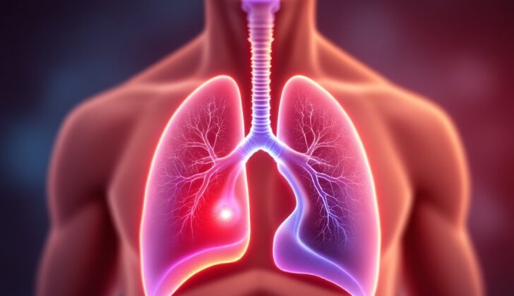
What is Mediastinal Cancer?
The mediastinum is a space in your chest that separates your lungs from other parts of your body. Generally, we can think of the mediastinum as divided into three main areas: the front, the back, and the middle. The boundaries of this space include the opening at the top of your chest, the muscle at the bottom of your chest that helps you breathe (the diaphragm), your spine at the back, your breastbone at the front, and the spaces around your lungs at the sides. This space contains your heart, the main blood vessel carrying blood away from your heart (the aorta), your food pipe (esophagus), a gland in your upper chest (thymus), and your windpipe (trachea).
The growth of cancer and the seriousness of its symptoms depend on how it behaves within this complex network of organs. Cancers can grow in the mediastinum either from organs located there or from organs that pass through the mediastinum as they develop. They can also spread there from cancers that start in other parts of the body.
To diagnose cancer in the mediastinum, doctors typically use a procedure called a mediastinoscopy, where they collect cells from the mediastinum while you’re under general anesthesia. This allows them to identify what kind of tumor is present. The treatment for cancers in this area depends mainly on the type of cancer, where exactly it’s located, how fast it’s growing, and what symptoms it’s causing.
What Causes Mediastinal Cancer?
There are various types of cancers that can occur in the mediastinum, which is the area in the middle of your chest between your lungs. The outlook for each type of cancer depends on how invasive it is, how likely it is to spread, and how resistant it is to treatment. The mediastinum is divided into three parts: anterior (front), middle, and posterior (back), and different types of tumors can occur in each area.
In the anterior mediastinum, thymic carcinomas are a type of cancer that begins in the epithelial cells of the thymus (a small organ in your chest that makes white blood cells). These are rare but can be quite aggressive and spread early. Other tumors that can occur in this area are thymomas, which also begin in thymic epithelial cells. We believe thymomas play a role in the development of the immune system, leading to its association with certain immune-based diseases.
Germ cell tumors, which are most commonly found in the ovaries or testicles, can also occur in the anterior mediastinum. They are believed to show up in these locations due to abnormal migration of germ cells during the development of the embryo. Teratomas, another type of tumor that can occur in this area, come from embryonic cells but have a low chance of occurring here.
Lymphomas, which are cancers of the immune system, can also arise in the mediastinum. There are various types and some originate elsewhere and spread to the mediastinum.
In the middle mediastinum, we can find parathyroid adenomas, which are benign tumors of the parathyroid glands. These glands regulate the calcium in your body. These tumors can spread to this area.
In the posterior mediastinum, most tumors are linked to the nervous system, made from the cells that form nerves. These tumors are typically found in children and can grow quite large before causing any symptoms. Some of the common tumors include Schwannomas, neurofibromas, and neuroblastomas.
Treatment for all these tumors typically involves surgical removal, and sometimes, additional treatments like chemotherapy or radiation are used. Sometimes the exact treatment strategy depends on the type of tumor, its stage, and other factors.
Risk Factors and Frequency for Mediastinal Cancer
Mediastinal cancers, which are generally rare, usually occur in people aged 30 to 50. However, they can originate from any tissue located in or passing through the mediastinum and can happen at any age. Children typically have cancers in the back of the mediastinum, while in adults, these are generally found at the front. In terms of gender, mediastinal cancers occur at a similar rate in both men and women, but rates could be variable depending on the specific type of cancer.
Signs and Symptoms of Mediastinal Cancer
When it comes to diagnosing mediastinal cancer (a type of cancer that occurs in the area between the lungs), doctors usually rely on a detailed medical history and a physical examination. The symptoms and physical signs can vary, depending on the exact type of cancer, where it’s located, and how aggressive the tumor is. In some cases, people may not have any symptoms at all, and the cancer is only discovered during a chest scan for another reason.
If symptoms are present, they’re often due to the cancer pressing on surrounding organs or structures, or they could be related to paraneoplastic syndrome, a rare condition that can occur with some cancers. Here are some symptoms that might be observed:
- Cough
- Shortness of breath
- Chest pain
- Fever
- Chills
- Night sweats
- Hoarseness
- Unexplained weight loss
- Swollen lymph nodes
- Coughing up blood
- Wheezing or stridor (a high-pitched wheezing sound resulting from turbulent air flow in the upper airway)
In certain cases, mediastinal cancer can also lead to abnormalities in other body areas, such as germ cell tumors causing testicular masses. As a result, it’s extremely important for the doctor to take a comprehensive medical history, including a complete review of symptoms, as well as conducting a thorough physical examination. It’s worth noting that malignant (cancerous) masses are generally more likely to cause symptoms.
Testing for Mediastinal Cancer
Mediastinal cancers, or cancers located in the area of the chest that separates the lungs, can show a wide array of symptoms. Depending on where the cancer is located and what it’s made of, it helps to determine what type of cancer a patient may have. Sometimes getting a biopsy, or small tissue sample, can be risky and not the best option for some diseases. To diagnose and assess mediastinal cancers, some common tests are performed:
With a chest x-ray, this is typically the first step in finding a mediastinal mass, or cancerous growth. It can help identify if the mass is on the anterior (front), posterior (back), or medial (middle) side of the mediastinum. It might be used to evaluate a patient with symptoms due to the mass or if the mass is spotted accidentally. However, chest x-rays don’t provide enough detail to fully characterize, or describe the specifics of, mediastinal cancers.
A chest computed tomography (CT) scan is usually done if a mass is seen on a chest x-ray. This scan uses X-rays but can provide more detailed images and includes the use of a dye that’s injected into the veins to improve visibility. This can help to know the exact location of a mass and if it’s contained or invasive. The CT scan can identify the mass based on specific features like the presence of water, air, fat, calcium, soft tissue, and blood vessels. In many cases, a CT scan is enough for diagnosis.
Magnetic resonance imaging (MRI) can be done if the CT scan results are not clear. MRI uses a strong magnetic field and radio waves to generate images and can identify the precise composition of mediastinal masses. Special MRI techniques can help to differentiate normal tissue from cancerous tissue and even provide information about tiny structural and metabolic differences that are useful for diagnosis.
The definitive diagnosis of mediastinal cancer is usually done by a procedure called mediastinoscopy with biopsy. This procedure involves collecting a tissue sample from the mediastinum under general anesthesia to determine the type of mass present. A small cut is made under the breastbone, and a small sample of tissue is removed to check for the presence of cancerous cells. This test helps doctors to figure out the type of cancer present.
Endobronchial ultrasound (EBUS) and endoscopic ultrasound (EUS) have become the preferred diagnostic procedures for most mediastinal pathologies. These procedures involve the use of an ultrasound probe attached to a flexible tube that’s passed through the mouth or nose and into the chest. This allows doctors to visualize and access most of the lymph nodes in the chest. These procedures are usually done on an outpatient basis under sedation or general anesthesia and are generally safe with minimal complications.
Basic blood tests can help doctors to differentiate between lymphomas and other types of cancers. In some cases, tumor markers can help confirm a suspected diagnosis. These include β-hCG, which is associated with germ cell tumors and seminoma; LDH, which could be elevated in patients with lymphoma; and AFP, which is associated with malignant germ cell tumors.
In cases where a primary testicular germ cell tumor is suspected to have spread to the mediastinum, palpation, or physical examination of the testicles, is not enough. An ultrasound of the testicles should be done in all patients as part of the diagnostic workup.
Treatment Options for Mediastinal Cancer
The way doctors treat cancers in the mediastinum (the space between the lungs) largely depends on the type of cancer, where it’s located, how aggressive it is, and the symptoms it’s causing. Let’s look at the treatments for some of the most common mediastinal cancers:
Thymic Cancers:
For thymic cancers (which occur in the thymus, a small organ in the chest), doctors usually perform surgery first, then follow that with radiation or chemotherapy. Before surgery, they’ll perform tests to check how well your heart and lungs are working. When patients have tumors that are invading structures that can be easily removed (like the lining of the chest cavity, lining of the heart, or nearby lung), surgery is the first treatment. After the operation, doctors send the removed tissue to a lab to see how advanced the cancer is. This will help them decide if you need radiation therapy or chemotherapy after surgery.
There are different types of surgery your doctor might use. These include minimally invasive methods that involve smaller incisions and shorter recovery time. If the cancer has spread and complete removal isn’t possible, treatment options involve chemotherapy and radiation. Your doctor might even try to remove as much of the cancer as possible before using radiation. Regular follow-ups using imaging tests such as CT scans are recommended.
Teratoma:
Teratomas are a type of germ cell tumor that can be treated effectively with complete surgical removal. If the tumor isn’t fully removed, it could come back. In certain cases where the tumor is immature or mixed with other types of cancer, additional treatments such as chemotherapy or radiation may be required.
Lymphomas:
Lymphomas, a type of cancer in the lymph nodes, are generally treated with chemotherapy and radiation. The specific treatment varies depending on the type of lymphoma, how advanced it is, and whether it has spread to other parts of the body. In some cases, doctors use a combination of different drugs to treat the lymphoma. Some patients might need a biopsy before starting radiation to confirm the presence of lymphoma.
Neurogenic Tumors:
Neurogenic tumors are cancers that begin in nerve cells. These are generally treated with surgery. And keeping track of the size of the tumor and whether it’s located in the spine is crucial before surgery. Different types of surgeries, including minimally invasive ones, can be used depending on the tumor’s size and location.
Mediastinal Germ Cell Tumors:
The treatment for mediastinal germ cell tumors depends on the type of tumor. This may include chemotherapy, surgery, radiation, or a combination. Smaller tumors might require radiation, while larger ones could be treated with a combination of drugs followed by surgery. Regardless, continuous monitoring and frequent check-ups are crucial.
The optimal treatment for these cancers is not yet fully defined, and survival rates can vary widely. Some of the factors that may affect survival include how long the patient receives chemotherapy, whether additional anticancer drugs are used, and the timing of the surgery.
What else can Mediastinal Cancer be?
There are various medical conditions that might cause discomfort or concern in the chest area. These include:
- Retrosternal and intrathoracic goiters: These are types of overgrown thyroid glands. The primary version is separate from the regular thyroid, and the secondary version extends into the chest area. They can cause compression of the windpipe, which can lead to a condition called tracheomalacia. The best treatment for these goiters is surgical removal.
- Tuberculosis: This condition can lead to inflammation and enlargement of lymph nodes in the chest area. It’s common in kids and in developing nations, and might cause symptoms similar to other disorders. It can lead to various complications, including inflammation and constriction of the heart lining and nerve damage.
- Aneurysm: These are abnormal bulges in blood vessels that might distort imaging results and appear to be masses. People with a condition called Ehler-Danlos syndrome type IV can be particularly prone to aneurysms. If these cause symptoms, they may need to be surgically removed.
- Bronchogenic cysts: These cysts often occur in the mid-chest and are due to abnormal development of the windpipe and lung branches during fetal development. If they don’t cause symptoms, a wait-and-see approach may be recommended. But surgical removal is suggested in young people without symptoms to prevent potential complications like infection, bleeding, and malignancy.
- Pleuropericardial cysts: These are abnormal fluid-filled sacs that often develop in the chest area. They can occur after examining or operating the heart. Most do not cause symptoms or become cancerous. Aspiration, or drawing out the fluid, is not recommended as it can lead to infection. But, if they do cause symptoms such as chest pain or shortness of breath, surgery is necessary. They might lead to potential complications like bleeding, irregular heart rhythms, and compression of the heart.
In all cases, medical professionals will advise on the best treatment approach based on individual circumstances.
What to expect with Mediastinal Cancer
Your chance of getting better depends on what’s causing the problem and how well the treatment works.
Possible Complications When Diagnosed with Mediastinal Cancer
Patients with mediastinal masses, or large lumps in their chest cavity, risk having their airways blocked, causing severe difficulty in breathing. This situation is sometimes referred to as critical mediastinal mass syndrome. If these patients are required to undergo a biopsy or surgery that involves general anesthesia, the risk of the collapse of their windpipe (trachea) or bronchial tubes increases. Doctors often recommend avoiding muscle relaxants, sedatives, and paralytic agents in such cases. They instead advise using a technique called positive end-expiratory pressure, which acts as an “air pressure splint” to keep the airway open. Some doctors have favored using heart-lung machines or artificial lung support devices for these patients.
Tracheomalacia is another condition involving the respiratory system. This is when the trachea, or windpipe, collapses excessively when breath is expelled because the elastic fibers in the tracheal wall are damaged. The collapse of the trachea wall narrows and shortens the airways, obstructing airflow. This can occur due to long-term external pressure on the trachea from cancers, but it can also be seen in noncancerous conditions like cysts, abscesses, inflammation of the body’s large blood vessels, chronic lung disease, or inflammation of cartilage. Diagnosis is made via a bronchoscopy, which is a test that allows your doctor to look at your airway. Doctors typically first try non-surgical treatments like continuous positive airway pressure. Depending on the patient, surgical methods such as repairing the trachea and bronchial tubes, making an incision in the neck to open up the trachea (tracheostomy), or using artificial scaffolding in the trachea (stenting) could be necessitated.
Common complications:
- Blockage of airways
- Collapse of the trachea or bronchial tubes
- Difficulty breathing
- Tracheal wall collapse and narrowing of the airway
- External tracheal pressure due to chronic diseases.


