What is Melanocytic Nevi?
Pigmented skin lesions, often known as freckles, include a variety of types. These range from solar lentigo, which is a sun-related spot; congenital nevi, which are birthmarks; mucosal nevi, which are moles on the mucus membranes; and special types of moles found on the palms and soles. We will discuss how to tell apart the different kind of moles found on the feet from a type of skin cancer known as acral lentiginous melanoma.
Melanomas on the sole of the foot, known as plantar melanomas, are typically diagnosed late. As a result, they often don’t respond as well to treatment and carry a significantly higher risk of death compared to melanomas found elsewhere on the body.
What Causes Melanocytic Nevi?
Severe sunburn during childhood is highly linked to a greater chance of developing melanoma, a type of skin cancer, later on. On the other hand, gradual sun exposure due to work (occupational exposure) doesn’t seem to increase this risk. These results support the idea that the risk of getting melanoma is mainly affected by heavy, irregular sun exposure that overwhelms the body’s immune system and destroys abnormal skin cells known as melanocytes.
Sunburn in the teenage years can harm the immune system for life and disrupt the normal process where abnormal, potentially cancerous cells are destroyed by the body. Genetics also play a role in the development of melanoma, potentially explaining cases of melanoma that occur in parts of the body not typically exposed to the sun. In fact, roughly 1 out of every 10 cases of melanoma is thought to be connected to inherited gene faults.
Risk Factors and Frequency for Melanocytic Nevi
The growing number of skin cancer cases, specifically melanoma, is highly connected to increased exposure to ultraviolet (UV) light. Melanoma is relatively rare but is becoming more common worldwide. Despite more awareness and screening, death rates have not significantly dropped. In the United States, melanoma is the fifth most common cancer in men and the seventh in women. The number of cases has doubled over the last decade.
- In a study reviewing data from 1992 to 2004, researchers found that the number of melanoma cases (across all thicknesses and socioeconomic groups) is increasing.
- The rise is from 4.7 to 7.7 cases per 100,000 older men and 5.5 to 13.9 cases per 100,000 women.
- Although skin cancer can occur anywhere on the body, only 5% of melanomas are found on the foot.
- Skin cancer on the foot, or pedal melanoma, is fairly rare in people with white skin.
- However, among East Asians and people with dark skin, acral lentiginous melanoma (a type of skin cancer that typically occurs on the palms of the hands, soles of the feet, or under the nails) is the most commonly diagnosed subtype.
Signs and Symptoms of Melanocytic Nevi
People might notice a change in a mole on their foot, but melanomas on the palms or soles are often only detected during a detailed examination. The palms and soles have unique features, like a lack of hair follicles and oil glands but many sweat ducts. They are also thicker and their regular patterns are distorted due to walking and grasping pressures.
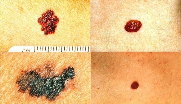
During a medical check-up, the doctor will examine the mole for asymmetry, irregular borders, color changes, a diameter greater than 6 mm, elevation, or change over time. Dermoscopy, a method that uses a special magnifying lens to check skin lesions, can further enhance the accuracy of diagnosis by 20%.
Remember, a thorough check of the soles, spaces between toes, and nails are essential parts of a complete lower extremity examination.
Testing for Melanocytic Nevi
When examining a coloured (pigmented) skin spot, doctors look at several features, also known as the A through E characteristics. Additionally, they utilise specific patterns observed through a tool called a dermoscope to aid in the examination. These patterns can indicate whether the skin spot is likely harmful (malignant) or not harmful (benign).
There are three patterns that usually indicate the skin spot is benign, or not harmful. The first one, known as a parallel furrow pattern, shows straight lines of colour and is considered benign around 93.2% of the time. The second one is the fibrillar pattern, which includes pigmented furrows and fine cross-hatched strands. The third is a lattice-like pattern of colour. Both of these are understood to be benign as well.
On the other hand, a parallel ridge pattern, which shows wide spread pigmentation across the skin ridges, usually suggests a type of skin cancer called melanoma about 93.7% of the time. If a normally benign parallel furrow pattern is disrupted, your doctor might recommend a biopsy, which is a procedure to remove a small sample of the skin spot for further examination.
Treatment Options for Melanocytic Nevi
An atypical mole, also known as an atypical nevus, found on the sole of your foot can be examined through a biopsy procedure. This involves using a tool, called a punch biopsy tool, to take a small sample of the skin. If a more extensive initial removal of skin is chosen, a small area of surrounding normal skin as well as some of the underlying fat tissue will also be removed. If the biopsy finds a type of skin cancer known as melanoma, a larger surgical operation is planned. This will involve the removal of a larger area of skin surrounding the melanoma – the size of this area depends on the thickness of the tumor. This procedure may unfortunately result in significant tissue loss, sometimes requiring amputation.
In most cases, small areas on the sole, called plantar areas, of the foot where melanoma is removed are usually left to heal naturally. Once the initial healing is complete, plastic surgery can help restore the foot’s ability to bear weight. A specialized form of surgery, known as Mohs microsurgery, can be used to minimize tissue loss. It’s important to have a well-experienced cancer care team to improve treatment outcomes.
The Melanoma Staging database, which is a classification system for melanoma based on the experiences of over 38,900 melanoma patients, is used to guide the treatment. This system considers the size and spread of the primary tumor, whether the cancer has spread to the regional lymph nodes or other parts of the body, and other factors such as the rate at which the tumor cells are dividing. The system also gives survival rates for different stages of melanoma. For instance, patients with primary lesions less than 0.5 mm thick have a 10-year survival rate of 96% which decreases to 54% if the lesions are 4.01 to 6 mm thick.
The system also identifies whether the cancer has spread to regional lymph nodes, which significantly affects long-term survival. A procedure called sentinel lymph node biopsy is recommended for tumors at or above 0.75mm thick. If the biopsy is positive, meaning it found cancer cells, a more complete lymph node dissection is recommended. Following this, a drug called interferon-alpha is often given to improve the chances of survival for patients who are expected to live more than 10 years.
Some therapies that target our immune response have shown promise in fighting melanoma. One such drug, Ipilimumab, can stimulate an anti-tumor response in our immune system and has shown to extend survival. However, it may worsen pre-existing immune disorders. Antibodies that enhance immune response, such as nivolumab and pembrolizumab, have shown to be more effective than conventional cancer drugs in clinical trials for various advanced tumors, including melanoma. However, the survival rate for plantar melanoma remains relatively low, especially for larger lesions.
What else can Melanocytic Nevi be?
When looking at different types of skin conditions, several can be considered. These include:
- Atypical mole
- Basal cell carcinoma
- Café au lait spots
- Cockade nevus
- Cutaneous melanoma
- Nevi of Ota and Ito
- Nevus spilus
- Nodular lesions
- Pyogenic granuloma


