What is Melanoma of the Head and Neck?
Melanoma is a type of cancer that develops from cells called melanocytes. Melanocytes, which are cells that give skin its color, can start to grow out of control and can show up sporadically or develop from an already existing abnormal spot or mole. Because these color cells come from a part of our body called the neural crest, melanomas can also appear in other areas where neural crest cells exist, such as the brain and digestive system.
About 10% to 25% of melanomas are found in the head and neck region. They are most commonly seen on the back of the head and the skin of the cheek. Other frequent places include the face (40 to 60%), scalp (14 to 49%), neck (20 to 29%), and ear (8 to 11%). Where the melanoma occurs is particularly important when it’s in the head and neck area because some locations can have more severe outcomes. Also, the place of the melanoma can affect how it’s treated compared to when it’s in other parts of the body.
What Causes Melanoma of the Head and Neck?
Frequent exposure to sunlight, particularly its ultraviolet (UV) light, is the most significant factor behind the development of melanoma, a type of skin cancer. The UV light can cause chemical reactions and damage in the DNA of skin cells, leading to the formation of pyrimidine dimers – pairs of certain DNA components (cytosine and thymine) that shouldn’t normally be together.
The UV light can be divided into two types: UVB and UVA. While UVB (290 to 310 nanometers) does the most damage to DNA, UVA (320 to 400 nanometers) is actually more common in natural sunlight. UVA can also harm DNA by causing oxidative DNA damage, which essentially means that it contributes to the destruction of DNA through stress.
Current sun protection creams and lotions usually have what’s called an SPF rating, which mostly considers UVB light. However, it’s no less crucial to protect your skin from UVA light, and for that, there are certain products available that can assist.
People tend to use sunscreen more, which puts them at a higher risk of getting melanoma as most products primarily focus on blocking UVB. Tanning beds, which produce mostly UVA light and sometimes UVB light, also add to the risk of melanoma, especially if used frequently.
Additionally, people who live in places with more sunshine throughout the year have a higher risk of getting melanoma.
Certain personal traits such as having blonde or red hair, blue eyes, and skin type I or II (identified through Fitzpatrick classification as skin most prone to sunburn) can increase melanoma risk. Having more melanocytic nevi (moles) or a family history of melanoma or pancreatic cancer could indicate a genetic mutation that increases the chance of developing the disease.
People of lower socioeconomic status could be at higher risk due to less awareness about the condition and an underestimation of its risk, which often results in the disease being more advanced when finally detected.
Large congenital nevi (moles larger than 20 cm), or Atypical mole syndrome (a condition caused by a defective gene that results in abnormal moles and increases melanoma risk), are other factors that can increase the risk. If relatives have melanoma or abnormal moles, the chance of getting melanoma gets even higher.
Finally, a hereditary condition called Xeroderma pigmentosa, which prevents the proper repair of DNA damage, leads to an increased risk of all types of skin cancers.
Risk Factors and Frequency for Melanoma of the Head and Neck
Melanoma is a type of skin cancer that is most commonly found in areas with lots of sunlight and among people with light skin. It is the sixth most common cancer in the United States. It’s more frequent in men, where it ranks as the fifth most common cancer, and women, ranking seventh. For white women aged 25 to 29, it’s the most common cancer, and the second most common for those between 30 to 34 years old. This type of cancer is more common in white people compared to Asians or Black people.
- In 2010, around 68,000 new cases of melanoma were reported in the United States, leading to 8,700 deaths.
- About 30% of melanoma cases are located in the head and neck.
- The incidence of melanoma has been increasing by 5% each year, and mortality has been increasing by 2% per year.
- The increase in melanoma is quicker than any other cancer except for lung cancer in women.
- Melanoma is the third most deadly type of cancer.
Signs and Symptoms of Melanoma of the Head and Neck
Melanoma is a type of skin cancer that can appear on different parts of the body. The outcome for people with melanoma can change based on where the cancer is located. When the disease affects the head and neck, for instance, it tends to be worse than in other places. The seriousness of the disease can be different depending on whether it’s found on the scalp, ear, cheek, or neck, with the scalp being the most dangerous location.
During a checkup, the doctor should carefully examine all the lymph nodes in the neck. These are small, bean-shaped structures that produce cells to combat infections and disease. The parotid gland, which contains lymph nodes and is located near the ear, also needs to be checked. This is particularly important when the melanoma is found on the front part of the scalp, temple, or cheek. For melanoma on the back part of the scalp or behind the ear, the occipital lymph nodes found at the back of the neck should be thoroughly examined.
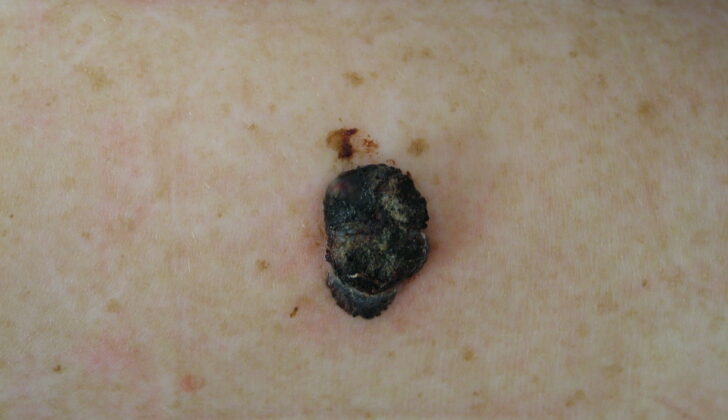
People with melanoma will often have a new skin need to be aware of any new skin changes such as a new marking or a mole that changes. However, a form of melanoma without the typical dark pigment, known as amelanotic melanoma, may appear pink, red, purple, or the same color as your skin. It is crucial to familiarize yourself with the common characteristics of melanoma, usually summarized by the acronym ABCDE:
- A: Asymmetry
- B: Irregular border
- C: Color variations, especially red, white, and blue tones in a brown or black lesion
- D: Diameter greater than 6 mm
- E: Evolution of the mole(s), which is the most important factor when diagnosing melanoma. This could include a mole that has recently changed in color or size or if there any signs of regression.
For advanced or aggressive melanoma, further physical examination might be required. This might include checking for signs like ulceration (breaks in the skin or surface of an organ), nodularity (small lumps or nodes), and satellite lesions (spread of cancer cells from the original tumor to other parts of the body).
Testing for Melanoma of the Head and Neck
In the case of melanoma (a type of skin cancer) appearing in the head and neck region, a biopsy is always the first course of action. A biopsy is a medical procedure that involves removing a small sample of the suspicious skin to examine it in detail. The doctor will need to determine not only if melanoma is present, but also how deeply it has invaded into the skin tissue. Because of this, they avoid using a shave biopsy – a technique which only removes a superficial layer of skin – as it does not provide this depth information. Instead, they opt for an excisional biopsy, which uses a cut to remove the entire abnormal area along with a tiny rim (2 millimeters) of normal-looking tissue around it.
There are multiple ways to carry out a biopsy, and the doctor might decide to use a different technique if the patient is also undergoing lymphoscintigraphy. This is a type of imaging test that maps out the lymph nodes to check if the melanoma has spread. While an excisional biopsy can interfere with the drainage of lymph fluid in the area, an incisional or punch biopsy would not. These methods only remove part of the lesion and leave some behind on the skin, which can be more tolerable for the patient.
To get an accurate measure of the depth of the melanoma, the biopsy will need to be performed through the thickest part of the lesion. The sample might also be sent to the lab for a full blood count and a detailed analysis to check proteins and liver function. Among these lab tests, doctors often look at the levels of lactate dehydrogenase, a substance that can be elevated in advanced melanomas.
Imaging tests like chest x-rays or CT scans with contrast are used to assess the spread of the disease. Chest x-rays give an overview of the chest area, while CT scans offer a detailed look at the head and neck area.
Additionally, if the melanoma is deeper than 4 millimeters, ulcerated (broken skin), consists of multiple lesions, or if it has recurred (come back after a primary lesion was previously treated), a more extensive search for possible metastasis (spread) is needed. This involves conducting a more comprehensive set of scans like CT scans of the chest, abdomen, and pelvic regions, MRI scans of the brain, and PET CT scans, which are highly accurate in revealing any hidden cancerous cells.
Treatment Options for Melanoma of the Head and Neck
Melanoma, a type of skin cancer, has different treatment approaches depending on how thick the tumor is, commonly measured in Breslow depth. The distance around the tumor that needs to be surgically removed, also known as the excisional margin, varies accordingly. For tumors less than 1 millimeter thick, a margin of 1 centimeter is normally used. For those between 1 and 2 millimeters, a slightly larger margin is preferred. For those above 2 millimeters, or if the tumor is ulcerated (meaning the skin has broken), a margin of 2 centimeters is suggested.
How deeply the surgeons go during the removal process may also vary, particularly for parts such as the face, scalp, or ear. For the face, the surgery usually goes as deep as the muscles that control facial expressions. For the scalp, the surgery ends at a layer of tissue covering the skull. In case of tumors in the ear, part or whole of the ear may be removed depending on the size of the tumor and the presence of additional smaller tumors in the vicinity. Tumors in the ear canal could require more extensive surgery involving the temporal bone bisecting the skull.
There is what’s called a sentinel node, which is the first node to receive lymphatic drainage from the tumor, and plays a critical role in managing melanoma. A biopsy of this node is often performed to see if the cancer has spread there. The presence of melanoma in this sentinel node can affect the overall survival and disease-free survival rates. Hence, doctors often forgo neck dissection, a surgery involving the removal of lymph nodes in the neck, if the sentinel node biopsy comes back negative. On the other hand, if the sentinel node is found to be positive, a neck dissection is necessary.
Doctors generally recommend a biopsy of the sentinel node for melanomas with a Breslow depth of 1 millimeter or more. In some cases, if the depth is between 0.75 and 1 millimeter, a biopsy might be suggested taking into consideration certain adverse prognosis factors such as ulcerations, lymphovascular invasion, extensive regressions, the patient’s age, a high mitotic rate (indicating how quickly the cells are dividing and spreading) and how deep the tumor has reached into the skin layers.
For melanomas in certain areas like the front of the scalp, face, temple or the ear and with evidence of spread in the region, the patient might require a surgery called a superficial parotidectomy which involves removing part of the salivary gland near the ear. However, for melanomas on the chin, neck or without evidence of regional spread, a parotidectomy is typically not needed.
For managing melanoma medically, doctors might use different types of medications, for instance, drugs that help stimulate the immune response against the cancer cells like Interleukin-2, or drugs that target specific molecules in the cancer cells. Also, some cases might require radiation therapy. While melanomas are known to be resistant to radiation, this form of treatment can be used to manage multiple positive nodes or macroscopic extranodal extension (cancer spread beyond the lymph nodes). It is a good option for older patients, nonsurgical candidates, and patients with brain metastasis.
What else can Melanoma of the Head and Neck be?
When it comes to skin lesions, it is important to identify them accurately in order to provide appropriate treatment. These can range from benign (non-cancerous) melanocytic lesions to severe conditions like melanoma. Here are some examples:
- Benign melanocytic lesions such as blue nevus, Mongolian spots, and melanocytic nevus
- Seborrheic keratosis
- Melanoma
What to expect with Melanoma of the Head and Neck
The prognosis, or how likely a person is to recover or have the condition worsen, for melanomas, a type of skin cancer, depends on various factors.
The location of the melanoma is significant, especially if it’s in the head and neck area, where the prognosis is not as good compared to other body parts. It’s worth noting that the scalp has the worst prognosis.
It’s also crucial to highlight the stage of the disease at the time of diagnosis. In simple terms, the stage helps doctors determine how much the melanoma has spread.
For Stage I Melanomas, a person typically has a more than 90% survival rate over five years.
In Stage II Melanomas, the five-year survival rate drops and varies between 45% and 77%.
For Stage III Melanomas, the five-year survival rate drops further, ranging between 27% and 70%.
In the case of Metastatic Disease, where the melanoma has spread to other parts of the body, the five-year survival rate is usually less than 20%.
Recovery from Melanoma of the Head and Neck
After being diagnosed with melanoma, a type of skin cancer, it’s very important for patients to have regular check-ups. This is essential from both a surgical perspective, to see how well they’re recovering from any procedures, and a dermatological perspective, to monitor the condition of the skin.
Preventing Melanoma of the Head and Neck
After being diagnosed with melanoma, a type of skin cancer, it’s important for patients to stay out of the sun as much as possible and always take protective measures. Exposing your skin to the sun increases the chance of developing more melanoma cases, so it’s crucial to be cautious and maintain your skin’s health.


