What is Oral Melanoma?
Oral melanoma is an unusually harmful type of cancer that grows quickly and acts very aggressively. This type of cancer makes up 0.2% to 8% of all melanoma cases and 1% to 2% of all mouth-related cancers. Compared to other types of melanoma, oral melanoma has the lowest 5-year survival rate, likely because it’s often discovered too late. Oral melanoma can appear in many forms, but it’s usually seen as a dark patch, spot or small, round growth. These could have different shades of grey, red, purple, or areas that have lost color. There have also been reports of this cancer appearing without any color.
The causes, risk factors, and why this disease develops are still not entirely clear and are an area of ongoing research. The best way to diagnose oral melanoma is through a biopsy, a procedure where a small sample of tissue is removed for testing. If oral melanoma is confirmed, the main course of treatment is an extensive surgery to remove the cancer. Other treatment options can include radiation therapy, chemotherapy (where drugs are used to kill cancer cells), and immunotherapy (which uses the body’s own immune system to fight cancer).
What Causes Oral Melanoma?
Exactly why people get oral melanoma isn’t well-known. Unlike skin melanoma, which is typically caused by sun exposure, oral melanoma isn’t linked to exposure to UV light because the inside of your mouth is protected from it. Most of the time, oral melanoma just appears without warning, though about 37% of cases are preceded by pigmented lesions (spots where the skin has darkened), which last from a few months to a few years.
Chronic irritation from dentures, infections, and smoking have all been suggested as potential risks factors. However, it’s not yet clear if there’s a direct cause-and-effect relationship.
Risk Factors and Frequency for Oral Melanoma
Oral melanoma is a very rare and serious type of cancer. It makes up only a very small percentage of all melanomas and oral cancers. What makes oral melanoma interesting is that, unlike skin melanoma, its occurrence rate has remained consistent over the years.
This rare disease tends to develop more frequently as people age. Typically, it appears between the ages of 40 and 70, with the average age of people diagnosed being 60. When it comes to gender, there doesn’t seem to be a significant difference. Some studies say it’s more common in females, others in males, or some even say there is no difference. As for ethnicity, it’s less common in white people and more common in Japanese, black, and Indian populations.
- Oral melanoma is a rare and aggressive type of cancer.
- It represents only up to 8% of all melanomas and 2% of all oral cancers.
- Unlike skin melanoma, the rate of oral melanoma cases hasn’t changed much over time.
- This disease usually occurs in people between 40 to 70 years old, with an average age of diagnosis being 60.
- Neither males nor females are significantly more prone to it.
- Oral melanoma is less common in white individuals and more common in Japanese, black, and Indian populations.
Signs and Symptoms of Oral Melanoma
Oral melanomas, or cancers that start in the mouth, are more aggressive and dangerous than their skin counterparts. Nearly one-third of individuals with oral melanoma already have cancer spread to their lymph nodes at the time of diagnosis, probably because this cancer doesn’t usually cause symptoms until it has advanced.
The majority of oral melanomas are first found on the upper jaw, particularly in the hard roof of the mouth, gums, or the ridge of bone in the mouth that holds teeth. There’s also a less common form of oral melanoma that can occur on the tongue. Most appear on what looks like healthy mouth tissue, but up to a third begin as a pigmented area – a condition known as melanosis.
Oral melanomas can look quite different from case to case. They might appear as spots, patches, or lumps, and can be a variety of colors: brown, black, gray, red, or purple, or even lighter or without color. The lesions are often asymmetrical with irregular edges, and sometimes there may be more than one. It’s not uncommon to see other smaller cancerous spots around the main tumor. In fact, up to a third of oral melanomas are ulcerated, or have a break in the skin or surface.
A typical presentation of oral melanomas contains three key features:
- A dark brown or black flat or slightly raised patch
- A light brown spot-like feature
- A central area with a lumpy or nodular appearance
Another form of this cancer, called amelanotic melanomas, lacks pigment and accounts for about a third of all oral melanomas. These can be difficult to identify correctly because they may be mistaken for benign, or non-cancerous, tumors or squamous cell carcinoma, a different type of skin cancer.
Testing for Oral Melanoma
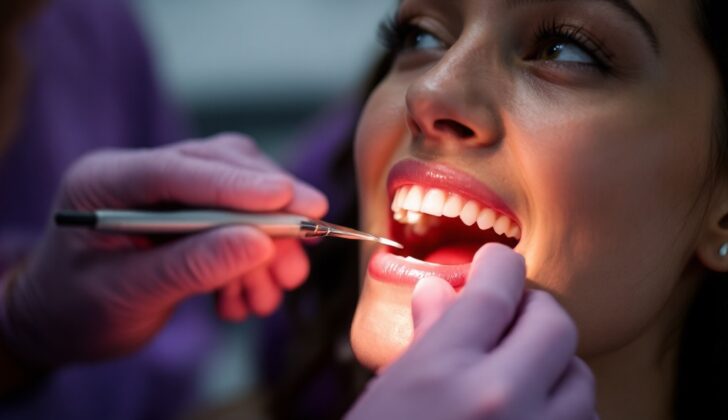
Early detection is very important for oral melanomas as they develop rapidly and generally have a grim outcome. Still, we don’t currently have specific criteria to assist in diagnosing this condition in the mouth. Clinical check-ups can make use of tools such as dermoscopy, a method where a special magnifying tool is used to examine the skin. However, the intricate nature of the mouth’s tissues and uneven surfaces can make this tricky.
The most reliable way to diagnose oral melanomas is through a biopsy. This is a medical procedure where a small sample of tissue is taken to be carefully examined in a lab. If the questionable area is large, an incisional biopsy, which involves cutting out a small piece of the thickest part, is done. If the lesion is small, an excisional biopsy, where the entire lump is removed, is chosen.
Lastly, to correctly determine the stage of the tumor, an ultrasound or CT scan of the head, neck, and the thoracoabdominal regions (chest and abdomen) might be required.
Dermoscopy is one technique used to visually assess the skin conditions. It typically reveals irregular pigmentation with false network-like structures, the presence of scar-like features, and a blue-white haze often associated with melanoma. Other features include uneven structures, irregular edges, and a sudden stop of a net-like pattern on the skin. Additionally, it might reveal atypical blood vessels, distinct points, and small rounded structures. These visual clues can help doctors in diagnosing melanoma.
Treatment Options for Oral Melanoma
Surgery is usually the primary way to treat oral malignant melanoma, a type of cancer found in the mouth. Doctors strive to radically remove the entire melanoma, ensuring that there’s no remaining disease within the margins of the excision.
After surgery, additional treatments such as radiation, chemotherapy, and immunotherapy are sometimes used. These treatments are meant to help manage the disease and prevent it from returning.
Melanoma is often not very sensitive to radiation – that’s the traditional understanding. However, some researchers have observed improvements and better control of the disease locally (where it started) and overall survival increase in patients who have undergone radiation therapy.
Chemotherapy – using drugs like platinum analog, nitrosoureas, and dacarbazine – as well as immunotherapy with interleukin-2 (IL-2), a protein made by white blood cells to regulate immune responses, haven’t shown a strong reaction in terms of disease management.
For melanomas that have spread to other parts of the body (metastasized) and have a specific genetic change called a c-kit mutation, a targeted therapy using imatinib has been beneficial. Furthermore, medications dabrafenib and vemurafenib are used in cases where there’s a specific genetic mutation (a change in DNA of the melanoma cells) called BRAF.
What else can Oral Melanoma be?
When diagnosing oral melanoma, a type of cancer, doctors need to make sure it isn’t actually something else. This cancer can sometimes look similar to different types of marks or discoloration in the mouth. One common example is an ‘amalgam tattoo’, which is a gray-blue or black spot in the mouth that can often be confused with oral melanoma. A helpful tool for doctors to tell the difference might include dermoscopy, a skin surface microscopy.
Here are some other conditions that might be mistaken for oral melanoma:
- Focal Oral Pigmentations:
- Amalgam tattoo (a patch of color caused by an outside source)
- Melanoacanthoma
- Melanotic macules
- Melanocytic nevi (a type of mole)
- Diffuse Oral Pigmentations:
- Physiological/racial pigmentations (natural color differences)
- Smoker’s melanosis (discoloration caused by smoking)
- Drug-induced hyperpigmentation (color change triggered by medication)
- Postinflammatory hyperpigmentation
- Systemic Diseases:
- Peutz–Jeghers syndrome
- Laugier–Hunzikerdisease
- Leopard syndrome
- Carney complex syndrome
- McCune-Albright syndrome
- Diseases of the adrenal gland
In each case, doctors would conduct tests and check the patient’s history to confirm the correct diagnosis.
What to expect with Oral Melanoma
Oral melanoma, a type of cancer in the mouth, is often diagnosed at later stages and is associated with a poor outcome. The estimated survival rate five years after diagnosis is only 25.5%.
About one-third of patients have cancer cells spread to their lymph nodes at the time of diagnosis, a process known as lymph node metastasis. This happens because the head and neck region has a rich supply of blood vessels and a network for lymphatic drainage, making it easier for the disease to spread.
The presence of a specific protein called bcl-2 can indicate a better outcome for this type of melanoma. However, abnormal levels of the p53 protein and the absence of p16 protein are linked to a worse outcome.
After surgery to remove the cancer entirely, there is a 20% chance that the disease will come back. Moreover, the disease may recur even up to 11 years after surgery. Cancer can spread to the lymph nodes of the neck, lungs, liver, and brain.
Possible Complications When Diagnosed with Oral Melanoma
The complications associated with the surgical removal of melanoma include loss of tissue, slow healing process, the need for grafting, and the requirement of artificial replacements or prosthesis. Further, dry mouth, a fungal infection called candidiasis, and a condition leading to nasal-sounding speech are potential side effects.
Complications After Surgery:
- Loss of tissue
- Slow healing process
- Requirement for grafting
- Prothesis needs
- Dry mouth
- Candidiasis (fungal infection)
- Hypernasal speech
Preventing Oral Melanoma
Identifying and treating oral mucosal melanomas (a type of skin cancer that occurs in the mouth) early on can greatly improve the chances of overcoming the disease. While we don’t have definite ways to prevent it, some medical professionals advise that regular self-examinations of the mouth could help spot unusual, coloured or colourless patches early.
For a thorough self-examination, you’ll need a mouth mirror, a standard mirror, and good lighting. You should also use a piece of gauze to gently hold your tongue, allowing you to see all areas inside your mouth more clearly.
People who have had oral melanoma are at a higher risk of having it again, so continual monitoring is necessary throughout their life.


