What is Retinoblastoma?
Retinoblastoma is a rare kind of eye cancer usually found in children. It occurs in about 1 in 18,000 births. While it is a fairly rare kind of cancer, it is actually the most common eye cancer in children and accounts for about 3% of all cancers found in children. After uveal melanoma, another type of eye cancer, retinoblastoma is the second most common eye cancer.
In specialized care centers, the survival rate for this condition is up to 95%, with most patients maintaining their sight, but this rate can be lower in developing countries.
Under the microscope, retinoblastoma cells, known as retinoblasts, have certain characteristics. They are basophilic, which means they stain blue, have hyperchromatic nuclei, indicating that the center of the cells is very densely packed, and scanty cytoplasm, meaning there isn’t a lot of cell fluid. Most of these cells are undifferentiated, or immature, but some are more mature and form structures known as rosettes.
The tumor can grow inward into the gel-like substance inside the eye called vitreous (endophytic), or seed throughout the eye, or it can grow outward under the retina, which is the layer at the back of the eye (exophytic). It can also present a mixed picture of these types.
The cancer can also invade the optic nerve, which is the cable connecting the eye to the brain, spreading into the area around the brain and even into the brain itself. It can also spread to other parts of the body such as the regional lymph nodes, liver, lungs, bones, and brain.
What Causes Retinoblastoma?
Retinoblastoma, a kind of eye cancer, occurs due to a change or “mutation” in a specific gene known as the RB1 gene, which is found on a specific area of chromosome 13. The RB1 gene works to control the growth of cells in our body, and when it doesn’t function correctly, a tumor can form in the eye. If both copies of your RB1 gene have mutated, there’s a strong likelihood you could develop retinoblastoma.
If a child develops retinoblastoma in both eyes (bilateral), it’s usually due to these mutations being present in the body from birth, which is often referred to as a “germline” mutation. It’s important to note, though, that only 5% of people with retinoblastoma have a family history of it, meaning it’s most commonly a random occurrence. In fact, 95% of cases come out of the blue, with no known cause or family history.
Now, let’s talk about the two main types of retinoblastoma: heritable and non-heritable.
A “heritable” retinoblastoma happens when one of the gene copies in every cell of the body is changed. If the second copy of the gene also mutates, it can result in the body’s cells becoming cancerous. As this mutation is present in all body cells, children with heritable retinoblastoma often develop tumors in both eyes, or in several places within one eye. This type also carries a higher risk for other types of cancers later in life, such as bone cancer (osteosarcoma), skin cancer (melanoma), and others. These other cancers usually appear during specific ages and are more likely if the original eye tumor was treated with a method known as external beam radiation.
On the other hand, “non-heritable” retinoblastomas usually only affect one eye (unilateral) and are not passed down in families. People with non-heritable retinoblastoma don’t have a higher risk of developing other types of cancers. In cases where the tumor is in one eye and there’s no family history of the disease, it’s usually a non-heritable retinoblastoma. The risk of developing non-heritable retinoblastoma for siblings or children of a patient is extremely low, at roughly around 1%. Most of the time, when retinoblastoma occurs in just one eye, it’s this non-heritable type.
Risk Factors and Frequency for Retinoblastoma
Retinoblastoma is a type of eye cancer that mainly affects children, making up 3% of all childhood cancers. It’s the second most common type of eye cancer. Every year, 300 new cases occur in the US. It affects boys and girls alike and usually shows up before a child turns three. The number of cases varies from place to place. For instance, in Mexico, there are six cases per million people, while in the US, there are four cases per million. However, India and Africa have the highest number of cases.
- Retinoblastoma is a common eye cancer in children, accounting for 3% of all childhood cancers.
- It’s the second most common eye cancer, affecting around 1 in every 14,000 to 20,000 live births.
- In the US, there are 300 new cases every year.
- Boys and girls are equally likely to get this disease.
- Most cases (90%) occur in children under the age of three.
- The number of cases differs in different regions, with India and Africa having the highest incidence.
Signs and Symptoms of Retinoblastoma
Retinoblastoma is a type of eye cancer that usually occurs in children within the first year of age when it affects both eyes, and within the first three years when it affects only one eye. Having a family history of eye cancers can increase the likelihood of developing this condition. The symptoms for retinoblastoma vary and can include:
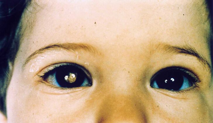
- Leukocoria, which is a white or whitish color seen in the pupil (this is the most common symptom found in 60% of cases)
- Strabismus, or squinting (this symptom makes it important to have a thorough eye examination for all children who squint)
- Painful, red eye, possibly associated with an increase in eyeball size (buphthalmos) or glaucoma
- Inflammation around the orbit of the eye, which might look like pre-septal or orbital cellulitis
- Visible growth outside of the eye
- Loss of vision
- Limited eye movement
- In rare cases, metastatic disease which is spread to lymph nodes, liver, lungs, brain, or bones
The signs upon examination may include the following:
- A whitish, dome-shaped growth in the retina with calcification (known as an intraretinal tumor)
- An endophytic tumor, which shows as a whitish growth in the jelly-like substance (vitreous) within the eye
- An exophytic tumor, which presents as a whitish growth under the retina resulting in retinal detachment
- Blood accumulation in the front part of the eye (hypopyon)
- Strabismus, which is an abnormal alignment of the eyes, commonly known as cross-eyed or squint
- Blood in the part of the eye between the cornea and iris (hyphema)
- Inflammation of the eye
- Different colours in the two eyes (iris heterochromia)
- Piercing of the eye globe (globe perforation)
- Abnormally protruding eyeball (proptosis)
- Cataract, clouding of the eye lens
- Glaucoma, a condition causing damage to the optic nerve due to high eye pressure
- An unequal size of pupils (anisocoria)
Testing for Retinoblastoma
If your doctor suspects you have retinoblastoma, a type of eye cancer, they might perform several tests to confirm this diagnosis. The simplest test is red reflex testing using a device called a direct ophthalmoscope; this device allows easy detection of leukocoria, a symptom where the pupil of the eye appears white instead of black.
For a more detailed examination, your doctor might recommend an examination under anesthesia. This would allow accurate measurement of the corneal diameter (the front, transparent part of your eye), check eye pressure, use a handheld slit lamp to inspect the front part of your eye, examine your retina (the light-sensitive layer at the back of your eye), measure your eye’s focusing power, and document all these findings.
An ultrasound might be conducted to measure the size of the tumor and observe any calcifications (areas of the tumor that have hardened due to buildup of calcium). This test also helps to rule out conditions similar to retinoblastoma, such as Coats disease, a rare eye disorder.
Wide-field photography can also be used for analyzing and documenting the condition of your eye. This information aids in managing retinoblastoma.
Your doctor may also suggest a CT scan, which would help detect calcifications. However, due to the risk of radiation exposure, a CT scan is typically not the first option for diagnosis.
Magnetic Resonance Imaging (MRI) is another imaging technique used to study the optic nerve (the nerve that transmits visual information from the eye to the brain), identify any spread of the cancer outside the eye, look for pineoblastoma (a rare type of brain cancer), and to exclude similar diseases.
You might also need to undergo a systemic assessment, which involves a physical examination, an MRI of the orbit (eye socket) and brain, a bone scan to check for cancer spread, a bone marrow aspiration (a procedure where a small amount of bone marrow is taken for testing), and a lumbar puncture (a procedure where a small amount of spinal fluid is taken for testing).
Lastly, your doctor might order genetic studies of blood samples and tumor tissue from you and your relatives to identify any genetic factors contributing to the condition.
Treatment Options for Retinoblastoma
Treating retinoblastoma, which is a type of eye cancer, involves teamwork from various healthcare professionals including an eye doctor, a pediatric cancer specialist, a specialist who studies diseases in the eye, a geneticist, and other healthcare providers, along with the child’s parents. Several different strategies are used to manage retinoblastoma.
Chemotherapy, which involves powerful drugs that kill cancer cells, is the primary treatment strategy. These drugs are typically delivered through a vein and may include medications like carboplatin, etoposide, and vincristine. The choice of drugs and the number of treatment cycles depend on the severity of the retinoblastoma. Sometimes, single or double drug treatments are used and have given good results in certain patients, like as a bridging therapy to sidestep more aggressive treatments. In some cases where the disease has spread to the jelly-like substance that fills the eye, a drug called intravitreal melphalan is used, though it carries a minor risk of cancer spread beyond the eye. After chemotherapy, additional treatments like cryotherapy (freezing therapy) or TTT (heat therapy applied through the pupil of the eye) can be used to ensure maximum control over the tumor.
TTT is mainly used after chemotherapy to consolidate the treatment effect. However, it can also be used alone. It works directly on the cancer cells but can also increase the effectiveness of chemotherapy.
Cryotherapy, specifically a method called the triple freeze-thaw technique, is an option for tumors located in the front half of the eye that haven’t spread too deep or into the jelly-like substance in the eye.
Brachytherapy, which involves applying radiation directly to the tumor, is used for front-of-the-eye tumors that haven’t spread into the vitreous and aren’t responding to chemotherapy.
As much as possible, external beam radiotherapy, which involves directing radiation at the tumor from outside the body, is avoided, especially in heritable retinoblastoma (retinoblastoma that can be inherited). While retinoblastomas respond well to radiation, it can unfortunately lead to side effects like cataract, nerve damage, retinal damage, and stunted orbit growth.
Enucleation, or removal of the eye, is done when the cancer has spread to the front part of the eye, caused new blood vessels to grow that increase eye pressure, invaded the optic nerve, or if it takes up more than half of the vitreous. It’s also done when chemotherapy hasn’t worked, and in cases of diffused retinoblastoma where it has a high risk of coming back due to poor visual prognosis. When performing enucleation, it’s important to minimize manipulation and cut about 10mm of the optic nerve. Thanks to recent advances, a long segment of the optic nerve can now be removed while the surgeon can directly see it.
If the cancer has spread outside the eye (extraocular extension), additional chemotherapy for 6 months is given after enucleation if the cancer has spread behind the tough white covering of the eye or severely spread into the vascular layer of the eye. If the tumor has grown to the cut end of the optic nerve during enucleation, or if it has grown through the white wall of the eye, then external beam radiation is delivered.
After treatment, careful and regular follow-up is needed to catch any recurrence or new tumor growth early, especially in patients with the inheritable form of the disease.
What else can Retinoblastoma be?
When doctors try to diagnose retinoblastoma, they take into account that other conditions can cause similar symptoms. These could include:
- Persistent anterior fetal vasculature
- Persistent posterior fetal vasculature
- Coats disease
- Retinopathy of prematurity (a vision problem in babies)
- Toxocariasis (an infection caused by roundworms)
- Uveitis (inflammation of the eye)
- Vitreoretinal dysplasia (an abnormal development of the retina)
- Coloboma of the choroid and optic disk (a hole in parts of the eye)
- Posterior cataract (a type of cataract)
These conditions should be considered and tested for in order to help reach the right diagnosis.
What to expect with Retinoblastoma
Patients with intraocular retinoblastoma, a type of eye cancer that starts in the retina, have a high chance of overcoming the disease, especially if they have access to up-to-date healthcare facilities. The survival rate of this condition is over 95% in developed countries. However, the prognosis can be worse if the cancer extends beyond the eye, either through the sclera (the white part of the eye) or by invading the optic nerve.
Patients who survive bilateral retinoblastoma, meaning the disease affects both eyes, have a higher chance of developing other types of cancer later in life. The time interval before the development of a second cancer is usually about 9 months. External beam radiotherapy, a type of radiation treatment, can reduce this time interval and increase the risk of developing a second cancer within the first 30 years of life.
The most common type of second cancer to develop after retinoblastoma is a sarcoma, a type of cancer that can occur in various tissues. Unfortunately, patients who develop a sarcoma have a survival rate of less than 50%.
Possible Complications When Diagnosed with Retinoblastoma
If retinoblastoma, a type of eye cancer, isn’t treated, it can lead to several serious health problems. Here is a list of potential complications:
- Detachment of the retina – the light-sensitive layer at the back of the eye
- Retinal necrosis – death of retinal tissue
- Orbital invasion – the cancer spreads to the eye socket
- Optic nerve invasion – the cancer reaches the nerve that connects the eye to the brain
- Blindness
- Intracranial extension – the cancer spreads to the brain
- Secondary neoplasms – new cancers develop
- Metastasis – the cancer spreads to other parts of the body
- Tumor recurrence – the cancer comes back after treatment
- Temporal bone hypoplasia – underdevelopment of the bones near the side of the skull
- Cataract – clouding of the eye’s lens
- Radiation neuropathy – nerve damage caused by radiation therapy
- Radiation retinopathy – damage to the retina caused by radiation therapy


