What is Somatostatinoma?
Somatostatinoma is a very rare type of tumor, specifically known as a neuroendocrine tumor (NET), which affects about 1 in 40 million people. It makes up less than 5% of all NETs that occur in the pancreas (pNETs). This type of tumor was first identified in 1977 in a 46-year-old woman and it was located in the head portion of the pancreas.
This tumor develops from special cells in the pancreas known as delta cells. Its main component is a hormone called somatostatin, but it may also contain small amounts of other hormones found in the pancreas such as insulin, glucagon, gastrin, and vasoactive intestinal polypeptide, which all help regulate various body functions.
In most cases (between 56 and 70%), the tumor remains within the pancreas. Among these cases, 36% are found in the head of the pancreas, 14% in the body, and 32% in the tail. Other places where this tumor can be found include the duodenum (19%), which is the first part of the small intestine, the ampulla of Vater (3%), which is where the bile and pancreatic ducts release their secretions into the duodenum, and the small bowel (3%).
In very rare cases, this tumor has been found in places outside of the gastrointestinal tract, like the lungs, kidneys, and thyroid.
What Causes Somatostatinoma?
Somatostatinoma, a type of tumor, can occur on its own or in combination with skin and nervous system disorders, the most common of which is multiple endocrine neoplasia type 1, which accounts for 40 to 50% of cases. Other disorders that are often associated with it include neurofibromatosis type 1 (NF1), and Von Hippel-Lindau syndrome.
Additionally, changes in specific chromosomes have been linked to the development of this disease. Examples of these changes are the loss of certain genes on chromosomes 11q and 6q, and an alteration to chromosome 3p. These changes to the genetic material have been associated with the onset of somatostatinoma.
Risk Factors and Frequency for Somatostatinoma
Somatostatinoma, a medical condition, typically appears in people between the ages of 40 and 60. It affects both males and females equally.
Signs and Symptoms of Somatostatinoma
Non-functional tumors often don’t show any symptoms. But as the disease progresses, they can cause abdominal pain, vomiting, yellowing of the skin and eyes (jaundice), and fatty stool (steatorrhea). This happens due to the tumor’s pressure on the bile and pancreatic drainage systems. On the other hand, functional tumors show symptoms due to the effects of somatostatin, a hormone that slows down the function of certain body systems.
- Almost 70% of symptomatic cases have gallstones (cholelithiasis).
- Around 60% have diabetes mellitus.
Occasionally, patients might exhibit a trio of symptoms: diabetes mellitus, gallstones, and fatty stool. This combination is known as the inhibitory syndrome because somatostatin suppresses insulin, cholecystokinin (a hormone that facilitates digestion), and pancreatic enzymes required for digestion.
In physical examinations, signs like yellowing skin (jaundice) could hint at obstructive jaundice. Abdominal tenderness might point towards acute cholecystitis, which is an inflammation of the gallbladder. People suffering from Neurofibromatosis Type 1 (NF1) might also have particular skin markings like ‘café au lait’ spots, nerve tumors (neurofibromas), and armpit freckling.
Testing for Somatostatinoma
Diagnosing somatostatinoma, a type of tumor, early on can be difficult. This is because non-functional tumors (ones that do not produce hormones) often don’t show any symptoms. Functional tumors (ones that do produce hormones) can present signs that are general and might appear to be other health issues. These non-functional tumors are usually found unexpectedly or when investigating unclear digestive tract symptoms.
If you have a functional tumor, you might see an increase in fasting serum somatostatin levels (higher than 14 mmol/l). Somatostatin levels might also increase with other cancers such as thyroid cancer, lung cancer, pheochromocytoma, and paraganglioma. The 24-hour urine level of 5-hydroxy indole acetic acid (5-HIAA), which is the breakdown product of serotonin, can be used as another test. But, the 5-HIAA level can be affected by different foods and medications.
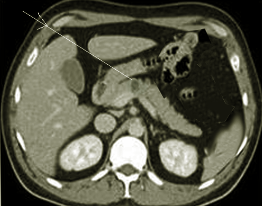
Somatostatinoma usually gets diagnosed later, when tumors are larger and easier to see with imaging techniques like CT, MRI, or MRCP. Multiphasic contrast-enhanced CT abdomen is the first test of choice because it is non-invasive and widely available. MRI is getting used more frequently where pNETs have a low signal density on T1-weighted images and high signal density on T2-weighted images. MRI also has a higher ability to detect small liver metastases. Procedures such as esophagogastroduodenoscopy (EGD), endoscopic retrograde cholangiopancreatography (ERCP), or endoscopic ultrasound (EUS) can be performed to take a closer look inside and take a sample of the tumor to confirm the diagnosis. EUS is very sensitive to detect lesions as small as 0.5 cm and is best for lesions in the head of the pancreas and lesions of the duodenal wall.
Treatment Options for Somatostatinoma
The treatment for a tumor depends on how well the tumor is working, if it can be operated on, and how much it has spread (staging). The best way to completely get rid of the tumor is through surgery, where the main tumor and any lymph nodes that it has spread to are removed. If nearly 90% of the tumor can be cut out, then the procedure is done with the hope of curing the patient.
In cases where the tumor has spread locally or to nearby regions like the liver and lymph nodes, two types of surgeries may be used. One is called tumor debulking, which reduces the size of the tumor. The other is called hepatic resection, where a part of the liver is removed. These procedures help alleviate symptoms caused by mechanical blockages or the large size of the tumor.
The type of surgery done depends on where the original tumor is located. Some common procedures include pylorus-preserving proximal pancreaticoduodenectomy (a surgery that removes a part of the pancreas and small intestine while preserving the opening from the stomach to the small intestine), distal pancreatectomy with splenectomy (a surgery that removes the tail of the pancreas and the spleen), and Whipple’s procedure (a surgery that removes the head of the pancreas and the first part of the small intestine).
Grade 3 pNETs tumors are usually widespread at the time of diagnosis, making them hard to operate on.
If the tumor has only spread to the liver, it can be treated with a procedure to remove the area of the liver with the tumor, or a treatment called selective hepatic transcatheter arterial embolization (TAE) which blocks blood flow to the area. Studies have shown that aggressively removing liver metastases (areas where the cancer has spread) can significantly increase long-term survival.
In more complex cases where the disease has spread to multiple sites or can’t be operated on, other treatments might be used. These can include ablative procedures (which destroy tissue), like radiofrequency ablation (using heat to kill cancer cells), laser-induced thermotherapy (using lasers to heat and destroy cancer cells), TAE, transcatheter arterial chemoembolization (injecting chemotherapy into the blood vessels to block blood flow and kill cancer cells), and selective internal radiotherapy (injecting radioactive material to kill cancer cells). These treatments are often combined with systemic chemotherapy to shrink the tumor.
What else can Somatostatinoma be?
When someone experiences symptoms due to a blockage in the bile ducts, doctors need to rule out caused by other conditions that can also lead to similar symptoms. These conditions can include:
- Gallstones in the bile duct (choledocholithiasis)
- Bile duct cysts (choledochal cyst)
- A condition that causes inflammation of the bile ducts (sclerosing cholangitis)
- Bile duct cancer (cholangiocarcinoma)
- Fluid-filled sacs in the pancreas (pancreatic pseudocysts)
- Pancreatic cancer (pancreatic adenocarcinoma)
Additionally, symptoms such as fatty stools (steatorrhea) and a particular type of diabetes that requires insulin (insulin-dependent diabetes mellitus) could indicate the presence of a hormone-producing tumor called a somatostatinoma. However, similar symptoms can also be seen in chronic pancreatitis and other types of pancreatic tumors, so these conditions need to be considered as well.
What to expect with Somatostatinoma
The 5-year survival rate for patients with a specific type of tumor called pancreatic or periampullary somatostatinoma varies. If the disease is caught early and hasn’t spread outside the pancreas (localized disease), it ranges from 60 to 100%. However, if the disease has spread to other parts of the body (metastatic disease), the 5-year survival rate is between 15 to 60%.
Several factors affect the patient’s survival rate. These include the size of the tumor, how abnormal the cancer cells are (degree of cytological differentiation), how much of the tumor is left after surgery (residual tumor post-resection), and how far the cancer has spread (extent of metastasis).
For patients who underwent tumor removal surgery, the 5-year survival rates for stages I, II, III, and IV were 89.9%, 82.6%, 75.8%, and 56.9%, respectively. Each of these stages represents the growth and spread of cancer, with stage I being the earliest and IV being the most advanced.
In a study looking back at patients with liver metastases (cancer that has spread to the liver), those who also had bone metastases (cancer spread to the bones) had significantly lower median survival spans of 15.4–62.1 months, compared to 18.2–166.3 months for those without bone metastases.
Possible Complications When Diagnosed with Somatostatinoma
The potential negative effects on the whole body include conditions like diabetes mellitus, gallstones, anemia due to deficient iron, enlarged red blood cells, and a lack of fat-soluble vitamins like A, D, E, and K. These deficiencies can lead to problems such as night blindness, weakened bones, and ease of bleeding. Diabetes mellitus is typically a mild condition, and a life-threatening complication called ketoacidosis is uncommon. It is managed with a special diet, oral drugs that lower blood glucose levels, or small amounts of insulin.
Even if a patient doesn’t show signs of gallstones, doctors often choose to remove the gallbladder as a preventive measure during the initial surgical exploration. This is due to the high occurrence of gallstones among these patients, particularly if they need somatostatin analogs treatment.
In terms of localized complications, the tumor can block the flow of bile, leading to excess bilirubin – a waste material – in the blood (called obstructive hyperbilirubinemia). It can also cause bleeding in the digestive tract.
- Diabetes Mellitus
- Gallstones
- Iron deficiency anemia
- Macrocytic anemia (enlarged red blood cells)
- Deficiency in vitamins A, D, E, and K
- Night blindness
- Osteopenia (weakened bones)
- Bleeding
- Obstructive hyperbilirubinemia (excess bilirubin)
- Gastrointestinal bleeding
- Possible preventive removal of the gallbladder
Recovery from Somatostatinoma
It is usually recommended that patients have follow-ups for at least 10 years if they’ve had a surgical removal of certain type of tumors in the digestive system, namely gastroenteropancreatic NETs, which are either well-differentiated or moderately differentiated in stages I to III. This recommendation comes from a global team of experts from different fields. The methods they suggest for follow-ups include imaging tests and biomarker testing, which help to monitor any changes in the body.
After surgery, the follow-ups will typically involve imaging scans (like CT scans of the stomach region or MRI) once a year for the first three years, and then once every one to two years. This applies specifically to pancreatic and midgut NETs, which are types of tumors found in the pancreas and part of the small intestine.
If a person’s Ki-67 mitotic index is above 10%, or if cancer cells are found in the lymph nodes, it’s usually advised to have these follow-ups more often. However, if the pancreatic tumors are at stage I or grade 1, or if the midgut NETs were discovered by chance at stage 1 or grade 1, follow-ups aren’t always needed.
Biomarkers, which are substances that can indicate the presence of cancer, are not normally recommended for regular monitoring. However, the European Neuroendocrine Tumor Society (ENETS) suggests doing imaging tests at the start and then every two years for grade 1 tumors, and after 3 months and then yearly for grade 2 to 3 pNETs, which are a type of tumor in the pancreas.
Preventing Somatostatinoma
It’s crucial for both your doctor to be knowledgeable and for you, the patient, to be informed about your disease for timely diagnosis and early start of treatment. Trustworthy online sources like the American Cancer Society offer information based on scientific evidence that both doctors and patients can use to understand the details of your health condition.


