What is Buphthalmos?
Buphthalmos is a term that comes from a Greek word meaning “ox-eyed.” It refers to an uncommon condition where the eye is unusually large from birth. Interestingly, even though this condition was documented as early as 400 BC by ancient doctors like Hippocrates, it was not associated with too high eye pressure until after the 19th century when tools to look inside and measure the eye’s pressure were invented.
Nowadays, the term “buphthalmos” is used to refer to the noticeable enlargement of an eye that is observed at birth or shortly after. This tends to be due to uncontrolled glaucoma during early childhood, a condition where the fluid pressure inside the eyes is too high.
What Causes Buphthalmos?
Buphthalmos is a condition that most often happens because of a disease babies are born with called primary congenital glaucoma. This disease increases the pressure in the eye. Other conditions that can increase eye pressure in early childhood can also lead to buphthalmos. These include Sturge-Weber syndrome, neurofibromatosis, and aniridia.
Risk Factors and Frequency for Buphthalmos
Buphthalmos, a type of primary congenital glaucoma, is a condition that affects an average of 1 in 30,000 newborns. However, the prevalence of this disease is significantly higher in communities such as the Slovak Romani population, South India, and Saudi Arabia. Remarkably, primary congenital glaucoma is a major reason for childhood blindness in these regions. Specifically, the highest occurrence of this condition has been noted in Slovakia and Saudi Arabia, with 1 in 1,250 and 1 in 2,500 live births being affected, respectively.
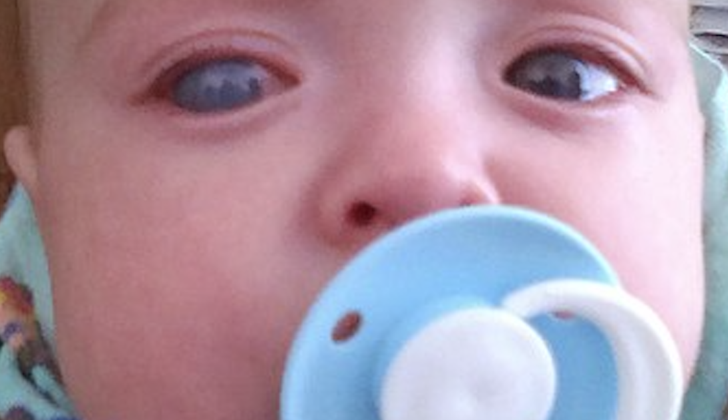
Signs and Symptoms of Buphthalmos
Buphthalmos, often resulting from primary congenital glaucoma (present at birth) or primary infantile glaucoma (starting after birth up to 3 years), is a condition where the eyeball becomes enlarged. Typically, if glaucoma develops after the age of 3 years, buphthalmos is not seen. This explains why juvenile-onset glaucoma (from 3 years to teenagers) doesn’t usually come with buphthalmos.
Classic symptoms of congenital and infantile glaucomas may include excessive tearing, sensitivity to light, and irritability. Parents might spot a cloudy cornea or an increase in cornea size. Medical professionals would observe an enlarged cornea diameter, an increase in eye size, and a deep anterior chamber (the space in the eye behind the cornea). They might also see corneal edema and signs of ruptures in Descemet’s membrane, known as Haab’s striae, through corneal examination. Anothere signs may include high eye pressure and possible optic disc cupping.
Some other conditions that could lead to buphthalmos include:
- Aniridia: This is defined by complete or partial underdevelopment of the iris. It is passed down through families (autosomal dominant inheritance). The glaucoma is thought to be caused by a closed angle in the eye.
- Neurofibromatosis type 1: This condition is recognized by multiple nerve skin tumors, light brown skin spots, iris Lisch nodules, and armpit/groin freckling. It is hereditary with a dominant trait. The glaucoma is considered to be due to developmental anomalies of the anterior chamber angle.
- Sturge-Weber syndrome: This syndrome is depicted by a birthmark on the face and meningeal angiomas. Glaucoma and associated angle anomalies can be seen in 60% of such cases. Anomalous angle development and increased episcleral venous pressure are suggested causes of glaucoma.
Testing for Buphthalmos
When examining patients suspected of having buphthalmos, a condition characterized by an abnormal enlargement of the eye, doctors often need to carry out the evaluation under anesthesia. This process may involve several types of examination:
Refraction using streak retinoscopy: This test checks how well your eyes respond to light. It helps the doctor to figure out the right prescription for glasses or contact lenses, if they’re needed.
Corneal Examination: The cornea of your eye will be observed carefully. The doctor may use calipers, a tool that measures size, to see if the cornea’s diameter is larger than usual. A diameter larger than 12mm is considered abnormal. They might also check for corneal edema, a swelling in the cornea caused by an increase in fluid pressure inside the eyeball.
The doctor may look for breaks in the Descemet’s membrane, the inner layer of the cornea. These breaks, called Haab’s striae, are usually sideways or parallel to the edge of the cornea.
The doctor will also examine the front part of your eye to check for other possible causes of buphthalmos, such as aniridia (a rare condition where the iris is missing), and Lisch nodules that are related to neurofibromatosis, a genetic nerve disorder.
The pressure inside your eye may be measured in the first few minutes of anesthesia to avoid any inaccuracies in the results. This is critical in diagnosing buphthalmos as the condition is often linked to an increased intraocular pressure.
Gonioscopy, an examination of the front part of your eye with a special lens, could reveal certain abnormalities associated with buphthalmos, such as an abnormal position of the iris or unusual blood vessels and tissues in the eye.
An ophthalmoscope, a tool to see the back of your eye, helps the doctor check for optic disc cupping. This condition, a sign of certain types of glaucoma, causes the optic nerve’s head to sink back into the eye. It’s worth noting that with the right treatment, optic disc cupping may be reversible in many cases of primary infantile glaucoma, a common form of buphthalmos.
In cases where the cornea is opaque and hard to see through, an ultrasound biomicroscopy may be conducted to examine the structures at the front of the eye. In addition, a genetics consult could be recommended in certain cases.
Treatment Options for Buphthalmos
The main purpose of the treatment for Buphthalmos, a condition characterized by enlarged eyes due to a rise in inner eye pressure, is to reduce this pressure. This is done to prevent further cloudiness of the eye’s outer layer (cornea) and damage to the optic nerve, which can lead to loss of vision. The best treatment for Buphthalmos is usually surgery. Medications can also be used to manage the pressure in the eye and to clear the cornea before an operation.
Medication Treatment
The use of certain medicines can be part of the treatment. These include eye drops such as beta-blockers, carbonic anhydrase inhibitors, and prostaglandin analogs.
Surgical Treatment
There are several types of surgeries that can be performed:
– Goniotomy: This method involves making openings in the trabecular meshwork, a network of tiny channels in the eye, to help reduce resistance to fluid outflow.
– Trabeculotomy: In this procedure, a tiny probe is used to cut open the trabecular meshwork, which also helps increase fluid outflow from the eyes.
– Trabeculectomy: This involves removing a part of the trabecular meshwork and another part of the eye called Schlemm’s canal to create a new pathway for fluid drainage, which is usually beneath a partial thickness scleral flap.
– Combined trabeculotomy and trabeculectomy: This method takes out a small part of the outer white part of the eyes (sclera) after performing a trabeculectomy.
– Glaucoma drainage implants: These are small devices implanted in the eye to help fluid drain.
– Cyclodestructive procedures: This is a group of surgical techniques that aim to reduce the production of eye fluid.
After the surgery, the doctor should correct any existing vision problems and address any issues related to lazy eye (amblyopia). It’s necessary for these patients to have their eye pressure checked regularly for the rest of their lives.
What else can Buphthalmos be?
There are several conditions that can alter the appearance of the eye in a way that looks like buphthalmos. A thorough eye exam is required to ensure that these other diseases are not causing the abnormal look. Here are some conditions that doctors would typically check for:
- Aniridia
- Coat’s disease
- Dysplasia of retina
- Endophthalmitis
- Inflammatory cyclitic membrane
- Neurofibromatosis type 1
- Persistent hyperplastic primary vitreous
- Retinoblastoma
- Sturge Weber syndrome
- Toxocariasis
What to expect with Buphthalmos
If a person has buphthalmos, a condition characterized by an abnormal enlargement of the eye, due to untreated congenital glaucoma, they usually have a very poor outlook for their vision. Vision loss caused by this type of glaucoma is serious and cannot be reversed. However, if these patients undergo surgery early on, they are more likely to have better visual results.
Indeed, if treated early, the majority of cases generally do not show any worsening of the glaucoma within the first year of treatment. Despite this, across their lifetime, almost half of the patients may see further progression of their condition. This emphasizes the importance of regular eye check-ups throughout the patient’s life.
Possible Complications When Diagnosed with Buphthalmos
When children have a high intraocular pressure (IOP), which is the pressure inside the eye, it can cause an eye condition known as buphthalmos. This condition can lead to various complications related to high IOP such as damage to the optic nerve (glaucomatous optic neuropathy), injury to the clear, front surface of the eye (corneal damage), lazy eye (amblyopia), and vision defects that cause blurry or distorted vision (refractive error).
Reducing the high IOP as soon as possible might prevent these complications. However, certain issues like lazy eye and refractive errors would still necessitate separate treatments. Lastly, injuries to the cornea might be permanent and could require future surgical intervention.
Possible complications related to high IOP include:
- Optic nerve damage
- Corneal damage
- Lazy eye
- Blurry or distorted vision
- Potential need for future surgery
Preventing Buphthalmos
It’s really important that doctors and parents learn about buphthalmos, which is a crucial sign of a serious eye condition called congenital glaucoma. This condition can cause permanent vision loss if not treated in time. A recent study showed that almost all of the time, parents were the first ones to spot the signs of this condition in their child and then take them to a doctor. This shows just how critical it is for parents to be aware of unusual eye symptoms.
Parents need to know that large eyes in their child might actually be a sign of a serious eye condition. If they notice this, they should take their child to an eye specialist as quickly as possible. Providing the right education for parents and doctors about the early signs and symptoms of congenital glaucoma can help prevent vision loss from this condition.


