What is Congenital Diaphragmatic Hernia?
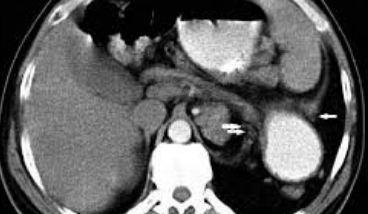
Congenital diaphragmatic hernia (CDH) is a condition that happens when there’s a development issue in the diaphragm, causing parts of the abdomen to push into the chest cavity. The hernia can be of different types based on where the issue in the diaphragm is. Bochdalek hernias are the most common (70% to 75%), occurring when there’s a problem in the back part of the diaphragm, usually on the left side and less commonly on the right. Morgagni hernias account for 20% to 25% of the cases, occurring when there’s a defect in the front part of the diaphragm. Central hernias are relatively rare and make up 2% to 5% of the cases. Defects on both sides are exceedingly rare and usually indicate a severe prognosis. It’s important to note that an acquired diaphragmatic hernia caused by trauma isn’t covered here.
What Causes Congenital Diaphragmatic Hernia?
The exact cause of congenital diaphragmatic hernia (CDH), a birth defect of the diaphragm, isn’t entirely known. However, it’s believed that a mix of genetic factors, environmental influences, and possibly nutrition may contribute. CDH can appear on its own or in conjunction with abnormalities in other organs.
Various chromosomal anomalies and gene mutations have been linked to CDH. The most common chromosome-related complications associated with CDH are trisomy 18, 13, and 21, and less commonly, Turner syndrome. Other disorders, such as Pallister-Killian syndrome, 8p23.1 deletion syndrome, Fryns syndrome, and Cornelia de Lange syndrome have been linked to CDH as well.
The condition can also be associated with exposure to certain harmful substances during pregnancy, such as mycophenolate mofetil, allopurinol, and lithium. Recent research points towards disruptions in the retinoid-signaling pathway, a biochemical communication route within cells, as a potential contributor to CDH.
Risk Factors and Frequency for Congenital Diaphragmatic Hernia
The occurrence of CDH, a condition linked to birth, is estimated to be around 1 to 4 cases per every 10,000 live births. This rate might be slightly different according to different population groups. In one study, it was found to be slightly more common in boys, however, other studies did not find this to be true.
Signs and Symptoms of Congenital Diaphragmatic Hernia
With improvements in prenatal scans, nearly two-thirds of congenital diaphragmatic hernia cases, a birth defect of the diaphragm, are identified before a baby is born. How this condition shows itself after birth depends on the size of the hole in the diaphragm.
Large diaphragmatic hernias often become noticeable at birth, indicated by problems with respiration, a blue skin color, reduced breath sounds on the side with the hernia, heart sounds that seem to come from the wrong place, and a hollow-looking belly. In contrast, small hernias might not be recognized right away, with symptoms like mild breathing issues and difficulties with feeding indicating their presence.
It’s important to note that a congenital diaphragmatic hernia can be just one aspect of a broader genetic syndrome. It might also be associated with issues affecting other organ systems, leading to a complex mix of birth defects such as heart defects, spinal cord defects, intestinal problems, and kidney anomalies.
Testing for Congenital Diaphragmatic Hernia
If a baby has a condition called Congenital Diaphragmatic Hernia (CDH), it is usually detected during a standard scan done between the 18th to 24th week of pregnancy. This condition can be noticed if the ultrasound shows parts of the stomach in the chest area and if the heart seems to be pushed to the opposite side of the hernia. But if the CDH is not very severe, it might not be noticed until later. This might happen if the hernia is not very large and there are no stomach parts having moved into the chest.
CDH that affects the right side of the diaphragm can be challenging to diagnose since the liver is similar in appearance to lung tissue on an ultrasound scan. In these cases, a more specialized type of ultrasound, called a color Doppler ultrasound, can help identify the presence of blood vessels from the liver in the chest.
Fetal Magnetic Resonance Imaging (MRI) has been increasingly used to assess the severity of CDH and detect any associated problems. The overall chance of survival and outcome in CDH is determined by factors like the underdevelopment of the lung (lung hypoplasia), the location of the liver, and any existing problems. For instance, the lung area to head ratio, a measure of the lung volume, is a crucial factor.
Based on a study, if this ratio is more than 1.35 in fetuses with CDH, it is associated with 100% survival, while a ratio of less than 0.6 contributes to no survival. However, as more infants with CDH are surviving, this ratio is more indicative of overall health challenges rather than mortality. The lung-to-head ratio varies with the baby’s age during pregnancy, hence the observed-to-expected ratio (O/E LHR) might be a better predictor of survival. In some cases, a ratio of less than 15 is associated with 100% mortality, but in cases where the ratio is more than 45 in left-sided CDH, the anticipated survival is more than 75%. An MRI can also assess total lung volume, known as O/E TFLV, which has also been found to have prognostic value.
CDH can complicate pregnancy if the amount of amniotic fluid (polyhydramnios) increases due to pressure on the esophagus, which affects the fetus’s ability to swallow. This could result in premature birth and adds to the health challenges and mortality associated with CDH. Considering the high risk of premature birth, medicines called antenatal corticosteroids are sometimes recommended. Lastly, due to an increased risk of chromosomal issues and related genetic conditions, genetic testing is advised for all CDH cases.
Treatment Options for Congenital Diaphragmatic Hernia
Once doctors confirm that a baby has a condition called congenital diaphragmatic hernia, they will monitor the baby regularly to ensure their wellbeing. For moderate to severe cases, some hospitals in the US offer a special treatment for eligible patients. The treatment involves blocking the baby’s windpipe with an inflated balloon around the 26th to 30th week of pregnancy. This process encourages the baby’s lungs to grow by accumulating lung fluid. However, this procedure also affects the production of a substance called surfactant that helps the lungs function properly. As a result, the balloon is removed around the 33rd to 34th week of pregnancy to help produce surfactant. This treatment might cause premature birth, but studies have shown that these babies generally have better survival rates compared to babies who don’t receive this treatment. Nonetheless, these babies will need further treatment after birth.
Babies with this condition should ideally not be delivered before their due date, i.e., the 37th week of pregnancy. Research has shown that babies with this condition who are born at full term, that is, the 40th week, have a much better chance of survival. So, if there are no complications, it’s best to let the pregnancy reach the 39th week. It’s also highly recommended that the babies are delivered in a hospital that specializes in treating this condition and where a treatment called extracorporeal membrane oxygenation (ECMO) is easily accessible.
Upon birth, a tube is typically placed in the baby’s nose that goes to the stomach to decrease pressure in the stomach and intestines. If the baby shows signs of respiratory distress, immediate intubation should be done to secure the airway. The rest of the medical care is based on standard neonatal resuscitation guidelines.
When it comes to ventilating these infants, the goal is to avoid injury to the lungs by maintaining gentle ventilation strategies. In some cases, a different type of ventilation called high-frequency ventilation may be needed. However, this is usually the last resort if regular methods don’t work.
Many of these babies will develop a condition known as pulmonary hypertension (PH) due to abnormal changes in their lung vessels. PH can be suspected based on symptoms like low oxygen levels, blue coloration of the skin, or greater oxygen saturation before it passes through the heart than afterward. It requires a test called echocardiogram, which looks at the heart on a screen. As for the treatment, it involves optimizing ventilation settings, maintaining normal blood pressures with certain medications if needed, and using medicines that increase blood flow in the lungs when necessary.
Surgery is one of the traditional ways to treat this condition. That said, surgery needs to be timed properly. Ideally, it should be delayed for at least 48 to 72 hours after birth to allow the baby’s lungs to adapt and improve. Those with severe PH may have their surgery postponed until PH is under control. In certain cases, ECMO, a treatment that uses a machine to take over the work of the heart and lungs, is used when all other treatments don’t work.
Historically, surgeons made an incision in the baby’s abdomen to treat this condition. Nowadays, less invasive methods are being used where possible. However, if the hole in the diaphragm is too large, it might not be possible to fix it directly. The doctors might need to use a patch, often made from synthetic material, a part of the abdominal or chest muscles, or a biological material to cover the hole. However, these procedures may increase the risk of future recurrence of this condition.
What else can Congenital Diaphragmatic Hernia be?
When doctors try to diagnose a congenital diaphragmatic hernia (CDH), they also consider other conditions that may appear similar. These could include lung abnormalities such as congenital cystic adenomatoid malformations, bronchopulmonary sequestrations, bronchogenic cysts, bronchial atresia, and teratomas.
One of the key findings that separate CDH from these conditions is the appearance of belly organs inside the chest on an x-ray. Before birth, signs like the presence of stomach bubble and intestines’ movement inside the chest when seen on an ultrasound can lead towards a diagnosis of CDH.
However, it can be tricky to distinguish between CDH and diaphragmatic eventration while the baby is still in the womb. The latter condition occurs when the diaphragm, the muscle that separates the chest from the abdomen, is intact but thin and raised.
What to expect with Congenital Diaphragmatic Hernia
The outcome of congenital diaphragmatic hernia, a birth defect in which an opening in the diaphragm allows the organs in the abdomen to move into the chest, can be influenced by several factors. These include how premature the baby is at birth, the size of the diaphragm opening, the development of the lungs, the level of blood pressure in the lungs, and the existence of other anomalies.
Over the years, improved strategies for aiding breathing, better management of high lung blood pressure and enhanced surgical methods have led to improved survival rates for babies with this condition. Nowadays, statistics show that 60% to 70% of babies with this hernia survive.
Possible Complications When Diagnosed with Congenital Diaphragmatic Hernia
Babies born with a birth defect known as a congenital diaphragmatic hernia often deal with a wide range of long-term health issues, even after their infant years. These could include:
- Respiratory problems like chronic lung disease, needing home oxygen, aspiration pneumonia, high blood pressure in the lungs, and blocked airways.
- Gastrointestinal issues like aversion to oral feeding, acid reflux, and poor growth.
- Neurological and emotional challenges including developmental delays, behavioral issues, and sensorineural hearing loss.
- The hernia coming back, which can happen months or years later, especially in babies with large defects.
- Orthopedic deformities like indented chests, uneven chests, and scoliosis.
All of these health problems may significantly impact the quality of life for these children. However, a recent study from Sweden found that, on the whole, children born with congenital diaphragmatic hernia have a good overall quality of life in terms of health.
Preventing Congenital Diaphragmatic Hernia
Patients born with a congenital diaphragmatic hernia need regular check-ups over a long period due to possible complications. It’s crucial that not only the patients but also their parents understand the importance of these regular doctor’s visits. Seeing a pediatrician regularly helps prevent health issues before they start. Additionally, consulting with specialized doctors helps manage any complications, minimizing the impact on the patient’s daily life. The American Academy of Pediatrics has put together a comprehensive guideline that outlines the necessary steps for these infant’s follow-up care.


