What is Craniosynostosis?
Craniosynostosis happens when the flexible joints in a baby’s skull, known as sutures, fuse together too early. Normally, these joints help the baby’s head fit through the birth canal during childbirth and allow the brain to grow afterward. If one or more of these sutures close before they should, it changes the shape of the skull. The skull grows in the direction where it finds least resistance, which is usually in a direction perpendicular to the closed suture. This results in an unusually shaped skull. The fused sutures can also increase the pressure inside the skull (known as intracranial pressure) which can affect the child’s breathing, nervous system, and overall development.
What Causes Craniosynostosis?
Craniosynostosis, a condition affecting the seams in a baby’s skull, can be categorized in two ways. If it involves only one seam, it’s termed as ‘simple’. But if it involves several seams, it’s ‘complex’. Another way to classify it is by noting if it is related to a syndrome like Apert, Crouzon, or Pfeiffer (syndromic), or if it’s the only issue being faced (non-syndromic).
Risk Factors and Frequency for Craniosynostosis
Craniosynostosis, a condition affecting the skulls of newborns, is relatively rare. It occurs in only 1 out of every 2000 to 2500 live births, but its occurrence has increased over time. Various factors can contribute to its development, including environmental conditions like maternal smoking and exposure to harmful substances in the womb, and genetic factors such as inherited mutations. Interestingly, even though a significant 20% of all cases are due to genetic causes, half of these are new mutations that weren’t inherited.
The majority of cases, around 75%, are classified as non-syndromic craniosynostosis. The other 25% are syndromic, in which other symptoms or disorders are also present. The way we classify the condition also depends on which part of the skull is affected.
- The sagittal synthesis is affected in 55% to 60% of cases.
- The coronal synthesis is affected in 20% to 25% of cases.
- The metopic synthesis is implicated in about 15% of cases.
- The lambdoid synthesis is affected in only 3% to 5% of cases.
Doctors usually identify craniosynostosis in the first year of a child’s life.
Signs and Symptoms of Craniosynostosis
To diagnose a cranial condition, doctors use careful physical examination and take a comprehensive medical history. They’d ask questions about things like family history of abnormal head shapes, exposure to harmful substances while in the womb, problems during pregnancy, or any developmental delays.
The physical examination helps the doctor understand if the sutures in the skull have fused prematurely and if there are other signs that might indicate a syndrome rather than an isolated case, such as different birth defects and unusual physical characteristics.
Checking the skull is important. This includes examining the head from all angles, measuring its size, and calculating its proportions to evaluate whether any sutures are fused. The doctor would also touch the scalp to feel for any irregularities, blood vessels on the skull, and examine the soft spots on the baby’s head.
Depending on the severity, the patient may show other symptoms caused by the abnormal shape of the skull, which should also be monitored. Eye tests should be carried out to check for signs of increased pressure inside the skull, and the doctor would examine the patient for possible breathing difficulties and feeding problems.
Cranial conditions can vary as follows:
- Scaphocephaly or dolichocephaly: features a long, narrow head with a more pronounced forehead.
- Anterior plagiocephaly: causes a flat forehead on the affected side, misaligned eyes in x-rays, uneven forehead, and a deviated nose.
- Posterior plagiocephaly: causes an uneven forehead and back of the head, the ear on the affected side appears lower, the head when viewed from above looks trapezoid.
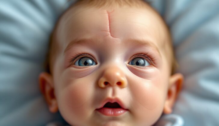
- Trigonocephaly: manifests as a narrow and pointed forehead, triangular head when seen from above, and unusually closely spaced eyes.
- Brachycephaly: characterized by a short skull, with a flat forehead and back head, whereas the frontal bone tends to be elongated and prominent.
- Oxycephaly or turricephaly: is due to the fusion of all or most of the cranial sutures.
Different syndromes can also cause these conditions, such as:
- Apert: affects a certain suture and causes physical characteristics like facial deformities, unusually widely spaced eyes, a beaked nose, small jaw leading to crowded teeth, abnormal fusion of fingers or toes, and potentially mild to moderate intellectual disabilities.
- Crouzon: can affect multiple sutures and causes facial deformities as well with normal intelligence.
- Pfeiffer: traits include abnormal skull shape, widely spaced eyes, small upper jaw, abnormally broad thumbs, toe abnormalities, and hearing loss.
- Muenke: affects a certain suture frequently and includes symptoms like facial anomalies, unusually large head, and hearing loss.
- Kleeblattschadel: known as the cloverleaf deformity and occurs with simultaneous fusion of two sutures producing a tri-lobar-shaped head and associated with excessive fluid in the brain.
Proper diagnosis is important as it guides appropriate medical intervention to avoid complications associated with untreated craniosynostosis.
Testing for Craniosynostosis
While diagnosing a condition typically involves a doctor’s clinical examination, sometimes additional tests like radiologic imaging are needed for further evaluation and confirmation. Multiple methods are used to assess the situation.
The most accurate of these methods is the CT scan with 3D reconstruction, which allows to thoroughly inspect all sutures. However, due to a potential risk of radiation, this method should be used after careful consideration. Plain x-rays are a more budget-friendly option and can be used for babies with a lower risk of craniosynostosis, but they are not as precise.
Magnetic Resonance Imaging (MRI) is slightly less precise compared to CT scans, but it’s still a very effective method. Doctors usually prefer to use it for children when any brain anomalies are revealed in CT scans.
Ultrasound is another affordable technique. It’s particularly useful for watching and checking sutures during each exam, though it can only be used with open fontanelles. Its effectiveness largely depends on the experience of the technician performing the ultrasound.
Recently, new methods such as GRASE (gradient-and-spin-echo), a type of MRI, have been used. GRASE improves the view of the boundary between bone and soft tissue, making cranial sutures stand out more.
Whenever doctors come across craniosynostosis in association with other syndromes, they typically recommend genetic testing. They particularly look for FGF receptor genes, which are most commonly associated with these syndromes. Examples of these include FGFR2 and FGFR3, as well as certain transcription factors (TWIST, MSX2). As research progresses, we have come to understand the genetic causes of these syndromes more effectively. To date, 57 genes have been identified as probable causes of craniosynostosis, with the most common ones being those previously mentioned.
Treatment Options for Craniosynostosis
The treatment for craniosynostosis—a condition in which one or more of the fibrous joints in a baby’s skull prematurely fuse—often depends on the specific type a patient has. Simple, non-syndromic cases are typically treated with surgery, but not urgently. On the other hand, certain syndromic types—those linked with other health issues—may require quicker surgical action due to complications involving the airway, vision, and the nervous system. In less severe, unilateral cases, an initial try might involve remodeling helmets, a non-invasive method that helps the skull grow into a more natural shape.
The surgery itself varies depending on the patient’s age and individual condition. Two approaches are commonly used:
* Endoscopic suturectomy: This method is most suitable for patients under six months old. At this young age, the bones in the skull are softer and easier to adjust with an endoscope (a thin tube with a light and camera at the end). Benefits of this approach are faster recovery time, reduced blood loss, and shorter surgery duration compared to open surgery. However, postoperative care usually involves the use of a remodeling helmet for four to six months.
* Open craniotomy: This technique is applied to patients over six months old due to their bones becoming stiffer and harder to shape using an endoscope. Despite being a more invasive procedure, open surgery grants a better reconstruction of the skull, reducing the need for helmet use after the operation.
The primary goal of craniosynostosis surgery is to give the brain enough room to grow and develop normally, while also improving the child’s physical appearance. The most suitable time for correction tends to be between six to twelve months old, assuming there are no signs of increased pressure inside the skull or issues with the airway. This period aligns with the infant’s most rapid phase of head and brain growth.
In some situations, further interventions may be needed, particularly for patients with syndromic craniosynostosis.
What else can Craniosynostosis be?
Positional plagiocephaly is a condition that can be easily confused with another called craniosynostosis. However, in positional plagiocephaly, there is no early fusing of the skull’s joints. Instead, it causes the baby’s head to have a parallelogram shape and the ear and head are moved towards one side. This condition is also characterized by a flattened spot at the back of the baby’s head on one side and a raised spot on the opposite side.
Popularity of the “back to sleep” campaign, which encourages parents to put babies to sleep on their backs to reduce the risk of sudden infant death syndrome, has led to a rise in the number of cases of positional plagiocephaly. Despite the unusual shape, this condition is purely a cosmetic issue and doesn’t affect the baby’s brain development. There’s no need for surgery – it can be managed simply by changing the baby’s sleep side regularly. For some, a specially designed helmet might be used to gently reshape the baby’s head. It’s very important, though, that parents should continue placing the baby on his back for sleep.
What to expect with Craniosynostosis
If craniosynostosis, a condition that affects the growth of a child’s skull, is not treated, it can hinder the child’s development. This is because it limits brain growth and can cause damage due to increased pressure inside the skull. The extent of the developmental delay depends on the type of craniosynostosis. For example, children with a type called sagittal synostosis are less likely to have learning disabilities than those with other types like metopic, uni-coronal, or lambdoid synostosis.
Spotting developmental issues early on and making sure the child gets the right help can prevent or limit negative effects on learning and cognitive abilities.
When surgery is performed in a timely manner, the outcomes are typically excellent. The child is likely to grow and develop relatively normally. Regular check-ups are necessary to catch and promptly address any new fusions in the skull or unusual head growth. This is especially important for children with syndromic craniosynostosis, a particular type of the condition that’s often more complex.
Possible Complications When Diagnosed with Craniosynostosis
Surgical treatments may lead to a range of complications. These include postoperative hyperthermia (which is the most common), infections such as meningitis, seizures, subgaleal hematoma (a swelling with a collection of blood), subcutaneous hematoma (a pooling of blood beneath the skin), and cerebrospinal fluid leakage.
It’s important to note that the risk of complications goes up if another surgery is needed. It also increases with open craniotomy (a surgical procedure to open the skull). On the other hand, the risk of complications is lower with an endoscopic approach (a less invasive surgery using a tube with a light and camera).
In cases where there’s a lot of blood loss, the situation can become very serious. In fact, the likelihood of death or serious health issues can reach as high as 50%.
Common Complications:
- Postoperative hyperthermia
- Infections
- Seizures
- Subgaleal hematoma
- Subcutaneous hematoma
- Cerebrospinal fluid leakage
Preventing Craniosynostosis
The only way to prevent syndromic craniosynostosis, a condition that affects the skull’s shape, is through genetic counseling. On the other hand, positional plagiocephaly, another condition affecting the shape of a baby’s head, can be prevented by switching the side the baby sleeps on regularly.


