What is Double Aortic Arch?
A double aortic arch is the most common kind of abnormal formation in the main artery system of the body. This condition occurs when the aorta and its branches, or non-functioning fibrous sections, completely wrap around and squeeze the windpipe or food pipe. If not addressed, it can seriously harm the patient and can potentially result in sudden death due to blocked airways. The first known case of this condition was recorded in 1737 after the patient’s death, but the first successful surgical repair wasn’t performed until 1947 by Robert Gross.
What Causes Double Aortic Arch?
The development of the aortic arch, a crucial part of our heart, is a complicated process that takes place between the second and seventh weeks of a baby’s development in the womb. During this time, six pairs of arches develop and then fade away in a particular order, leaving behind components that become important blood vessels. This process forms the different variations of the aortic arch.
The first two sets of arches fade away earlier, leaving behind what becomes the maxillary, hyoid, and stapedial arteries. The third set turns into the common carotid arteries and a small part of the internal carotid arteries. The fourth set creates two aortic arches, one on each side of the body. By the fifth week of development, typically, the right-sided arch fades away, leaving a normal aortic arch on the left side.
However, sometimes, this process doesn’t go as planned. If both the right and left-sided arches stay, resulting in a double aortic arch. Or, if the left-sided arch fades away instead of the right one, it leads to a single, right-sided aortic arch. There are three types of double aortic arches: a dominant right-arch with a smaller left-arch, a dominant left-arch with a smaller right-arch, and balanced aortic-arches.
The fifth set of arches doesn’t play a major part in the formation of blood vessels. On the other hand, the front part of the sixth set develops into the pulmonary arterial trunk and the ductal artery, important parts of our cardiovascular system.
Risk Factors and Frequency for Double Aortic Arch
The incidence rate, or how frequently, a double aortic arch occurs is not well known. It’s estimated that about 1% of vascular rings, a certain type of heart problem, are found to be a double aortic arch. In fact, up to 55% of patients who have surgery to fix vascular rings have this condition. It equally affects all genders and races. Also, about 12.6% of these cases are found in people who have other heart problems like ventricular septal defect, tetralogy of Fallot, and other complicated heart conditions present at birth.
- A double aortic arch doesn’t favor a specific gender or race.
- About 12.6% of cases are found in patients already diagnosed with other heart problems.
- There is a connection between a double aortic arch and chromosome 22q11 deletion, trisomy 21, and other syndromes in roughly 20% of cases.
- There is a 2 to 3 times increase in finding a double aortic arch during routine first-trimester ultrasounds, especially with babies conceived using in-vitro fertilization, compared to data after birth.
Signs and Symptoms of Double Aortic Arch
A double aortic arch is a heart condition that can show different symptoms depending on how it affects the person’s overall health. Typically, children who have a double aortic arch are diagnosed earlier than those with other vascular issues, like a right-sided aortic arch.
It’s not uncommon for this condition to be diagnosed by accident during routine medical imaging for other health issues. Sometimes it’s detected during fetal ultrasounds, or even when dealing with unrelated medical emergencies like a foreign object being swallowed or an aortic dissection in adults.
The symptoms experienced by the patient largely depend on how the double aortic arch is pressing on their windpipe or esophagus. This could result in:
- Respiratory failure shortly after birth
- Episodes of apnea (temporary stopping of breathing)
- Life-threatening or unexplained events
- Noisy breathing, including stridor (a high-pitched, wheezing sound caused by disrupted airflow) or wheezing
- Turning blue due to lack of oxygen (cyanosis)
- Constant coughing
- Repeated lower respiratory tract infections
- Asthma symptoms resistant to treatment
- Choking or regurgitation
- Persistent difficulty swallowing (dysphagia)
- Failure to thrive, meaning a child’s growth is not as expected for their age
- Limited ability to exercise due to symptoms
On a physical examination, a doctor might not find anything unusual. However, signs like slow growth, difficulty tolerating food, abnormal breathing sounds, symptoms of respiratory distress, and signs of infections in the lower respiratory tract could suggest the presence of a double aortic arch.
Testing for Double Aortic Arch
If your doctor suspects a double aortic arch, they’ll likely use certain tests to diagnose it. Lab tests won’t typically confirm this condition on their own, but they can be useful for recognizing potential additional health issues. For example, genetic testing can help find underlying conditions, and a full blood count test can help diagnose respiratory infections.
However, to truly identify a double aortic arch, imaging tests are the key methods doctors use. The first test they’ll usually run is an echocardiography. This test lets them analyze the shape of the aortic arch and its branches and see if there are other problems with the heart.
Even though your heart’s electrical activity, as measured by an electrocardiogram, is usually normal with a double aortic arch, the electric currents can be abnormal when other structural heart problems exist.
Options like a simple chest X-ray or a barium swallow test can show certain signs of a double aortic arch, like a missing aortic knob or dents in the esophagus or trachea, but these tests are usually not critical for diagnosis.
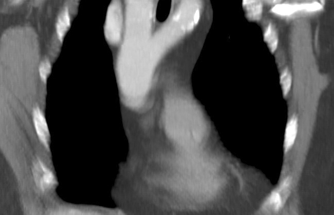
Rather, the best tests for diagnosing a double aortic arch are advanced imaging techniques like computer tomography or magnetic resonance imaging. These methods provide a clear picture of the aortic arches, their branches, location, the tissue around them, and if there’s any blockage in the trachea or esophagus. These imaging tests are also valuable for planning surgery, and usually come after an echocardiography.
Lastly, a bronchoscopy is often done before surgery to examine the airway and rule out other causes of respiratory symptoms. It can also confirm and gauge the level of airway compression. This test is not typically used after surgery to determine the success of unblocking the airways.

Treatment Options for Double Aortic Arch
Surgery is usually the main step in treatment for those patients showing symptoms like difficulty breathing or swallowing because their windpipe or food pipe is being compressed. The same treatment is applied as an additional remedy for patients having other heart and lung abnormalities.
The objective of this surgery is to alleviate any pressure on the windpipe and/or food pipe by removing parts of the lesser arch. This is a complex process conducted under general anesthesia where doctors access the patient’s chest through a single exam. It should be noted that heart-lung machines, which are often used in other heart surgeries, are not involved in this operation.
Typically doctors would go through the fourth rib space to enter the chest cavity. Once they’ve carefully drawn the lung aside and opened up the membrane that protects the chest cavity, the doctors will spot and protect important structures such as the food pipe, windpipe, and various nerves located in the vicinity.
Before the doctors start making any incisions on the arch, they pause the blood flow to ensure adequate blood supply to major parts of the body such as the head and lower body. The medical team then gradually removes the section of the arch and sews up the incision while making sure no bleeding occurs. A tube might be placed to drain out any potential fluid build-up or residual bleeding after the operation.
What else can Double Aortic Arch be?
The doctor would look into some possible conditions that could be causing the patient’s symptoms. Here are some examples:
- Laryngomalacia, a condition that usually improves with age and involves the softening of the tissues of the larynx (voice box)
- Tracheomalacia, a rare condition where the windpipe (or trachea) is weaker than usual
- Bronchiolitis, an inflammation in the bronchioles, the smallest airways of the lungs
- Viral induced wheeze, a condition where a viral infection leads to wheezing
- Lower respiratory tract infection, referring to infections in the lungs or below the voice box
- Subglottic stenosis, a narrowing of the airway below the vocal cords
- Gastro-oesophageal reflux disease (GORD), a digestive condition where stomach acid flows from the stomach back up into the food pipe
These are only some of the possibilities, and each of these conditions will require different treatments. Hence, it’s important for the doctor to arrive at the correct diagnosis.
What to expect with Double Aortic Arch
People who have had a double aortic arch repair typically have a very good outlook, without any major impact on their physical activity or overall lifestyle. They also don’t have an increased risk of developing heart rhythm problems or dying suddenly. Women who have undergone this procedure can generally choose to become pregnant without any concern. However, they are advised to have regular fetal echocardiography, which is a kind of ultrasound, especially if there is no significant remaining obstruction in the airway.
Possible Complications When Diagnosed with Double Aortic Arch
Complications after surgery are rare, but they can happen. Some of these might include difficulty tolerating food and lingering symptoms related to the airways, like coughing, breathing difficulties, strange high-pitched sound when inhaling (stridor), and wheezing. There’s also the possibility of rare under-the-skin injuries during surgery. For example, damage to the phrenic nerve, recurrent laryngeal nerve, or thoracic duct could lead to paralysis of the diaphragm, vocal cord paralysis, or fluid accumulation in the chest (chylothorax) respectively. Very rarely, the formation of an abnormal connection between the aorta and esophagus (aorto-oesophageal fistula) and wearing away of the esophagus (oesophageal erosion) can occur after surgery.
Possible Post Surgery Complications:
- Difficulty tolerating food
- Persistent coughing
- Breathing difficulties
- Stridor (high-pitched sound when inhaling)
- Wheezing
- Paralysis of the diaphragm (due to phrenic nerve injury)
- Vocal cord paralysis (due to recurrent laryngeal nerve injury)
- Chylothorax (fluid accumulation in the chest due to thoracic duct injury)
- Aorto-oesophageal fistula (abnormal connection between aorta and esophagus)
- Oesophageal erosion (wearing away of the esophagus)
Preventing Double Aortic Arch
After a diagnosis is made and while waiting for surgery, it’s essential for parents to be informed about certain symptoms. If these symptoms appear, they should immediately reach out to a healthcare professional. This advice is crucial because there’s a risk that a serious and potentially fatal breathing problem could develop.
Healthcare providers have to be tactful with this advice to avoid increasing the family’s already high levels of worry and stress.
Parents should also be made aware that their child might continue to have noisy breathing for up to a year after the surgery. This condition could be due to the weakening of the windpipe and/or the branches of the lungs resulting from the presence of the vascular ring—a condition where abnormal blood vessels create pressure on the windpipe.


