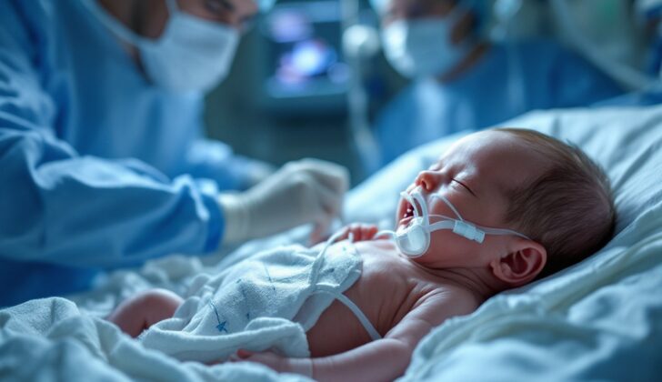
What is Esophageal Atresia?
The esophagus is a muscular tube that carries food from the back of your mouth and throat, which we call the pharynx, to your stomach. It develops from an early cell layer when you’re still in your mother’s womb, forming parts of your body like the pharynx, esophagus, stomach, and the lining of your digestive and respiratory systems. While you’re still developing as a fetus, the windpipe, known as the trachea, and the esophagus come from the separation of a joint tube. If this tube doesn’t separate or develop completely, it can lead to two rare disorders known as a tracheoesophageal fistula (TEF) and esophageal atresia (EA).
Before birth, babies with EA may cause their mothers to have too much amniotic fluid, mostly in the final trimester of pregnancy, which can be a hint at this condition. Additionally, almost half of the babies with TEF/EA may also be born with other birth defects grouped under two terms: VACTERL, which stands for vertebral defects, anal atresia, cardiac defects, TEF, renal anomalies, and limb abnormalities, or CHARGE, short for coloboma, heart defects, atresia choanae, growth retardation, genital abnormalities, and ear abnormalities.
Once the baby is born, signs that they may have EA include excessive slobbering, coughing or choking, and inability to pass a tube from the nose to the stomach. If there’s a TEF associated, the air from the windpipe can pass through to the stomach causing it to inflate with air. If a baby shows these signs, it is critical to investigate quickly for EA and TEF, and refer them to a care facility equipped to deal with children’s operations for further examination.
What Causes Esophageal Atresia?
Esophageal atresia, with or without a connected tracheoesophageal fistula, occurs when the upper part of the gut, known as the foregut, fails to properly separate or fully develop. A tracheoesophageal fistula is an abnormal connection between the windpipe and the esophagus.
This happens because a small offshoot from the early-stage lung, known as the embryonic lung bud, doesn’t divide as it should due to issues with the interaction between epithelial and mesenchymal cells. These are two types of cells that play a key role in the development of organs and tissues in the body.
While some genes, such as Shh, SOX2, CHD7, MYCN, and FANCB, have been linked to the development of esophageal atresia, the exact cause is still not completely understood and is likely due to a variety of factors. Patients can either have esophageal atresia/tracheoesophageal fistula on its own or it may be a part of a syndrome like VACTERL or CHARGE.
Risk Factors and Frequency for Esophageal Atresia
EA is a birth defect that affects the upper part of the digestive system. It occurs in roughly 1 in 2500 to 1 in 4500 births around the world. Specifically, in the United States, out of 10,000 live births, about 2.3 babies have this condition. It’s also worth noting that this condition tends to be more common in babies born to older mothers.
Signs and Symptoms of Esophageal Atresia
About a third of babies with esophageal atresia/tracheoesophageal fistula (EA/TEF), diseases that affect the food tube and windpipe, can be spotted before birth. The main telltale sign seen during pregnancy scans is excess amniotic fluid, found in about 60% of such pregnancies. When diagnosed prebirth, parents can be advised of what to expect afterwards and plan for the birth to happen where there are specialist medical teams available.
However, many cases go undetected until after birth. Once babies with EA are born, they quickly show signs like high amounts of secretion, leading to choking, difficulties with breathing, or turning blue during feeding. With Type C to E EA, the connection between the windpipe and lower part of the food tube leads to a bloated stomach visible on a chest x-ray. Babies with Type A and B EA won’t have swollen bellies because there’s no connection from the windpipe to the lower part of the food tube. Additionally, babies with TEF can have contents of the stomach flow back into the windpipe, causing aspiration pneumonia and breathing problems. Sometimes, if the connection is small in EA Type E cases, the diagnosis could be delayed.
Testing for Esophageal Atresia
Esophageal atresia or EA is usually identified when a doctor fails to pass a small tube from the mouth into the stomach. If it stops and shows as coiled on a chest X-ray, the doctor might consider EA. For a more certain diagnosis, the doctor may use a technique where they inject a safe contrast material into the tube while monitoring it with a fluoroscope, a kind of real-time X-ray. It’s important to avoid using barium for this, as it can cause lung inflammation if it accidentally gets sucked into the lungs. Other helpful tools for diagnosis include esophagoscopy and bronchoscopy, which involve using a thin, flexible tube with a camera to visually examine the esophagus and trachea respectively.
If a newborn has EA and tracheoesophageal fistula (TEF, a condition where there is an abnormal connection between the esophagus and the windpipe), the doctor will also check for other conditions that could be associated, such as VACTERL and CHARGE abnormalities. These can appear in up to half of the newborns with these conditions. The checks may include an echocardiogram (an ultrasound of the heart), X-rays of the limbs and spine, a kidney ultrasound and a thorough physical examination of the anus and genitalia for any abnormalities. A study involving around 3,000 patients in the U.S. found that the most common abnormalities associated with EA/TEF were vertebral anomaly (25.5%), malformation of the anus (11.6%), congenital heart disease (59.1%), kidney disease (21.8%), and limb defects (7.1%). Almost one-third had three or more abnormalities and met the criteria for a VACTERL diagnosis.
Treatment Options for Esophageal Atresia
Once a baby is diagnosed with esophageal atresia (EA), a condition in which their esophagus doesn’t properly connect to their stomach, they will be intubated. Intubation involves placing a tube down the baby’s windpipe to help them breathe and prevent them from breathing food or fluid into their lungs. They’ll also have a catheter carefully placed to clear out any secretions in their mouth and throat. The baby will be given antibiotics and intravenous fluids, and they won’t be allowed to eat or drink anything (this is called NPO, or nil per os). Because the NPO status can last several weeks, the baby will receive total parenteral nutrition (TPN), which provides all their necessary nutrients through a vein.
Once the baby is stable in terms of blood flow and breathing, a pediatric surgeon should be consulted. The exact timing for surgery to treat EA, along with a related condition known as tracheoesophageal fistula (TEF) where there’s an abnormal connection between the esophagus and the windpipe, depends on the baby’s size. If the child weighs more than 2 kilograms, surgery is usually offered once any heart-related issues have been addressed. Babies weighing less than 1.5 kilograms are usually treated with a two-part approach where the abnormal connection is closed first, then the EA is repaired when the baby has gained more weight.
The surgeon has a couple of options for repairing EA and TEF: an open thoracotomy, or surgery involving a large incision in the chest, or video-assisted thoracoscopic surgery, which is a less invasive procedure. In either case, the abnormal connection is located and closed, and the parts of the esophagus are connected so they can function normally. If the ends of the esophagus can’t be connected without causing stress to the tissue, the Foker technique can be used. This involves gradually bringing the ends of the esophagus closer together using special stitches before the repair is done.
If there’s a very long gap in the esophagus that can’t be connected, the surgeon may use a piece of another organ, such as the stomach, colon, or part of the small intestine. In some cases, the abnormal connection can be closed through a high cervical incision in the neck, avoiding the need for chest surgery. Typically, a gastrostomy (feeding tube into the stomach) isn’t necessary unless the initial repair doesn’t work.
After surgery, the baby will be closely monitored in the neonatal intensive care unit (NICU). They’ll have a chest tube to help drain any fluids, and they’ll continue to be kept NPO with a nasogastric tube (a tube in the nose going to the stomach) to help remove secretions. After about 5-7 days, a special X-ray called an esophagogram will be done to check for any leaks in the repair. If no leaks are seen, the baby can start to feed orally. If there is a leak, the chest tube will collect the drainage. The chest tube will stay in place until the leak heals and/or the baby can tolerate oral feedings.
What else can Esophageal Atresia be?
When trying to diagnose the conditions known as esophageal atresia (EA) and tracheoesophageal fistula (TEF), doctors also need to think about other conditions that can have similar symptoms. These include:
- Laryngotracheoesophageal clefts (a rare birth defect where there’s a gap in the larynx and trachea)
- Esophageal webs or rings (extra tissue in the esophagus that makes it narrower)
- Esophageal strictures (a narrowed esophagus that can cause swallowing problems)
- Tubular esophageal duplications (a rare congenital formation of a second esophagus)
- Congenital short esophagus (a rare condition where the esophagus is shorter than normal)
- Tracheal agenesis (a birth defect where the trachea, or windpipe, doesn’t develop).
To help figure out the correct diagnosis, doctors use a range of imaging tests, including x-rays, CT scans, endoscopy (where a flexible tube with a camera at the end is used to look inside the body), and sometimes surgery.
What to expect with Esophageal Atresia
The future health outcome or prognosis for newborn babies with esophageal atresia (EA) and/or tracheoesophageal fistula (TEF) – these are birth defects that affect the tube connecting the mouth to the stomach (esophagus) and/or the windpipe (trachea) – is generally optimistic. Whether the baby has heart or chromosome issues will strongly impact their health outcome, more than the EA/TEF condition itself.
Typically, about 85%-90% of babies survive this condition. But survival rates can be lower if the baby also has heart anomalies alongside EA/TEF. Early death is usually associated with heart issues, whereas late deaths are more likely due to respiratory complications.
The distance between the two esophageal pouches, particularly if it’s large, can also affect the prognosis. All babies who have EA/TEF repair surgery can expect some problems with their gastrointestinal (digestive) and respiratory (breathing) systems, but these usually get better as the child grows older.
Possible Complications When Diagnosed with Esophageal Atresia
After surgery to repair an EA/TEF (esophageal atresia and tracheoesophageal fistula), some complications may occur. The most severe issue that can arise is the leakage of the esophagus, which is where the food pipe connects with the stomach. If it’s a minor leak, it can usually be managed with a chest tube and stopping food by mouth until it heals. However, if the leak is large or the connection between the esophagus and stomach disrupts, another surgery may be needed. This often involves the insertion of part of the stomach, colon, or small intestine.
Another potential issue is narrowing at the surgery site, which is typically handled progressive endoscopic procedures to widen the esophagus. Although it’s quite rare, the esophagus and the windpipe can form an abnormal connection, which can be corrected through another surgery.
Even when the surgery goes well, there can be non-surgical complications. Doctors usually recommend using medication to reduce stomach acid for at least a year following surgery because issues with esophagus movement can cause acid reflux and risk of inhaling food or fluids into the lungs (aspiration). An abnormal softening of the windpipe is commonly seen after this type of repair. It’s not unusual for newborns with EA/TEF to experience increased difficulty swallowing, more respiratory infections, and inflammation in the esophagus.
Because children with EA/TEF tend to get acid reflux more frequently, they also have higher rates of Barrett’s esophagus, which is a precancerous condition, and a greater risk of developing esophageal cancer later in life. Although there is some debate, some doctors recommend screening for esophageal cancer in these patients.
Common Complications:
- Esophageal leak
- Narrowing at surgery site
- Abnormal connection between esophagus and windpipe
- Increased stomach acid leading to acid reflux
- Softening of the windpipe
- Difficulty swallowing
- More respiratory infections
- Inflammation of esophagus
- Increased risk of esophageal cancer
Recovery from Esophageal Atresia
After surgery, newborns should be transferred to the neonatal intensive care unit where they will be monitored closely. They might need to have certain procedures done, like the insertion of tubes into their veins (invasive central catheters) or their windpipe (endotracheal tubes), and may need medication to increase their blood pressure (vasopressors). It’s important that these babies have frequent check-ups with their doctors because there is a possibility of complications like narrowing of the esophagus (esophageal strictures), difficulty swallowing (dysphagia), and insufficient weight gain or growth (failure to thrive).
Preventing Esophageal Atresia
If your child is diagnosed with esophageal atresia, a condition where the esophagus (food pipe) doesn’t develop properly, it is crucial for you as parents to know what to expect during your child’s recovery. You should be familiar with any possible complications that could arise.
Understanding the diagnostic process, or how the doctors figure out your child has this condition, will help. It’s also important to educate yourselves about how the doctors will treat this issue, the possible surgical procedures they might recommend, and what the recovery process might look like after the surgery.
Lastly, regular check-ups after your child is discharged from the hospital are crucial to make sure their recovery is going well.


