What is Hydrops Fetalis?
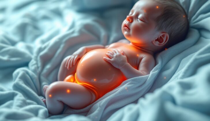
Hydrops fetalis is a condition that can affect unborn babies, causing an unusual buildup of fluid in two or more parts of their body, like the stomach cavity, lungs, and heart sac. Another way to define this condition is seeing fluid accumulation in two parts of the baby’s body, or a buildup in one part coupled with general body swelling.
In the past, doctors usually identified this condition when delivering a baby with extreme swelling, often stillborn. Nowadays, thanks to ultrasound technology, Hydrops fetalis can be diagnosed before birth. As the condition progresses, swelling is a common component, frequently accompanied by an abnormally large placenta and excessive amniotic fluid. Hydrops fetalis can result from numerous conditions that can severely affect the baby. It is classified into two types: immune and nonimmune. If it is associated with a reaction to the baby’s red blood cells, it is referred to as immune hydrops fetalis. Otherwise, it’s called nonimmune hydrops fetalis.
Immune hydrops, also known as erythroblastosis fetalis, happens when antibodies from the mother’s bloodstream pass through the placenta and react with the baby’s antigens, leading to the destruction of the baby’s red blood cells. Nonimmune hydrops, on the other hand, is caused by issues other than reactions between antigens and antibodies. It’s important to note that Hydrops fetalis is not caused by heart failure, rather it is due to an excess of blood volume and high blood vessel permeability in the fetus. In contrast, heart failure would be a very late result of a consistently overworked heart.
What Causes Hydrops Fetalis?
Hydrops refers to a medical condition linked to a wide range of issues, which are primarily split into two categories: immune hydrops fetalis and nonimmune hydrops.
Immune hydrops fetalis happens when the mother’s immune system cells attack the baby’s red blood cells. This can occur from an immune reaction called isoimmunization, which involves things like Rh factor incompatibility (when the mother and fetus blood types don’t match), the presence of different antibodies, and so forth.
Nonimmune hydrops accounts for around 80%-90% of hydrops cases. It happens when an illness, genetic disorder, or congenital malformation (birth defects) hampers the baby’s body’s ability to regulate fluid levels. This type of hydrops could stem from various factors such as:
– Heart-related conditions like irregular heartbeats, underdeveloped heart chambers, defects in the heart’s inner lining, and abnormalities in the baby’s pulmonary (relating to lungs) airways
– Genetic conditions such as Turner syndrome, Down syndrome, and Edward syndrome
– Disorders in the lymphatic system
– Infections in the mother like Parvovirus B19 (causes a rash illness), cytomegalovirus, and syphilis
– Metabolic (body’s chemical reactions) disorders
– Different types of tumors
– Mother’s health conditions: Diabetes and overactive thyroid (hyperthyroidism)
Other causes include urinary system-related conditions, complications that affect digestion, blood-related conditions, issues with red blood cell metabolism, and complications in the production and shape of red blood cells.
Risk Factors and Frequency for Hydrops Fetalis
The number of babies born with Nonimmune Hydrops Fetalis (NIHF) varies, with estimates ranging from 1 in 1500 to 1 in 4000 births. This wide range is due to differences in how the condition is defined, the populations studied, and how thorough the evaluations were. It also depends on whether pregnancies that were ended late (also known as late pregnancy terminations) were included in the count.
Since 1968, the use of a medicine called anti-D immunoglobulin has greatly reduced the number of babies born with a related condition called RhD alloimmunization. This has led to NIHF being the cause of nearly 90% of all cases of ‘hydrops fetalis’, a severe swelling in an unborn baby.
Signs and Symptoms of Hydrops Fetalis
Hydrops fetalis is a condition where abnormal quantities of fluid accumulate in two or more body compartments of a fetus. These can include areas such as the areas around the lungs (pleural effusion), in the belly (ascites), around the heart (pericardial effusion), and under the skin (skin edema). The more locations with fluid buildup, the worse the expected health outcome is for the newborn. Diagnosis is primarily accomplished using prenatal ultrasounds or post-birth medical examinations.
Other signs and symptoms of the condition include anemia, an enlarged placenta (placentomegaly), excessive amniotic fluid (polyhydramnios), and enlarged liver and spleen (hepatosplenomegaly). In regards to pleural effusion, mild cases may lead to breathing difficulties, whereas severe cases can result in underdeveloped lungs and serious respiratory or circulatory diseases. Rare but severe complications may involve pneumo or chylothorax.
Ascites, or build-up of fluid in the belly, can be an early sign of hydrops fetalis, sometimes visible as early as 20 weeks of gestation. Distinguishing this from other diseases is crucial. Large amounts of ascites can compress the bowel and stunt lung development. A specific measure of proteins in the fluid, called the serum ascites albumin gradient (SAAG), can help indicate the cause of the condition.
Anemia can be a result of a blood incompatibility between mother and baby, a viral infection, or genetic factors. Polyhydramnios, or an unusually large amount of amniotic fluid, can be caused by problems with fetal swallowing, kidney function, or intestinal blockage.
Enlarged placenta, or placentomegaly, occurs when there are disruptions in fluid balance within the body. This is usually seen in diseases that cause high output of blood from the heart like anemia and a certain type of tumor. A large placenta and polyhydramnios are considered indicators of how well a baby will do.
Enlarged liver and spleen (hepatosplenomegaly) can be diagnosed during the second and third trimester. This is generally associated with specific types of genetic diseases. Newborns may have blue skin and not respond well to oxygen therapy, suggesting heart disease. They may also have low muscle tone which might indicate they’ve been born with muscle diseases or a thyroid condition. Other signs might include large liver, heart muscle disease and abnormal facial features, skin inflammation, and hepatitis which suggest potential for infection from certain organisms.
- Abnormal fluid collections in two or more compartments
- Anemia
- Large placenta
- Excessive amniotic fluid
- Enlarged liver and spleen
Testing for Hydrops Fetalis
Hydrops fetalis, a severe swelling in the fetus, is usually discovered unexpectedly during routine check-ups during pregnancy. Depending on the cause, symptoms and future health outcomes can vary significantly. For instance, if the swelling is due to chromosomal abnormalities or gene changes, it’s typically found early on in pregnancy. On the other hand, if a heart issue is the cause, it might not be found until later in the pregnancy during the second or third trimester. Thus, thorough check-ups during pregnancy are critical if there’s any suspicion of hydrops fetalis.
There are several ways for doctors to check for signs of hydrops fetalis. The first detailed ultrasound is usually performed between 18 and 22 weeks into the pregnancy. Early signs such as an unusual buildup of fluid in the fetus’s abdomen and skin edema, which is unusual swelling of the skin over 5mm thick, might be seen in the baby’s head, neck, chest, and belly. These signs might indicate chromosomal issues or physical defects. Other common signs like excess amniotic fluid and placental swelling are commonly seen before the 20th week of pregnancy. However, symptoms like fluid in the fetus’s chest or heart sac are rarely seen before the 15 weeks of gestation.
Doctors should also check for certain infections in the mother such as TORCH and parvovirus B19. These infections can affect the fetus and might be related to hydrops fetalis. For instance, a parvovirus B19 infection is often linked with low red blood cell levels (anemia) and an unusual buildup of fluid in the fetus’s abdomen. Other infections like cytomegalovirus and toxoplasmosis can lead to birth defects like large ventricles in the brain, small head size, and unusually dense, hard feces.
Monitoring the fetus’s heart rate, the resistance to blood flow in the umbilical artery, and blood flow in the brain can also give clues about what’s causing hydrops. These factors can, for instance, help in identifying chromosomal abnormalities. In particular, a high MCA-PSV ratio (the ratio of peak blood flow speed to rest speed in the brain’s middle cerebral artery) of more than 1.5 can be a sign of anemia in the fetus.
In some cases, chorionic villous sampling (CVS) may be recommended. This is generally done if hydrops fetalis is diagnosed early, before 15 weeks of gestation. CVS can help detect genetic conditions and chromosome abnormalities that could be causing hydrops fetalis.
Lastly, monitoring alpha-fetoprotein (AFP) levels can be useful as an increased level could suggest bleeding between the fetus and mother, potentially leading to anemia and hydrops. Blood tests called direct and indirect Coomb tests can also help detect hydrops due to immune issues.
Treatment Options for Hydrops Fetalis
Treating infants with certain medical conditions can be complex and requires proper evaluation, ongoing monitoring, and specific resuscitation techniques to increase their chances of survival. Some conditions, like heart abnormalities and genetic disorders, are difficult to treat and have a poor prognosis.
If an ultrasound scan shows significant problems such as fluid accumulation, shifting of the heart, excess amniotic fluid, or swelling in the fetus, certain interventions might be carried out while the baby is still in the womb. These interventions could include draining excess fluid or doing a procedure to ensure the lungs develop properly. If the fetus has severe anemia or an irregular heart rhythm, measures such as a blood transfusion or medications to regulate the heart rhythm could be used, even while the baby is still in the mother’s womb. Indeed, such procedures may sometimes lead to the birth of a healthy baby, especially among babies infected with parvovirus B19. In a few isolated cases, tumors can even be removed surgically while the fetus is in the womb.
Once born, babies will typically need immediate attention to support their breathing, often requiring a procedure to help them breathe properly. If the baby has a buildup of fluid in their chest, abdomen, or around their heart, procedures can be done to remove this excess fluid. Severe anemia might be treated with a blood transfusion. The heart rhythm will need to be closely monitored, especially if the baby’s heart rate is too fast or irregular, and can be managed with procedures or medications as needed.
Despite all these interventions, the survival rate for these babies is unfortunately low, at around 10%, and some babies that do survive may have long-term issues affecting their brain development and cognitive abilities. Recurrence is most common in mothers whose babies have genetic abnormalities or a specific type of blood incompatibility. Consequently, autopsies and in-depth examinations of the placenta and genetic testing are typically conducted to determine the cause of these severe conditions.
What else can Hydrops Fetalis be?
If a baby is found to have hydrops fetalis, a condition characterized by severe swelling, doctors might consider a variety of different causes. These could include:
- Newborn hemochromatosis, a disorder causing too much iron in the body
- Congestive heart failure, when the heart struggles to pump blood efficiently
- TTTS, or Twin-Twin Transfusion Syndrome
- Hepatitis B, a viral infection of the liver
- Hypercalcemia, which is too much calcium in the blood
- Hypernatremia, which is too much sodium in the blood
- Hypoprothrombinemia, a bleeding disorder caused by low levels of a clotting protein
- Maternal diabetes, which is diabetes in the mother
- Fetal abdominal cysts, fluid-filled sacs in the baby’s abdomen
- A blocked bowel
- Blocked urinary system
What to expect with Hydrops Fetalis
The survival outlook for hydrops fetalis (a serious fetal condition where abnormal amounts of fluid build up in two or more body areas of a baby) mostly depends on the root cause, how far along the pregnancy is at diagnosis, when the baby is born, the degree of fluid buildup, and any in-utero treatments carried out. Babes with causes related to the chest and lung malformations generally have a good survival outlook. However, infants with chromosomal abnormalities, structural abnormalities, and certain genetic metabolic disorders may have a less favorable survival outlook.
Survival in hydrops fetalis is dependent on the root cause, the availability of treatments to tackle the fluid buildup, and the gestational age at birth, rather than the specific anatomical manifestations of fluid build-up in more than one body part. The cardiovascular profile score might be a useful marker in predicting survival in babies with hydrops and a high ratio of heart size to chest size (also known as a high cardiothoracic ratio).
When hydrops can be alleviated in babies who have access to fetal intervention for their underlying condition, and there is no preterm delivery, survival rates usually improve. Interestingly, babies who have very different disease processes and cardiac findings can all show the common anatomic feature of fluid in two compartments, suggesting an fundamental trigger of fetal distress yet to be discovered.
Possible Complications When Diagnosed with Hydrops Fetalis
The complications that can occur with hydrops fetalis include:
- Spontaneous miscarriage
- Death of the fetus before birth
- Birth occurring too early (preterm delivery)
- Deaths occurring during birth or shortly after (perinatal deaths)
- Deaths occurring during the first weeks of a newborn’s life (early neonatal deaths)
If an infant with hydrops fetalis survives, they may face conditions like:
- Rupture of the appendix
- Cystic hygroma, a fluid-filled sac that commonly appears on the neck
- Intestinal obstruction (blockage in the intestine)
- Severe delay in learning and development
Preventing Hydrops Fetalis
Hydrops fetalis, a serious condition involving fluid build-up in a fetus, often has a poor outlook if the unborn baby develops lung underdevelopment (pulmonary hypoplasia), heart defects, or if the condition is found before 24 weeks of pregnancy. If hydrops fetalis is diagnosed early in pregnancy and there’s no way to treat it, ending the pregnancy may be considered. This condition is linked to a very high risk of death before or shortly after birth, which can be between 50% to 98%.
The chances of a baby surviving depend on several factors, such as the cause of the condition, how early it starts, how far along the pregnancy is at the time of the baby’s birth, and whether there are fluid build-ups in the lung area (pleural effusions). As a rule, the earlier this condition starts, the worse the outlook for the baby. Specifically, the presence of fluid-build ups in the lung area and excessively amniotic fluid (polyhydramnios) before 20 weeks of pregnancy indicate a poor outcome, due to increased risks of underdeveloped lungs and premature birth, respectively. However, if the baby doesn’t have chromosomal abnormalities or major physical defects, their chances of survival improve.
This condition happening again in future pregnancies is rare, unless it’s related to a blood conflict between mother and baby (Rh incompatibility) or genetic abnormalities. Regular prenatal appointments using ultrasound tests every 1 to 2 weeks are crucial when hydrops fetalis is improving, and the pregnant woman is closely watched for mirror syndrome signs. Babies affected by immune hydrops (a type of hydrops fetalis caused by blood incompatibility between the expectant mother and the baby) are usually born around the 37th week of pregnancy or when the baby’s lungs are mature enough. There’s no solid proof that a cesarean section is better than vaginal birth. The birth is planned in a high-level medical facility with neonatal intensive care and a skilled newborn care team around, ready to provide immediate medical aid. After birth, newborns afflicted by this condition need proper care.


