What is Periventricular and Intraventricular Hemorrhage?
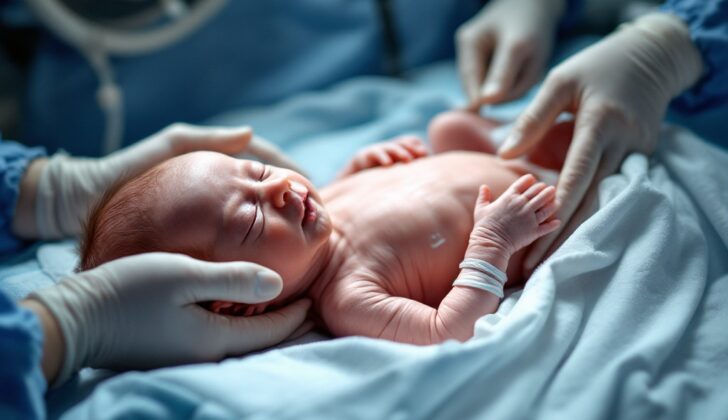
Periventricular-intraventricular hemorrhage (PIVH) is a health problem that affects newborn babies who are born prematurely. This condition happens when blood vessels in a part of the brain called the germinal matrix burst and bleed. This can spread into the ventricles of the brain, which are fluid-filled chambers, causing a condition known as intraventricular hemorrhage (IVH). If the bleeding is severe, it can fill a large part of the ventricle and spread to the surrounding brain tissue. Babies born before 33 weeks of pregnancy are the most at risk, as their germinal matrix is not yet fully developed.
This condition is a major risk for premature babies, to the point where it’s the second leading cause of death for them. It’s also one of the main reasons for brain damage in preterm babies with low birth weight. The most common reason for death among premature babies is a condition called hyaline membrane disease.
Intraventricular hemorrhage (IVH) was first identified in 1968 by Abraham Towbin. Ten years later, a team led by Papile created a way to classify PIVH based on a type of brain scan called a head computed tomographic (CT) scan. This method was later adapted to use ultrasound scans in 1984, mainly because these machines are portable and the scans can be done repeatedly.
The severity of the bleeding is classified into four grades from I to IV:
– Grade I: The bleeding occurs only within the germinal matrix.
– Grade II: There’s bleeding into the ventricles, but they do not expand.
– Grade III: The bleeding spreads into the ventricles, occupying more than 50% of them, causing them to expand.
– Grade IV: The bleeding reaches the surrounding brain tissue.
Grades III and IV are considered “severe IVH”.
What Causes Periventricular and Intraventricular Hemorrhage?
The main cause of PIVH, or bleeding within the brain of a premature baby, is the weakness of the blood vessels in a part of the brain called the germinal matrix, as well as less developed mechanisms that regulate blood flow in the brains of these young babies.
The blood vessels in the germinal matrix are different from those in other areas of the brain because this area has a higher metabolic need due to the fast replacement of its cells. The blood vessels are denser and wider in this area than other parts of the brain. In addition to this, these vessels are more round because they’re not fully developed yet.
PIVH can cause damage to surrounding brain tissue (white matter). This can happen due to pressure from swelling in the spaces within the brain (ventricles), and because the lining surrounding these spaces is weak. If these ventricles get too stretched, it can lead to damage of the nerve fibers within the white matter, cause swelling and trigger reactions from cells in this area. A similar process happens if the lining gets stretched and ruptured, which exposes the white matter to cells and products from the blood.
Transporting a newborn shortly after birth poses a major risk factor. Other factors like use of mechanical breathing support, high levels of carbon dioxide in the blood, chest complications like a punctured lung, respiratory distress syndrome, frequent insertion of breathing tubes, and unstable blood pressure also contribute to the risk of this type of brain hemorrhage.
Risk Factors and Frequency for Periventricular and Intraventricular Hemorrhage
Globally, the occurrence of Periventricular-Intraventricular Hemorrhage (PIVH) varies between 3.7% to 44.7%. Recent reports show an overall occurrence of 36.2%, with severe cases representing 7.1% of it. The breakdown of PIVH occurrence in premature babies by severity levels are: 17% for grade I, 12.1% for grade II, 3.3% for grade III, and 3.8% for grade IV. Remarkably, 50% of these cases occur on the first day of the newborn’s life, and by the third day, there are 90% of PIVH cases.
On the brighter side, the occurrence of PIVH has generally lessened since the 1980s. In Brazil, facts show a decrease going from 50.9% in 1991 to 11.9% in 2005. In the US, the count of preterm births has been steady at 10% over the recent years.
One interesting fact is that the occurrence of PIVH is influenced by the baby’s gestational age (the time baby was in the womb) and birth weight. For premature babies with a weight of 1000 grams or less, every extra week of gestational age reduces the likelihood of severe PIVH by 19%. There’s a 3.5% reduction in PIVH for every extra week of gestation up until 32 weeks. The total count of PIVH for all newborns between 22 to 28 weeks of gestational age is 32%.
- PIVH happens in 25 to 30% of newborns who weigh less than 1500 grams.
- It’s found in up to 45% of newborns who weigh less than 1000 grams.
Severity levels of III or IV PIVH is mostly found in patients with a weight less than 1000 grams or have a gestational age of 22 to 27 weeks. Babies older than 31 weeks in the womb or heavier than 1500 grams rarely develop a severity level III or IV PIVH (it happens in only about 2.7% of the cases).
Signs and Symptoms of Periventricular and Intraventricular Hemorrhage
Periventricular-intraventricular hemorrhage (PIVH) often gets discovered unexpectedly when conducting ultrasound screenings on infants born prematurely or with low birth weights. Infants with symptoms may show signs of neurological decline, breathing problems, sleep apnea, a bulging fontanelle (soft spot on the head), seizures, lack of activity, decreased responsiveness, or a dazed state.
Several factors that could make an infant more likely to have PIVH are tied to the mother’s pregnancy course:
- Pregnancy duration of 32 weeks or less
- Expecting mother did not receive steroids during pregnancy
- Expecting mother experienced bleeding during pregnancy
- Maternal chorioamnionitis (a bacterial infection of fetal membranes and amniotic fluid)
- Vaginal birth
Details about the infant’s birth may also increase the chances of PIVH in preterm babies:
- Birth weight under 1500 g
- Early sepsis infection
- Treatment-required low blood pressure
- Low oxygen level in blood
- High carbon dioxide levels in blood
- Breathing difficulties right after birth
- Use of a ventilation machine at birth
- Prolonged use of a ventilation machine
- Pneumothorax (collapsed lung)
- Low scores on the Apgar test at 1 and 5 minutes after birth
- Seizures
- Unusually wide ductus arteriosus, the blood vessel in a fetus’s heart
- Frequent suctioning of the newborn’s windpipe
- Surfactant use
- Platelet deficiency in the blood
Preterm babies with grade IV PIVH are usually lighter at birth and have a shorter gestational age than those with grade III. However, certain often-cited factors for PIVH don’t affect whether a grade III or IV appears, including low blood pressure, early-onset sepsis, a wide ductus arteriosus, surfactant therapy, pre-birth steroid use, maternal bleeding, maternal fever, delivery mode, Apgar scores, and premature rupture of membranes. Grade IV and III PIVH are more prevalent in infants born after the placenta detaches prematurely.
That being said, the usage of pre-birth steroids and having a cesarean section have been observed to reduce the occurrence of PIVH.
Testing for Periventricular and Intraventricular Hemorrhage
In preterm infants, or babies born too early, a condition called intraventricular hemorrhage (IVH), or bleeding in the brain, typically happens within the first three days after birth. In fact, 70% of these cases occur within the first 24 hours post-birth, and 95% happen within the first seven days.
The American Academy of Pediatrics has a specific recommendation for detecting IVH in these babies. Any baby born at or before 30 weeks of pregnancy should have a special form of ultrasound called a cranial ultrasonography by the time they are 7 to 10 days of age. If the results of this initial screening come back abnormal, the baby will need to have repeat screenings more often. If the results are normal, another follow-up scan should be taken when the baby is aged between 4 to 6 weeks. The baby should also have one more scan when they are aged around 36 weeks or before they leave the hospital. However, the usage of CT scans and routine MRI scans before the discharge is not recommended for IVH screening.
Brain magnetic resonance imaging (MRI) though, may be used in certain cases. Despite not being employed in the primary check-up, it can help identify other types of brain injuries and is used to further investigate any brain anomalies found on ultrasound. But it’s important to know that prenatal, or “fetal,” MRI does not offer any extra benefit compared to standard ultrasound or brain-specific ultrasound (neurosonography) for at-risk babies.
An easy daily check that doctors use to monitor the presence of hydrocephalus, or “water in the brain,” in an IVH patient is measuring the size of the baby’s head. If the head size increases rapidly, it could be a sign of hydrocephalus development caused by IVH.
Treatment Options for Periventricular and Intraventricular Hemorrhage
The main goal for preterm babies is to prevent early birth, if possible. If an early birth is expected, doctors usually give the mother medicine, including corticosteroids, that help the baby’s lungs to mature. The mother may also be moved to a hospital with specialized facilities to care for very small babies. Research has shown that these steps can help protect preterm babies from brain hemorrhage (bleeding in the brain).
When the early baby is born, doctors typically delay clamping the umbilical cord. Leading healthcare organizations, like the American College of Obstetricians and Gynecologists, support this practice. Waiting for about 30 to 180 seconds before clamping the cord has been shown to lower the risk of brain hemorrhage compared to immediate clamping.
After the baby is born, the focus is on avoiding low oxygen levels and fluctuations in the baby’s brain blood flow. Certain drugs, such as phenobarbital and indomethacin, have been used to achieve this. One review of 10 studies found that giving indomethacin to preterm babies lowered the occurrence of severe brain hemorrhages.
Once a brain hemorrhage has happened, there’s no specific treatment to stop the bleeding. But, implementing early measures can prevent this condition. These measures aim to keep the baby stable and prevent sudden changes in the baby’s cerebral (brain) blood flow and blood pressure. The measures include keeping the baby’s head in line with the body, ensuring proper breathing support, avoiding physical therapy maneuvers, maintaining constant blood pressure, and minimizing pain. These steps should be taken at least for the first three days after birth when the risk for brain hemorrhage is the highest.
There has been success in Brazil with a certain care approach, “care bundle”. This involves:
1. Keeping the baby lying on their back with the head in line with the body (if the position is wrong, it can affect the return blood flow in the neck veins)
2. Avoiding physical therapy maneuvers (these can cause changes in brain pressure and blood flow)
3. Suctioning the breathing tube only if necessary (this can change blood pressure, cerebral blood flow, and brain pressure)
4. Not collecting fluid from around the brain and spinal cord (this procedure can change heart rate and oxygen levels)
5. Not weigh the baby (handling the baby can cause changes in the heart rate, oxygen levels, and blood pressure)
What else can Periventricular and Intraventricular Hemorrhage be?
The process of diagnosing this condition is pretty straightforward as it mainly involves screening a preterm newborn specifically for this condition. It’s important to identify and treat sepsis as quickly as possible. Low blood sugar levels, also known as hypoglycemia, could drop the baby’s alertness level very rapidly.
What to expect with Periventricular and Intraventricular Hemorrhage
The possibility of recovery and risk of death is influenced by the severity of the injury. For instance, the mortality rates for grades I to IV are 4%, 10%, 18%, and 40% respectively. Also, any level of PIVH or bleeding in the brain can affect the mental development abilities of the child, with the chances of developing cerebral palsy ranging from 8% to 50% as the injury severity increases from grades I to IV. Infants born prematurely (less than 27 weeks) with a grade I or II PIVH typically do not show signs of developmental delays.
Premature infants with serious PIVH are at a higher risk of developing cerebral palsy, especially those who weigh less than 1000 grams at birth.
The effect of brain bleeding on the mental development of infants can depend on the extent of brain injury. A study called EPIPAGE revealed a correlation between brain bleeding (IVH) and negative neurological outcomes. It was found that cerebral palsy rates were around 33% in infants born between 24 to 26 weeks of gestation as compared to a mere 5% in infants born between 31 to 32 weeks of gestation.
Possible Complications When Diagnosed with Periventricular and Intraventricular Hemorrhage
One of the main complications that can arise after Periventricular-Intraventricular Hemorrhage (PIVH) is called posthemorrhagic hydrocephalus. This is a condition where the fluid in the brain (cerebrospinal fluid) doesn’t get absorbed properly or gets blocked. It often happens in preterm infants who underwent PIVH and show quickly increasing head size.
To detect the presence of hydrocephalus, infants’ head sizes are usually measured daily. Meanwhile, the amount of fluid in the brain can be monitored via ultrasound. In more severe circumstances, the child may need neurosurgery. Many factors affect the treatment strategy for hydrocephalus, but the severity of the IVH is the most significant one.
- Medical complications:
- Posthemorrhagic hydrocephalus
- Enlarging fontanel or ‘soft spot’
- Bradycardia – slow heart rate
- Treatment strategies:
- Subgaleal shunt placement
- Ventricular reservoir placement
- Ventriculoperitoneal shunt placement
Periventricular leukomalacia, or PVL, can be another outcome of PIVH. It’s basically damage to the brain’s white matter, often found using ultrasound or MRI scans. It can lead to reduced cognitive functioning in infants. The presence of free radicals and iron influx from ventricular blood contributes to this white matter injury, increasing the chances for PVL development.
PIVH can also trigger the development of compact clusters or cysts in the space around the ventricles of the brain, a situation that may primarily cause a delay in the neurodevelopment of children on recovery from PIVH.
The risk for cerebral palsy is also increased in premature children who suffered severe PIVH, particularly if they weighed less than 1000 grams at birth.
Seizures and cognitive disabilities are long-term complications of PIVH, especially when the condition is severe.
- Long-term complications:
- Periventricular leukomalacia (PVL)
- Neurodevelopmental delay
- Cerebral palsy
- Seizures
- Neurocognitive problems
Preventing Periventricular and Intraventricular Hemorrhage
It’s important for expecting mothers to understand how to manage and avoid risk factors during pregnancy to prevent an early or preterm birth. This includes stopping smoking and regularly attending prenatal care appointments.
Mothers should also know that most cases of Periventricular Hemorrhage (PIVH)—a brain bleed in premature babies—are symptom-free. Also, if it’s a Grade I or II, the outlook is usually good or excellent, with very few complications occurring. This should encourage them and reduce any unnecessary worry.


