Overview of Achilles Reflex
The reflex test for your deep tendons, specifically in your tricep (the muscle at the back of your upper arm), patellar (the tendon connecting your kneecap to your shinbone), and Achilles (the tendon connecting your calf muscles to your heel bone) was first introduced by doctors Wilhelm Heinrich Erb and Carl Friedrich Otto Westphal in 1875. Ever since, the Achilles heel reflex test, also known as the ankle jerk test, has become an important part in examining your lower body’s nerve function.
This reflex test involves a doctor gently tapping a specific area with a small hammer to test the response of your S1 nerve root, which is part of a network of nerves in your lower spine. Like all other deep tendon reflex tests, your doctor analyses your reflex reaction on a scale from 0 to 4, evaluating both your legs and comparing the reactions to each other.
These reflex tests offer doctors an understanding of how well your reflex arc, the pathway that controls your reflexes, is functioning. They also help in checking for any imbalances in the reactions when compared between your legs. By conducting these tests, doctors can make appropriate clinical decisions and determine if there are any issues involving nerve cells that control your muscles.
Anatomy and Physiology of Achilles Reflex
The Achilles tendon is located just above the back of your heel. It’s the biggest and strongest tendon in our body and connects a group of muscles in your calf (soleus and gastrocnemius) to the heel bone. A thin layer of tissue, called a paratenon, surrounds the tendon. This tissue helps reduce friction and also has blood vessels running through it.
Nerve signals in the Achilles tendon come mostly from S1 and S2 nerve roots of the tibial nerve. The Achilles tendon reflex is an automatic reaction to the tendon being stretched, like when a doctor taps your knee with a small hammer in a check-up. When the tendon is tapped, this stretches the muscles in your calf, causing sensors in these muscles to send a message via the spinal cord to the brain. The brain then sends a message back to the muscles, telling them to contract, producing the reflex.
During this process, the nerve message heading for the spinal cord also interacts with a second type of nerve cell, which plays a critical role in making sure that only the necessary muscles (your calf muscles, in this case) contract during the reflex. This is done by preventing the contraction of the front muscles of your lower leg.
Even though the major brain areas are not directly involved in this reflex reaction, they still play a role in adjusting the intensity of the reflex.
Physiologically, this reflex arc can be divided into an upper and lower motor neuron component. The upper part involves structures like the cerebral cortex that help in regulating the intensity of the reflex. Meanwhile, the lower part includes the nerve roots in the spinal cord and the nerves that feed into the muscles. For the Achilles reflex, the lower component includes the S1 nerve root and the tibial nerve.
Why do People Need Achilles Reflex
The Achilles reflex test is a part of a full body check-up, especially when the doctor needs to examine the nerves of the legs. This test is primarily used when there’s a possibility of an issue with the S1 nerve root, a nerve bundle that’s located in the lower part of your spine. It can help determine whether the problem is in the upper motor neurons (nerves that send signals from the brain to the spinal cord) or lower motor neurons (nerves that send signals from the spinal cord to muscles), and identify where the issue is specifically located.
Moreover, the test can also give insights about how the peripheral nerves are working. Peripheral nerves are the nerves outside the brain and spinal cord. This information can be crucial for some people, such as those living with diabetes or hypothyroidism, a condition where the thyroid gland doesn’t produce enough thyroid hormones. These conditions can affect nerve function, making this test particularly important.
When a Person Should Avoid Achilles Reflex
In simple terms, there aren’t any major reasons to avoid doing the Achilles reflex test, a routine health check. But, there might be occasional circumstances where the test needs to be postponed. This could be because of an injury caused by an accident or from other health conditions that make the test difficult for the doctor to carry out and the patient to go through.
Also, skin diseases could be another reason to avoid this test. For example, someone suffering from severe psoriasis, a skin condition that causes red, flaky, crusty patches covered with silvery scales, on the area to be tested could find this test uncomfortable.
Equipment used for Achilles Reflex
The Achilles reflex test uses tools called reflex hammers. There are four different kinds of reflex hammers that doctors use nowadays: the Taylor hammer, Queen Square hammer, Babinski hammer, and the Troemner hammer. But no specific type of hammer is considered better than the others. The choice usually depends on what the doctor prefers and what is traditionally used at their workplace.
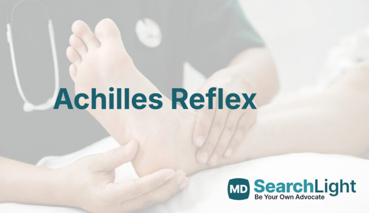
The doctor holds the reflex hammer by its handle, generally using their dominant hand’s thumb and forefinger to support most of its weight. The flat top part of the hammer is directed towards the patient’s Achilles tendon, which is located at the back of the ankle area. Other larger and more technologically advanced reflex hammers, like the pendulum hammer, can also be used to carry out the Achilles reflex test.
Who is needed to perform Achilles Reflex?
Any trained medical person, such as nurse practitioners, physician assistants, and doctors, can carry out the medical procedure. Nurse practitioners are nurses who have advanced training and can give special types of care. Physician assistants are medical professionals who can diagnose illness, treat conditions, and prescribe medication under the supervision of a doctor. Doctors, or physicians, have gone to medical school and can provide a variety of healthcare services. All these professionals have the required skills and training to perform the procedure.
Preparing for Achilles Reflex
The Achilles reflex test, a common check-up that looks at the reflex of your Achilles heel, does not require much preparation from you. During the test, you should be calm as you lay on an examination table, with your Achilles tendon region visible, from the back of your lower leg to the sole of your foot. The doctor conducting the test will have clean hands and a reflex hammer ready to conduct the exam.
How is Achilles Reflex performed
An Achilles reflex test is a medical procedure used to check the health of your nervous system, especially your spine. There are a few ways your doctor may do this.
The traditional way to perform this test is by lightly tapping the tendon located just above your heel, which is known as the Achilles tendon. For this traditional method, you will be asked to lie down on your back on an examination table, with your knee slightly bent and your hip turned outwards. The doctor will then slightly lift your foot from the bottom, and with the other hand, gently tap your Achilles tendon with a reflex hammer. This can easily be done with your leg hanging off the table or when the doctor holds your leg up.
In some cases, like for patients who cannot move around much, a different method called a plantar strike is used. For this method, you will lie down on your back with your legs stretched out. The doctor will then tap their fingers on the bottom part of your foot (near your toes), causing a slight tension that lifts your foot. This should trigger a reflex that moves your foot downwards. Research shows that this method produces the same results as the traditional Achilles tendon tap.
Another method doctors sometimes use is with a tool called a pendulum hammer. This is most often done when the patient is standing, which allows the doctor to assess how your reflexes change when you’re in different positions or performing different actions. However, this method is more often used in research studies and not in regular medical checkups.
Sometimes, if your reflexes aren’t clear or strong enough to be noted, doctors use a technique known as the Jendrassik maneuver. This involves you locking your fingers together and trying to pull them apart as the doctor evaluates your reflex. This action can help enhance deep tendon reflexes, making them easier to detect. This maneuver has been shown to visibly enhance the Achilles reflex in a majority of older patients who originally showed no signs of the reflex.
What Else Should I Know About Achilles Reflex?
The Achilles reflex test is a way for doctors to examine certain reflexes and responses in your body. It helps give insight into the condition of your nerves and can identify potential issues. The results from this test are rated on a scale from 0 to 4. If you get a 0, this means the reflex was not detectable. A 1 means the reflex was small or weaker than normal. A 2 indicates a reflex that is in the lower half of the normal range, while a 3 means a reflex that is in the upper half of the normal range. A rating of 4 denotes that the reflex was stronger than normal.
A ‘+’ or ‘-‘ can be added to the score to show whether the reflex was slightly above or below the given grade. A rating of 5 is used when a certain muscle reaction, called clonus, is observed for a long period after the muscle is rapidly stretched, often seen in the Achilles tendon.
Your doctor will compare each reflex to the same reflex on the other side of your body. This is crucial in assessing health because differences between the two could indicate a problem. If either side’s reflex is weaker or stronger than the other, or the response is asymmetrical, this could signal an abnormality and will likely lead your doctor to conduct further examination.
If the reflex is weaker than expected, called Hyporeflexia, it could mean there’s damage to the nerves that connect your brain and spinal cord with your muscles (lower motor neurons). This damage could result from a variety of causes, such as many different types of nerve disease and conditions like diabetes or a lack of adequate nutrition. Certain medications can also lead to Hyporeflexia as a side effect.
In contrast, if the reflex is stronger than normal, known as Hyperreflexia, this often means there’s an issue with the nerves that descend from the brain into the spinal cord (upper motor neurons). Damage can occur due to a variety of causes, like brain or spinal cord injury, stroke, multiple sclerosis, and other degenerative disorders. It might even result from spinal cord compression or cancer.