Overview of Bone Age
Many techniques can be used to measure how a person’s bones are aging.
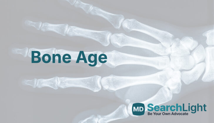
The first method involves getting an x-ray of the hand and wrist; this doesn’t expose you to a lot of radiation and is standard practice. Usually, this is done on the left hand because most people use their right hand more, and the left hand is less prone to injury or deformity.
The second method uses a book of reference images, called the Greulich and Pyle Atlas. It contains images of the left hand and wrist at different ages, up to 18 years for girls and 19 years for boys. This method is simpler and quicker than some others. But, it may not work as well for people with Asian backgrounds.
A third technique is the Tanner Whitehouse Method, which looks at 20 specific areas in the bones of your hand and wrist. Each area is given a score based on how mature it is, and these scores are added together. Although this method is a bit more time-consuming, it’s known to be more accurate.
The Gilsanz and Ratibin Atlas is a digital book, created by analyzing the size, shape, and density of certain growing parts of bone in healthy children. The images in this atlas are clearer than in the Greulich and Pyle Atlas, but the results tend to be similar.
Automated skeletal bone age assessments involve…
1. Cleaning up an x-ray image and removing unwanted parts.
2. Separating the parts of the hand and wrist we’re interested in from everything else in the image.
3. Analyzing those parts to determine bone age.
New software can calculate bone age using either the Greulich and Pyle Atlas or the Tanner Whitehouse Method, and it has been shown to work well for different ethnic groups.
Other techniques include measuring certain bones using x-rays, using specialized machines to produce an image of your bones using sound waves (ultrasound), getting a magnetic resonance imaging scan (MRI) of your hand and wrist, or looking at the growth centers of certain bones in your elbow. But, some of these techniques expose you to a bit more radiation, and others still need more testing before they can be used regularly.
Additional methods include looking at the ossification, or the process by which new bone tissue forms, of certain areas like the humeral head bump at the top of the upper arm bone, the clavicle or collarbone, the iliac bone in the pelvis, and the femoral head bump at the top of the upper leg bone. However, more research is needed to finalize these methods.
Each method has its pros and cons, and your doctor will choose the best one for you based on a variety of factors. The goal of all these methods is to assess bone age, which can provide important information about growth and development.
Why do People Need Bone Age
Bone age assessments, which evaluate how mature your bones are for your age, are often needed for a variety of reasons. They can be used to:
1. Diagnose and manage hormone-related disorders.
2. Evaluate disorders that affect body growth and cause an unusually tall or short stature.
3. Monitor the slow maturity development in various genetic disorders; and,
4. Check treatment effects in various developmental disorders.
Bone age assessments can also be important in legal situations, particularly when dealing with children and adults with bone malformations and skeletal deformities. For instance, they can be used to estimate a person’s age when no reliable birth records are available. This method could be useful in immigration cases, legal disputes, or even in competitive sports where age might be an issue.
In addition to these, bone age assessments can help calculate the final expected height of healthy children. In the case of children and young adults with bone-related problems, bone age assessment plays a significant role in managing orthopedic conditions such as:
– Slipped capital femoral epiphysis (SCFE), a condition where the ball at the top of the thigh bone slips off in a backwards direction.
– Early onset and teenage idiopathic scoliosis, a condition where the spine curves to one side.
– Limb length discrepancies, deformities, or conditions where the end part of the limb has stopped growing – the test helps predict the expected discrepancy in lower limb length and timing of a surgical procedure called epiphysiodesis that can stop or delay growth in one leg to allow the other leg to catch up.
Lastly, bone age assessments can be useful in the fields of anthropology, orthodontics, and jaw orthopedics.
Equipment used for Bone Age
The standard method for checking how bones are growing and developing uses plain X-rays. The downside of this approach is that it involves exposure to a small amount of radiation, and it’s not that great at imaging certain areas of the body, such as the collarbone.
Other options now available avoid these problems. For example, ultrasound (US) and magnetic resonance imaging (MRI) are methods that don’t involve any exposure to radiation. Another method, known as computerized tomography (CT), doesn’t just give a flat, 2-dimensional picture like an X-ray, but provides a 3-D image that can be more helpful for viewing certain structures and areas of the body.
What Else Should I Know About Bone Age?
To understand how quickly a child is growing or maturing, doctors often look at ‘bone age’. This means they examine certain bones via x-rays to see how mature they are. Which bones they look at depends on the child’s age:
– For children less than 18 years old, doctors check the left hand and wrist using an x-ray.
– For those aged 18-22 years, they examine the clavicle (collarbone), either using a CT scan or an MRI. Things like sexual, dental, and hand maturity are less helpful for this age group.
This bone age assessment is particularly important in certain orthopedic (bone-related) conditions.
When treating scoliosis (a condition where the spine curves to the side), doctors need to understand a child’s remaining potential for growth and development. They classify spinal growth into 3 phases:
– Phase 1: from birth to five years old
– Phase 2: from ages 5 to 10
– Phase 3: starts at age 10
The growth is fastest during Phases 1 and 3. This growth influences how quickly the curve in the spine is likely to worsen. There are different ways to track this, like looking at the bones in the fingers (phalanges), or looking at other elements of the spine. If the curve in the spine is more than 30° before the time of the fastest growth, the child might need surgery.
Bone age also helps doctors when looking at leg length discrepancies (when one leg is shorter than the other). They use these assessments to predict how much more the legs are likely to grow, and to plan any necessary surgeries. Similarly, for a condition called slipped capital femoral epiphysis (when the head of the thigh bone slips off the neck of the bone), understanding bone age can help doctors predict if the slip might occur in the other leg as well.


