Overview of Contrast Sensitivity
Contrast sensitivity is like how well you can see the details of something, especially really small stuff. It’s also like being able to tell the difference between shades of light and dark. Imagine looking at a black and white photo, and being able to see all the different grays in between. But it’s not quite the same as what you might know as “20/20 vision”. Usual eye tests, like the Snellen (the one with the big letters at the top and small at the bottom), check the clarity of your vision, but not your contrast sensitivity.
Even if you have perfect 20/20 vision, you might still struggle with contrast sensitivity. You might not realize how important it is, but losing it can be really upsetting. It’s like everything is there but just not clear enough. Contrast sensitivity was first accurately measured in the 60s, and it turned out to be something the eye does all on its own.
So, what exactly is contrast sensitivity? It’s how well you can see the difference between black and white. The smaller the difference you can see, the better your contrast sensitivity. It can be influenced by many things like how bright the light is, what you’re looking at, and even the shape of the object. Lots of eye conditions can affect your contrast sensitivity, like needing glasses, getting older, having cataract surgery, glaucoma, diabetes, nerve damage in the eye, and even a type of tumor called a pituitary adenoma.
Contrast sensitivity can be tested in various ways. There’s no one-size-fits-all test, but many techniques like the Arden grating or the Bailey Lovie chart can assess it. So, while this may sound complex, understanding contrast sensitivity can help doctors pinpoint what’s wrong with your vision, and find the best way to help you see clearly again.
Anatomy and Physiology of Contrast Sensitivity
The brain and eyes work together to help us see. According to Campbell and Green, our eyes have specialized visual channels for processing information about different spatial frequencies, or detailing in what we see. This suggests that the retina, or the light-sensitive layer at the back of our eye, is specialized in different areas. For instance, the fovea, the central point of the retina, is specialized for seeing high detail. Conversely, the peripheral vision, or the vision around the edge of our sight, is receptive to low frequency or less detailed visual information.
For simple patterns, both the central and peripheral vision areas offer the same sensitivity to contrast, but when a larger area is stimulated, the contrast sensitivity increases. The ability to see contrast can be reduced in diseases that affect the peripheral vision. Therefore, using low-frequency patterns can serve as a quick way of checking whether the peripheral vision is functioning correctly.
Scientists have identified independent mechanisms within our nervous systems that are highly sensitive to a limited range of detailing. They found that our brains can adapt to differences between our eyes, which helps us to recognize complex images.
According to the research, our vision is processed via two pathways in the ganglion cells (nerve cells in the retina):
– P cells, which handle high detail images,
– M cells, which handle higher contrast and speed but lower detail images.
The ganglion cells are categorized into p cells and m cells, which respectively relay information to the parvocellular (mostly detail-oriented) and magnocellular (motion and contrast-oriented) layers of the lateral geniculate body – a region of the brain involved in vision.
Researchers have also proposed a ‘Channel Theory’, suggesting that our cerebrospinal fluid, the fluid in our brains and spines, can respond differently to various detailing frequencies. The information about what we see is split into different detailing components, which are then passed on separately to different ‘channels’ in the brain, where they are processed and combined. This ‘channel’ system is thought to include 4 to 6 detailing frequency channels.
The ability of this system to detect certain frequencies depends on a variety of ganglion cells with different receptive fields – areas within which they can detect light. If the visual stimulus, or what we’re looking at, is smaller than the central receptive field of the ganglion cells, we only get a partial response. But if it is larger, the response from the ganglion cells is reduced.
Why do People Need Contrast Sensitivity
If you’re looking to work in certain professions, like the armed forces, as a driver, pilot, merchant navy member, laboratory technician, or medical professional, or if you’re considering a career in athletics, your eyesight will likely need to be tested. These jobs require good vision to perform tasks safely and effectively. However, certain medical conditions can negatively affect your eyesight, making these occupations challenging.
Vision loss can be qualitative, which means your eyesight isn’t as sharp or clear as it should be. This can occur due to diseases of the retina or optic nerve, or problems in the visual pathway, which is the route signals from your eyes take to reach your brain.
Glaucoma, or ocular hypertension, can cause vision loss. These conditions increase the pressure inside your eyes, which can damage your optic nerves. Retrobulbar neuritis, a type of optic neuritis, also affects the optic nerve and can occur as a result of, or along with, a condition called multiple sclerosis.
Amblyopia, often referred to as lazy eye, can cause poor vision in one eye. Diabetes can cause a condition called diabetic retinopathy, where high blood sugar levels damage the back of the eye. A pituitary adenoma is a benign tumor in your pituitary gland that could cause vision problems. Congenital dyschromatopsia, a color vision deficiency present at birth, can also cause difficulties in distinguishing colors.
Changes in vision can also be caused by a cataract, where the lens of your eye becomes clouded. Higher-order aberrations refer to visual defects, such as glare or halos around objects, that glasses or contact lenses cannot correct. Age-Related Macular Degeneration (AMD) causes gradual loss of central vision, while trauma to the eye can result in various types of eye damage.
Lastly, certain types of vision corrective surgeries like LASIK (laser vision correction) or PRK (photorefractive keratectomy) can also cause temporary vision disturbances.
Equipment used for Contrast Sensitivity
Measuring contrast sensitivity, or your ability to distinguish between shades of light and dark, can help your doctor understand how well you see. Think of this like how much detail you can pick up in a black and white photograph. If you can see a lot of detail, your contrast sensitivity is likely good. There are a few different methods to measure this; we’ll explain some here.
Two of the formulas doctors use are Michelson’s and Weber’s formulas. They rely on terms like “luminance,” which is the amount of light that comes off of an object, and “contrast,” or the difference in color or shading that makes an object distinguishable.
The process of measuring contrast sensitivity can be influenced by factors like the amount of light being reflected off a surface, the darkness of the surface, and the distance between light and dark areas.
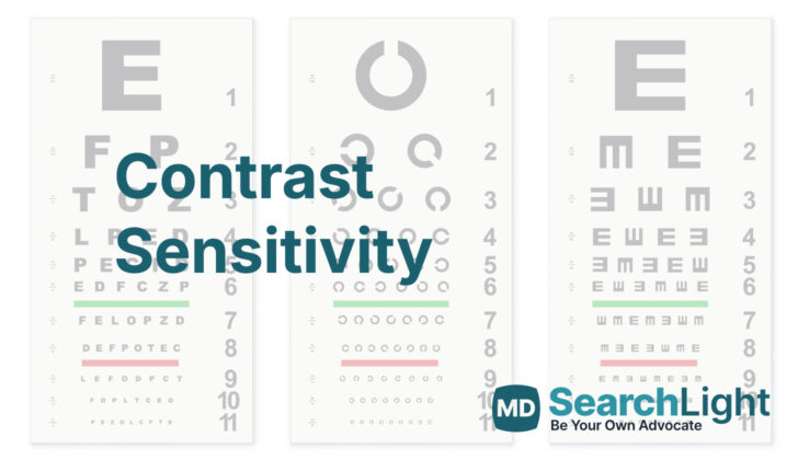
Various methods have been developed to measure contrast sensitivity. These often involve looking at patterns or letters to gauge how well you can differentiate between light and dark. Here are a few:
Arden Gratings: This test uses a booklet of seven pages with patterns whose contrast changes from top to bottom. You will be asked to identify the patterns from a certain distance.
Cambridge Low Contrast Gratings: This one involves a book of ten pages with patterns. You’re asked to identify which of two displayed pages has a pattern, starting with high contrast patterns and moving to lower contrast ones. Your score is based on how far you get before making mistakes.
Pelli-Robson Contrast Sensitivity Chart: This test uses a chart with large letters whose contrast decreases from top to bottom. Just like with an eye chart at a doctor’s office, you start by reading or identifying letters from the top down. The score is based on the last line where you can correctly identify most of the letters.
Mars Letter Contrast Sensitivity Test: This is similar to the Pelli-Robson test, but it involves a smaller chart viewed at a closer distance.
Vistech Chart: This test uses patterns of lines at different orientations that you have to identify.
Functional Acuity Contrast Testing (FACT): This test uses a chart with different patterns whose contrast varies. You’re asked to identify the orientation of the patterns.
Regan Low Contrast Sensitivity Letter Charts: This test uses three letter charts with different contrasts. You start reading from the top and continue until you can no longer identify the letters.
Spaeth Richman Contrast Sensitivity Test (SPARCS): This is a computerized test that measures your central and peripheral contrast sensitivity. It can be done online, which is convenient, especially for people who can’t read, as it uses patterns instead of letters.
Your doctor may use one or more of these methods to measure your contrast sensitivity, depending on your specific needs and their preferences.
Who is needed to perform Contrast Sensitivity?
Various healthcare professionals such as paramedics, eye specialists, optometrists, or technicians skilled in Contrast Sensitivity (CS) testing can help check your vision. CS testing measures how well you can see different levels of contrast and it’s scored in something called ‘log contrast’. A lower score means poorer vision.
For example, if you score 2, this indicates good vision, while a score of less than 1.5 flags some visual problems. A score of less than 1, however, indicates a visual disability.
Contrast Sensitivity can be of two types: Spatial and Temporal.
‘Spatial’ refers to how well your eyes can identify a pattern of stripes with varying levels of light and dark, at different sizes or ‘frequencies’. You would be shown a series of bars of light and dark (sine wave grating), and be asked what’s the least contrast level at which you can see the bars. The width of these bars is very important as it shows the ‘spatial frequency’ – a concept describing pairs of light and dark bars that make an angle of 1 degree when seen from your eye. A high spatial frequency means the bars are narrow, and low frequency means the bars are wide.
‘Temporal’ relates to how well you can see changes in an image over time, not in different positions in the image. Here, you would be shown a uniform image that changes over time, and your vision assessment would be based on how well you can track these changes. This is because our eyesight involves a system that not only sees stationary images, but can detect changes in an image over time too.
How is Contrast Sensitivity performed
When doctors check your eye sight, they often use a test developed by Dr. Snellen. However, this test only measures how well you can see things that are very clear and have high contrast. In everyday life, you don’t just see things in high contrast, but also in low contrast, such as when the light is dim or when colors are similar. Because of this, the Snellen test might not provide a complete picture of how well you can see. Testing a person’s ability to see things in lower contrast—known as contrast sensitivity—could give a better understanding about their overall vision.
Imagine your vision being like a music equalizer, with each frequency representing a different aspect of your vision. In this case, the frequencies would range from high to low—like bright daylight to a dim room. Doctors can plot this like a curve, with the frequencies going from left to right, and how well you see them (the sensitivity) going from bottom to top. This curve can then be used to assess your overall vision.
This curve is known as the ‘Contrast Sensitivity Function.’ Think of it as a graph that maps out the details of your vision. On one side of the graph is spatial frequency—how often something happens in a given space, like how many stripes are in a pattern. On the other side is contrast sensitivity—how well you can distinguish these patterns. Ideally, the graph should show high sensitivity across a range of frequencies for good eye visual function.
Also the ‘Modulation Transfer Function’ is another way to understand how the eye works. This function measures how well the eye can take in details and form an image out of them successfully. This is especially important in situations where the details are fine and closely spaced.
Another technique involves looking at a series of light and dark bars, which gives an idea of how sharp or blurry your vision is. One important feature here is the ‘spatial frequency.’ Just imagine if you are looking at a zebra, the ‘spatial frequency’ refers to how many stripes you can see within your line of sight. If the stripes are too close together and they appear blurred, then you have a lower ‘spatial frequency.’ If you can clearly see each stripe, then you have a higher ‘spatial frequency.’
There are mainly two types of these light and dark bars: The ‘Sine Wave Gratings’ and the ‘Square Wave Gratings.’ The ‘Sine Wave Gratings’ do not have clear boundaries, and they change gradually from light to dark. On the other hand, the ‘Square Wave Gratings’ have sharp boundaries with sudden changes from light to dark. The type of grating you can see better can tell the doctor more about your vision.
What Else Should I Know About Contrast Sensitivity?
Patients suffering from Contrast Sensitivity (CS) loss can have difficulties with their vision in certain situations. They may experience issues like seeing halos and glare around lights during the night, or they may require additional light to read or view objects clearly. Simple tasks like seeing clothes, dishes, and small objects, or noticing people’s facial expressions could become challenging. There can also be strain on their eyes, leading to the eyes feeling congested or uncomfortable.
The severity and impact of CS on a person’s vision can be influenced by several factors:
- Refractive errors: This refers to how your eye refracts or bends light. Only high-level details (high spatial frequencies) may become difficult to see; general viewing (low spatial frequencies) is usually not affected.
- Cataract: In early stages of cataracts, CS can be lowered, impacting the ability to see low-level details. Interestingly, this isn’t related to your overall vision (visual acuity). For example, following cataract surgery, contrast sensitivity was found to decrease significantly in a study by Brannan and colleagues.
- Age: CS tends to decline with age. From your twenties onwards, contrast sensitivity can lessen by around 10% every ten years. This average decline is similar to the full range of CS seen within a normal population of a specific age.
- Systemic disease: Certain diseases and health conditions can lead to reduced contrast sensitivity.
Contrast sensitivity can be used to assess visual function because it can decline in various eye conditions and other health problems. The relationship between the level of detail your eyes can see (spatial frequency), how intricately you can see in your visual field, the required contrast needed to see clearly, and the brightness of the object or light (stimulus luminance) is fully captured by CS. Using glasses with a yellow tint can help to improve CS as they can make objects appear sharper during the day. However, you should avoid them while driving to prevent glare. Glasses with an anti-reflective coating can be used to reduce glare. Moreover, yellow or copper tinted lenses can improve contrast and make it easier to see in a dimly lit environment.


