Overview of Electroencephalogram
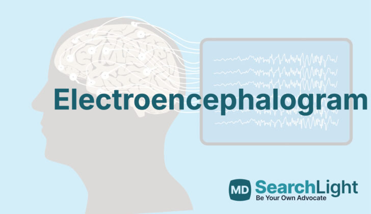
An electroencephalogram, or EEG, is a special test that helps doctors understand the electrical activity in the brain. While there have been many advances in how we can look at the brain, the EEG is still an important tool, especially when doctors need to study seizures.
The EEG is used mainly to check for seizures and conditions that can look like seizures. It can help figure out what kind of seizure someone is having, investigate patients who are in a coma in the intensive care unit, and examine certain brain abnormalities.
The fact that the brain has electrical properties was first discovered by an English scientist named Richard Caton in 1875. Around 50 years later, the German psychiatrist Hans Berger recorded the first human EEG.
Anatomy and Physiology of Electroencephalogram
The EEG (electroencephalogram) records brain activity through electrodes placed on the scalp. These electrodes pick up electrical signals produced by the neurons, or nerve cells, in the brain, specifically in an area of approximately 10 cm2. These neurons are mainly concentrated in certain layers of the cerebral cortex, part of our brain.
When brain cells communicate, they release chemicals that generate electrical signals. These signals can either excite or inhibit other brain cells, causing changes in their electrical balance. When many neurons in the same area synchronize, the summation of their electrical activity creates a sort of electrical field with positive and negative ends, which we call a dipole. The EEG records this combined activity.
The neurons within the brain link in complex ways, allowing for communication between the surface and deeper parts of the brain. During rest or relaxation, the EEG shows a pattern of rhythmic activity believed to be due to interaction between the brain’s surface (like the visual cortex) and its deeper structures (like the thalamus). But during active brain processes or a seizure, EEG displays different patterns, which can provide crucial information about the location and spread of abnormal brain activity.
EEG can capture different frequencies of brain wave activity, categorized as alpha (8 to 12 Hz), beta (13 to 30 Hz), theta (4 to 7 Hz), and delta (less than 4 Hz). Depending on one’s age and whether they are awake or asleep, the dominant waveform of the EEG can differ. For instance, slower waveforms become more prevalent in the later stages of sleep. Also, an adult at rest typically shows a distinctive frequency pattern of 8.5 Hz in the back of the head. Interpreting EEG activity also involves understanding certain benign or harmless variations and avoiding false-positive results.
Why do People Need Electroencephalogram
An electroencephalogram, or EEG, is a test that measures electrical activity in the brain. It’s used for various purposes, such as:
- Figuring out the type of seizure and where it starts in the brain. This information helps doctors provide the right treatment.
- “Sodium amobarbital”/”Wada test” that helps in understanding which parts of the brain are responsible for language and memory. This test is very useful when planning surgery for conditions like severe epilepsy.
- Managing seizures that don’t stop (status epilepticus) or inducing a therapeutic coma. Both situations require careful monitoring of the patient’s brain activity.
Besides these, an EEG is also used in other situations, including:
- When a patient’s mental state has changed due to reasons like a toxic reaction or a metabolic disorder that affects the brain. If the cause is unknown, an EEG can help doctors assess how serious the brain disorder is.
- When a person has fainted (syncope) or experienced loss of consciousness, and a heart-related cause has already been ruled out. In this case, an EEG can help determine if the fainting is due to a neurological cause.
- For patients in a coma in an intensive care situation, if they show continued confusion or reduced responsiveness. An EEG can provide clinicians with valuable insights in such cases.
- Understand the prognosis after a cardiac arrest, as an EEG can detect any damage to the brain from lack of oxygen.
Moreover, an EEG is helpful in identifying the delayed changes in the brain after a bleeding stroke (subarachnoid and intracranial hemorrhage), as well as in assessing the depth of anesthesia during surgery and in determining brain death.
When a Person Should Avoid Electroencephalogram
While there aren’t any strict rules that stop an EEG (a test that uses sensors to record the electrical activity of your brain) from being carried out, certain scenarios can make setting it up more difficult. For instance, if there’s recently been surgery in the head (a craniotomy) or if a person has any open wounds or damage to the skull, placing the test’s electrodes might be tricky.
An EEG is typically done and becomes necessary after looking into the person’s health history in depth, usually if there are worries about possible seizures or epilepsy. However, there are specific activities related to the EEG that might need to be avoided depending on certain existing health conditions.
For instance, a part of the test process may involve hyperventilation (rapid or deep breathing), which can be risky for people with certain health issues. Folks with a history of strokes, myocardial infarction (a heart attack), certain surgeries like transplants, acute respiratory distress syndrome (a severe lung condition), asthma, Moyamoya disease (a rare, progressive blood vessel disorder), and sickle cell anemia (a genetic disease where red blood cells cannot carry enough oxygen), might need to skip this part of the test.
Equipment used for Electroencephalogram
The basic tools used in an EEG, or brain wave test, are electrodes, an amplifier, and an EEG system which includes a monitor and processor. Electrodes, most commonly made of silver or silver-chloride, are used because they best pick up the electrical signals from your brain.
In the past, the information gathered by these electrodes would be recorded by a machine using pen and paper, kind of like how a seismograph records earthquakes. But nowadays, an EEG system will usually record this data electronically, and can take in information from over 128 channels at once. This allows for very detailed data to be collected quickly, with a sampling rate of over 10 kHz from all channels and a resolution of 24 bits at each amplifier.
Some other key elements in this setup include a special gel and salts. These are applied to your scalp before the electrodes are attached to help them better pick up the electrical signals from your brain. This helps make the recordings as clear and accurate as possible. Recently, dry electrodes have also become common, as they make the process of preparing your scalp for the EEG quicker and easier.
Who is needed to perform Electroencephalogram?
An EEG, or electroencephalogram, is a test that’s done by a specially trained technician or technologist. This person has completed the needed education and training and earned their certification to be able to perform this kind of test. After the EEG is done, a doctor who specializes in how the brain works, called a neurologist, will take a look at the results. This neurologist has also completed extra training in things related to EEGs and seizures, called epilepsy.
If the EEG has to be performed in special units in the hospital like the Intensive Care Unit or an epilepsy monitoring unit, there will be other staff on hand to help. This might include nurses, support staff, and monitor technicians, who can help to make sure everything goes smoothly and that patients are safe and well cared for during the test.
Preparing for Electroencephalogram
When getting an EEG (a test that records electrical signals in your brain) done at a healthcare center, patients are given clear directions before the test. It’s important not to use hair conditioners or other products that might interfere with the quality of the recording. This is because these substances can change the electrical resistance (impedance) between the recording electrodes and the scalp, which can affect the test results. Doctors ensure the scalp is cleaned properly so the electrodes can record your brain signals accurately.
Typically, the resistance value should be lower than 5 kohm (a technical term referring to electrical resistance). For patients in the Intensive Care Unit, special steps are taken to minimize any interference from different medical equipment and wires. Sometimes, it might be necessary to use physical restraints, not chemical ones, to make sure the EEG recording is done properly.
How is Electroencephalogram performed
An EEG, or electroencephalogram, is a test that is done in a calm setting with adjustable lighting. This test is typically conducted by a trained technician, using specific methods to place special sensors, known as electrodes, on your scalp. The 10 to 20 system is a commonly used method to place these sensors, with usually around 21 electrodes being used, including some control ones. After placing these electrodes, the technician ensures that they are working correctly and are able to pick up your brain signals with high accuracy.
Before starting the test, calibration is carried out – this involves recording a set signal and carrying out a biological calibration. During the EEG, you will be asked to do certain things that help provoke your brain into showing certain patterns. These can include opening and closing your eyes, breathing heavily, exposure to flashing lights and sleeping. Sometimes, you might even be asked to stay awake the night before the EEG – this technique is used because lack of sleep can make certain patterns more visible.
Once the test is done, the results are displayed specifying the differences in the brain electrical activity as picked up by the electrodes. The way these results are arranged and displayed is called montage. Different types of montages (like referential, bipolar, and Laplacian montages) are used to accurately identify and locate abnormal signals. The digital transformation of EEGs has made it easier to arrange and adjust these montages as per the needs of the test interpreter.
Referential montages show the electrical activity difference between a chosen electrode and other electrodes on your body. Bipolar montages show the difference in electrical activity between two nearby electrodes and they are very helpful in detecting differences in activity between the two halves of your brain. Laplacian montages, meanwhile, use a reference that is a weighted average of the potentials surrounding a particular area. This kind of montage is particularly useful for looking at localized discharges with minimum interference.
Possible Complications of Electroencephalogram
It’s very important to be thorough when assessing patients before we proceed with certain procedures, such as hyperventilation. If this is not properly done, some unexpected complications might arise. One of these complications is skin injuries that may occur over time with extended use of EEG (Electroencephalogram – a test used to assess the brain’s electrical activity) monitoring in units made specifically for monitoring epilepsy and in intensive care units. So, it’s crucial to take proper care when these sessions are ongoing for long durations.
What Else Should I Know About Electroencephalogram?
An EEG, or Electroencephalogram, is a test that helps doctors look into potential issues with the brain’s function. It’s often used with other tests to understand conditions that might affect our thoughts, feelings, and behaviors. It’s especially useful in cases where people experience seizures, changes in awareness or consciousness, certain sleep disorders, mental conditions caused by things like body imbalances or harmful substances, memory disorders, and stroke.
In addition, an EEG can predict the outcome for people who’ve had a severe lack of oxygen to the brain, suffered traumatic brain injuries, or are suspected of having brain death due to severe injury. It can also help understand the effects of certain drugs and check activity in the brain when a person is unresponsive or in a coma.
Furthermore, EEG helps doctors discern between actual seizures and other physical or psychological conditions that may look like seizures. Special techniques during an EEG can help the doctor to pinpoint the specific areas of the brain involved in seizure activity, or to find out if a person’s epilepsy is due to inherited factors.
Long-duration EEG tests that include video can record seizures when they happen, providing valuable information on the type and nature of the seizures. This is particularly useful when planning for surgery to cure seizures that are difficult to control with medication.
There are also more advanced types of EEGs that use special sensors, which are placed directly on the brain’s surface or deeper within the brain. These tests give a detailed picture of the electrical activity in the brain which can be helpful in certain medical situations.


