Overview of Evaluation of Visual Acuity
Understanding your vision and how healthy your eyes are is an important part of an eye doctor’s check-up. A detailed, 8-step eye exam involves checking the clarity of your vision, examining the pupil’s reaction to light, looking at the movement and alignment of your eyes, testing the pressure inside your eyes, testing your field of vision, examining the external part of your eyes, using a special microscope called a slit lamp to look closely at the front parts of the eyes, and examining the back parts of your eyes which include the retina and optic nerve.
The clarity or sharpness of your vision is called “visual acuity”. When your doctor is documenting your eye examination results, you will often see three Latin abbreviations: OU, OS, and OD. To break them down:
- OU stands for “oculus uterque” and means both eyes
- OS stands for “oculus sinister” and represents the left eye
- OD stands for “oculus dexter” and indicates the right eye
You may also see abbreviations like DVA (distance visual acuity), IVA (intermediate visual acuity), and NVA (near visual acuity). These denote how sharply you can see at a distance, at an intermediate level, and up close respectively. These might sometimes be combined with other prefixes to explain whether this is with correction (like glasses or contacts), without correction, or with both eyes.
Most numbers from a visual acuity test are based on a “normal” person’s vision at a distance of 20 feet or 6 meters. If your vision is listed as 20/20 or 6/6, this means that you can see as clearly as a person with normal vision at that distance. If your vision is recorded as 20/40 or 6/12, this means that what you can see clearly at 20 feet, a person with normal sight could see at 40 feet.
Visual acuity is a measure of the smallest object or detail you can recognize at a certain distance. To fully see an object, you must be able to distinguish its various parts. The smallest angle your eye can recognize a detail to separate it from the rest is called the minimum angle of resolution (MAR).
There are four types of visual acuity:
- Minimum (detectable) visible acuity: The smallest thing you can simply detect the presence or absence of.
- Minimum (separable) resolvable acuity: The smallest space between two things you can distinguish as separate. This tests how well your eye, and primarily the retina, can differentiate between two objects.
- Minimum recognizable acuity: The smallest part of an object that you can identify, like identifying a letter from a certain distance. Though 20/20 is often used as the standard, different age groups might have a sharper vision.
- Minimum discriminable acuity: This refers to the smallest change in position, size, or orientation of an image that you can notice. This impressive ability to detect alignment in objects brings us to the use of the term “Vernier acuity” named after Pierre Vernier, who devised a scale used in ship navigation.
Anatomy and Physiology of Evaluation of Visual Acuity
Minimum detectable visual acuity is a term that describes how well your eyes can distinguish the differences between an object’s brightness and its background. In simpler terms, it’s the smallest noticeable change in how different the object is from what’s around it in terms of light and dark.
Other measures of visual acuity are minimum resolvable acuity and minimum recognizable acuity, which look at how close together things can be before you can no longer tell them apart. These measures are influenced by factors like how close the light-sensing parts of your eyes are, any imperfections or distortions in your eyesight, and the size of your pupils.
When your eyesight is tested, you might be presented with a pattern of black and white stripes. Beyond a certain point, the stripes might just look gray or appear confusing, where the stripes seem to have different shapes or sizes. The maximum point where this occurs is defined by where the lighter and darker parts of the image align with the different light-sensing parts of your eyes. Usually, this point corresponds to a very specific visual angle.
There’s another measure called minimum discriminable visual acuity, which is also known as hyperacuity. This is a much more sensitive measure than what can be explained just by considering how close together the light-sensing parts of your eyes are. In fact, this measure can detect changes that are even smaller than the size of these light-sensing parts. Although the exact mechanisms behind hyperacuity isn’t fully understood, it’s thought that changes in the light-sensing components of your eyes or their optical properties might play a role. Furthermore, researchers believe that the brain’s processing of visual information is crucial for this type of acuity.
Why do People Need Evaluation of Visual Acuity
If you have an issue with your eye and go to an emergency room or visit your doctor’s office, they will conduct an eye exam. This exam will be recorded in your medical records. If you are seen by eye doctor or in the eye clinic of a hospital, they will also check your vision. This is known as a visual acuity test.
When a Person Should Avoid Evaluation of Visual Acuity
There are no specific reasons why a visual acuity test, which measures how clearly you can see, can’t be performed. However, several factors can influence the test and potentially lead to inaccurate results. Some of these factors may include:
– Eye diseases
– The brightness of the chart used during the test
– The visual contrast between different parts of the chart
– The size of your pupil
– The type of symbols (optotypes) used in the test
– Errors in the way your eye focuses light (refractive errors)
– The position of your retina (retinal eccentricity)
– How long you’re exposed to the test
– Difficulty with seeing all symbols clearly due to the effect of nearby symbols (crowding)
– The amount of light you were exposed to prior to the vision testing
– Your willingness to cooperate with the test or any functional eye diseases that might influence the test results.
Equipment used for Evaluation of Visual Acuity
Tests to measure how well you can see are categorized into three main types, and some of these tests need specific equipment.
Detection acuity tests help to figure out how well you can detect the smallest thing you can see. Examples of these types of tests include the Boeck candy bead test, Catford drum test, Dot visual acuity test, Schwarting metronome test, and STYCAR graded balls test.
Recognition acuity tests see how well you can identify what you’re looking at. Examples of these types of tests include the Bailey-Hall cereal test, Bailey-Lovie chart, Beale Collin picture charts, Early Treatment of Diabetic Retinopathy Study (ETDRS) chart, Landolt C test, Lea symbols chart, Lighthouse test, Lippman’s HOTV test, Sheridan letter test, Sjogren hand test, Snellen charts, and the Snellen E test.
Resolution acuity tests include tests like the Optokinetic drum and Preferential looking test.
When it comes to testing how well a person of a specific age can see, the options can change. Some tests for different age groups include:
For infants: Cardiff acuity cards, Catford drum test, Fixation test, OKNOVIS, Preferential looking test or Teller acuity cards test, Reflex response, and Visual evoked responses.
For kids aged 1 to 2 years: Boeck candy test, Sheridan ball test, Worth Ivory-ball test.
For kids aged 2 to 3 years: Coin test, Dot visual acuity test, Miniature toys test.
For kids aged 3 to 5 years: Landolt C, Lea symbols chart, Lippman’s HOTV test, Sheridan letter test, and Tumbling E.
There are also other tests to evaluate your vision like the Brückner test. This test can detect conditions like strabismus by using a direct ophthalmoscope to look at a red glow in both your eyes at once. If you have strabismus, you’ll have a brighter red glow in one eye that doesn’t align properly. This test can also help to pick up an uneven refractive error.
A relative afferent pupillary defect test uses a swinging flashlight in a dim room to spot any issues with your optic nerve or retina. This test involves shining a bright light on one of your eyes for a few seconds, then rapidly moving it to your other eye. If your other pupil dilates instead of getting smaller, it may have what’s called a relative afferent pupillary defect or a Marcus Gunn pupil.
The cover test can identify any lazy eye issues in a child who can’t speak yet. Such children will resist or cry when the good eye is covered, but show little to no reaction when the lazy or “amblyopic” eye is.
Who is needed to perform Evaluation of Visual Acuity?
The process of checking how well you can see involves many different specially trained people like hospital workers, technical staff, eye doctors and other healthcare providers. How accurately they can measure your vision can depend a lot on their training and skills.
Preparing for Evaluation of Visual Acuity
It’s really important that the patient understands what the eye test involves. This helps them cooperate fully and gives the best results. Sometimes, eye tests can feel a bit subjective since they depend on what the patient says they can see. If the doctor suspects that the patient either isn’t telling the truth (malingering) or is experiencing loss of vision that’s not related to the physical condition of the eye (known as functional vision loss), special tests can be used.
How is Evaluation of Visual Acuity performed
Visual acuity testing is crucial to assess how well you can see. One of the easiest methods is to use the Snellen chart. It assesses your distance vision. During this test, the doctor ensures that you stand the right distance away from the chart and tests both your eyes together and then each individually. The test results will show how well you can see both with and without glasses or contact lenses.
The results of these tests are often shown with the letters V, VA, or Va. Uncorrected visual acuity (UCVA) represents how well you see without glasses or contacts, while best-corrected visual acuity (BCVA) shows your vision with corrections. Near vision is often indicated by NV. Initially, distance vision is evaluated. By convention, your right eye is tested first, then your left eye, and then both eyes together.
There are various charts available for these tests. The charts can either be printed, shown on a computer screen, projected, or viewed with the help of a mirror. In small exam rooms, a mirror may be placed 3 meters away from the patient seat to mimic a distance of 6 meters. The chart is put on the wall in the inversion, so the letters will appear straight when viewed through the mirror.
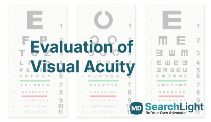
The most common chart, called the Snellen chart, was created by a Dutch eye doctor and has been in use since 1862. It consists of lines of letters that get smaller as you read down the chart. The top line has the largest letters, and the lines below have more letters that decrease in size. The letters and numbers on the chart represent how well you can see compared to an average person.
The letters on the Snellen chart are designed to take up a specific amount of space in your field of vision. For instance, the letters on the 20/20 line of the chart (the smallest line most people can read) are each 9 millimeters tall, and each has a specific thickness and spacing from the other letters. They are designed to be viewed from 20 feet (or 6 meters) away, which is considered to be “normal” distance vision.
While the Snellen chart is common, it’s not universally ideal due to its inconsistent size changes between lines and uneven amount of letters per line. One such patient category that faces this problem is people with Amblyopia. So, it’s crucial to realize that this type of testing isn’t flawless and sometimes additional tests may be needed for certain people to get an accurate vision assessment.
One alternate method of testing is the LogMAR chart, which overcomes some limitations of the Snellen chart. The LogMAR chart uses a strict mathematical formula to determine how well you can see, it has consistently spaced rows of letters for better accuracy. All the letters have similar recognizability, and there is an equal number of letters in each row which is determined by the British Standard test.
Overall, eye exams are crucial to measure how well you can see and to help detect any potential vision issues. There are various methods and charts available to conduct these tests, and your eye doctor will decide which one is best for you based on your specific needs.
What Else Should I Know About Evaluation of Visual Acuity?
Testing how well you see, a test known as visual acuity testing, can provide important information for doctors. It can highlight eye-related illnesses and help identify other health issues including neurological disorders.
Examining your visual acuity is done along with other eye tests. These include checking your pupils, measuring your eye’s internal pressure, testing your field of vision, refraction tests, a slit-lamp examination and a check of your retina. The best-corrected visual acuity, meaning your vision when you’re wearing corrective lenses, often changes as you get older. A study involving the eyes of 200 people aged 25 to 74 found that until age 64, healthy eyes typically have 20/20 vision or better. At 55-59 years, vision starts to decline, and after 60 years, the visual acuity is less than 20/15.
For babies and toddlers who can’t talk yet, different techniques are used to test how well they see. These can include displaying black and white lines in front of their eyes to see if they react (Optokinetic nystagmus testing) or showing them a striped pattern and a blank pattern to see which one they look at longer (Preferential-looking test, used in Cardiff Acuity Test and Teller Acuity Cards II testing). There’s also a technique where a child’s brain activity is measured in response to visual stimuli (Visual evoked response/potential testing).
Doctors often use a Snellen chart to test visual acuity. This chart uses lines of letters of different sizes that you read from a specific distance. The results from a Snellen chart test can be converted into a “logMAR” value, which is another measurement of visual acuity. There are tables to convert Snellen results into logMAR, but it’s important to note this conversion may inaccurately represent a person’s visual acuity, so it should be used with care.
It’s also been found that measuring visual acuity with a logMAR chart is twice as repeatable as with a Snellen chart. This is why the Snellen chart may not be as reliable as the logMAR chart, and why some suggest its use should be phased out. This is because the Snellen chart’s design makes it difficult to get precise measurements, meaning it may be less useful when comparing measurements over time, even with conversions to logMAR. Despite some limitations to the Snellen chart, it is still widely used.


