Overview of Fetal Monitoring
Continuous fetal monitoring, also known as cardiotocography, is a process used to keep track of a baby’s heart rate and the mother’s contractions while she’s in labor. Doctors think this information could be important because there appears to be an association between these two factors and how much oxygen the baby is receiving. The goal of this monitoring is to ensure both the mother and baby are safe, and to help doctors know if they need to step in and help.
Across much of the United States, it is common to perform continuous fetal monitoring on most women giving birth. However, there is some debate around this practice. Studies have shown that routine monitoring of the baby’s heart rate doesn’t lead to a substantial improvement in the health of most newborns or children as they grow. What is certain though, is that it increases the number of operative vaginal deliveries (forceps or vacuum extraction) or Cesarean sections.
Quite frankly, the jury’s still out concerning whether monitoring the baby’s heart rate can reduce the neonatal death rate, albeit there are fewer cases of newborn seizures when the baby’s heart rate is continuously monitored. Beyond this, we haven’t observed significant marked improvements in Apgar scores (a quick measure of a newborn’s health), incidents of nerve-related injuries, cerebral palsy, developmental delay, and NICU (Neonatal Intensive Care Unit) admissions.
Fetal monitoring isn’t just useful during labor; it can also help doctors check for placental abruption (when the placenta detaches prematurely) after a pregnant woman has suffered a traumatic injury to the abdomen. Additionally, it can assist in distinguishing between real and false labor. Obstetric care teams are typically expected to know how to perform and interpret continuous fetal monitoring, as it is considered particularly vital in high-risk pregnancies and for evaluating the baby’s condition outside of labor.
Anatomy and Physiology of Fetal Monitoring
Childbirth, or labor, is typically divided into three main parts. The first part starts with the beginning of labor and goes until the cervix (the lower part of the uterus that opens into the vagina) is fully dilated (opened) and thinned out. This part has two phases. The first phase, known as the latent phase, is usually slow and gradual, with the cervix opening up slowly. When the cervix dilates to about 6 centimeters, women are said to enter the second phase, called the active phase, where the opening of the cervix speeds up a lot. This first part of labor is also marked by an increase in the frequency and regularity of uterine contractions (when the muscles of the uterus tighten and then relax). However, the exact beginning of labor might not be clear, since contractions can happen off and on and cervical changes can progress very slowly in the later part of pregnancy.
The second part of labor happens when the cervix is fully open until the baby is delivered. The third part of labor takes place from the delivery of the baby until the placenta (the organ that supplies the baby with nutrients and oxygen during pregnancy) is fully delivered.
During the first two parts of labor, the baby’s blood circulation can be compressed as the baby changes position and as the uterus contracts. This can possibly lead to a lack of oxygen and/or too much acid in the baby’s blood, which could cause the baby distress and lead to poor outcomes. Therefore, doctors must closely watch the baby’s heart rate, fluctuations in the heart rate, heart rate increases, and decreases.
The baby’s heart rate and its fluctuations are impacted by changes in the activity of the baby’s central nervous system, the volume of blood in the baby’s body, and stimulation of certain receptors that help regulate blood pressure and that respond to changes in chemical conditions in the body like oxygen and CO2 levels.
An increase in the baby’s heart rate is related to stimulation of the baby’s central nervous system and periods of increased activity. Early decreases in the baby’s heart rate are associated with compression of the baby’s head, especially during contractions. When the baby’s head is compressed, it causes a reflex that slows down the baby’s heart rate. This reflex is so fast that the decrease in heart rate can be seen at the same time as the contraction. Altogether, an appropriate baseline heart rate, variability, increases, and early decreases in the baby’s heart rate are favorable signs of the baby’s neurological, autonomic (involuntary), and cardiovascular (heart and blood vessels) systems working properly.
Late decreases in the baby’s heart rate are thought to be caused by temporary lack of oxygen. This can happen because during contractions, the blood vessels in the uterus and placenta are compressed, reducing blood flow. Even though blood flow is temporarily reduced, oxygenation is usually fine in a normal labor. However, if the baby’s oxygen levels are already low or the decrease in oxygenation is severe enough, it can compromise the baby’s oxygen levels and lead to a lack of oxygen. An oxygen deficit is picked up by certain sensors in the baby’s body (chemoreceptors) that initiate a reflex to constrict the peripheral blood vessels (vasoconstriction), pumping up the blood pressure. This increased blood pressure triggers a reflex to decrease the baby’s heart rate. This whole sequence only starts when the baby’s oxygen levels are low enough and goes on in a step by step manner, so that the decrease in heart rate starts after the start of the contraction, and the lowest point of the decrease is shifted from the peak of the contraction.
Variable decreases in the baby’s heart rate are associated with compression of the baby’s umbilical cord. These can happen with or without a contraction. Once the baby’s umbilical artery (the blood vessel that carries oxygen-rich blood from the placenta to the baby) gets compressed, it blocks the blood flow, decreases oxygenation, and causes vasoconstriction, which leads to a relatively sudden drop in heart rate. Both late and variable decreases in the baby’s heart rate may suggest a risk of injury due to lack of oxygen and/or too much acid in the baby’s body, and should warrant further investigation, closer monitoring, and possibly intervention by the doctor.
These findings and their interpretation will be discussed further in the clinical significance section.
Why do People Need Fetal Monitoring
Fetal monitoring during labor, which is a way to track the baby’s heartbeat, is a topic doctors have varying opinions about. Some believe it’s very necessary, while others argue that there’s no clear evidence it improves outcomes, even though more medical interventions could be taken when it’s used. The American College of Obstetricians and Gynecologists (ACOG), however, recommends that fetal monitoring be used in high-risk pregnancies. Some examples of high-risk pregnancies can include conditions such as preeclampsia (high blood pressure during pregnancy), type 1 diabetes and growth problems for the baby while in the womb.
Aside from monitoring the baby during labor, doctors might also use fetal monitoring before labor starts, if they think the baby might not be doing well due to certain health problems. For instance, if a pregnant woman has had a belly trauma after being pregnant for 20 weeks, doctors might use fetal monitoring for 4 to 24 hours to check for a condition called placental abruption, where the placenta— which provides oxygen and nutrients to your baby—detaches from the womb too early.
When a Person Should Avoid Fetal Monitoring
There are no strict situations where external monitoring of a baby’s heart rate (FHR) can’t be done. But, it’s not usually suggested for pregnancies that are low-risk, or less likely to have complications. Situations where a sensor is attached directly to the baby’s scalp to monitor heart rate (known as fetal scalp electrode monitoring), are not recommended if the mother has HIV or active herpes infections, and if there is a chance the mother or the baby has conditions that affect blood clotting.
Equipment used for Fetal Monitoring
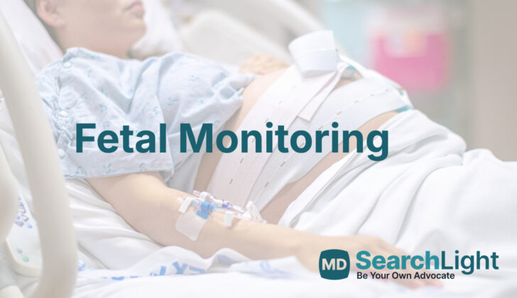
External fetal monitoring is a common method used to check on a baby’s well-being before birth. In this procedure, two sensors are placed on the mother’s belly. One sensor is positioned over the baby’s heart to track its heart rate, while the other is placed over the top part of the uterus, or womb, to track contractions. These sensors comprise of the ‘transducers’.
At times, if there’s a problem with the reliability of heart rate measurements due to interference from the mother’s own heart rate, a special electrode may be attached to the baby’s scalp. This is called a fetal scalp electrode.
There are also situations where external methods aren’t as effective; for example, if the mother is overweight. In such cases or when there’s a need for exact pressure measurements- like when labor is going slower than expected or has stopped, a device called an intrauterine pressure catheter may be used. This device measures the pressure inside the uterus and gives more accurate results. It’s known as ‘tocodynamometry’ when the intensity of uterine contractions is measured using this method.
Possible Complications of Fetal Monitoring
A research study from 2017 showed that women who had continuous monitoring of their baby’s heart rate during labor were more likely to have cesarean section births or use tools like forceps for vaginal deliveries, compared to those who had occasional listening of the baby’s heart rate.
More direct methods of monitoring offer extra risks. Invasive procedures, such as placing an electrode on the baby’s scalp or taking samples of the baby’s scalp pH, can raise the chance of transferring infections like herpes simplex virus (HSV) and human immunodeficiency virus (HIV) from mother to baby. These methods can also increase the likelihood of injury to the baby’s scalp, cause a blood collection under the scalp or inside the skull, choking hazards, and severe infection.
Placing a pressure measuring device inside the uterus is mostly related to tearing or separating the placenta due to improper positioning. Other serious complications like penetrating the uterus or getting the umbilical cord tangled have also been observed.
What Else Should I Know About Fetal Monitoring?
Fetal monitoring is a method used by doctors to check on the health of a baby before they are born. This can be done by measuring the baby’s heart rate (FHR) and observing other physical signs.
It’s normal for a baby’s heart rate to be between 110 and 160 beats per minute. This can vary based on various factors, such as how the baby’s heart is working and the mother’s overall health condition or medication use.
Doctors will track patterns in the baby’s heart rate over a 10-minute period. These patterns are referred to as variability and can be described as absent, minimal (less than 5 beats per minute change), moderate (6 to 25 beats per minute change), or marked (over 25 beats per minute change). Moderate variability is generally a good sign, as it suggests that the baby is getting enough oxygen and is not in distress.
Doctors also look for increases in the baby’s heart rate called accelerations, as well as decreases known as decelerations. Early decelerations are not a cause for concern and do not predict any positive or negative outcomes for the baby.
However, repeated late or variable decelerations could suggest potential risks to the baby’s health, especially if the variability is decreased.
Doctors classify these heart rate patterns into three categories. Category I patterns are reassuring and suggest that everything is normal. Category II patterns may indicate potential issues relating to the baby or the mother’s health. These patterns require further investigation and potentially some actions to aid the baby, like changing the mother’s position to improve the flow of blood to the baby, or introducing treatment for suspected conditions.
Category III patterns could suggest significant health concerns such as lack of oxygen or potential neurological injury to the baby. Things can move quickly in this scenario, with steps taken to help the baby and prepare for a possible emergency delivery.
At the end of the day, doctors use fetal monitoring to keep a close eye on the baby’s health. When they notice concerning patterns, they can take steps to identify any potential problems and work towards ensuring the best outcome for the baby and the mother.


