Overview of Hysteroscopy
The procedure known as hysteroscopy was first done on a patient in 1869, and it involved identifying and treating a growth in the uterus using a special instrument developed for looking into body cavities. As medical science improved, better tools and techniques were developed, and by the 1980s, it was possible to perform hysteroscopy in a doctor’s office. Today, advances in technology and the development of effective and safe practices have made hysteroscopy the preferred method for dealing with issues inside the uterus.
The key to doing hysteroscopy is choosing the right patient and having the right equipment. The process involves inserting a specially designed instrument into the uterus via the cervix and using a fluid to expand the uterus for better visibility. The type of fluid used depends on how the procedure is performed. Certain fluids are not suitable when electric current is used in the procedure due to the risk of transferring electricity outside the operating area. There are also limits on the amount of fluid used to avoid overloading or complicating the patient’s health, especially in the case of older patients or those with other health issues.
The kind of instrument used for the hysteroscopy is chosen based on the specifics of the operation. These instruments, known as hysteroscopes, come in different varieties and have different parts that allow the doctor to view the inside of the uterus from different angles. A special type of tool, known as an operative hysteroscope, is needed when surgery is involved.
Smaller, more advanced hysteroscopes have made it much easier to perform these procedures in a doctor’s office. For women with abnormal uterine bleeding, hysteroscopy can be an effective alternative to more invasive surgeries in some cases. It’s also used to diagnose fertility issues and safely remove leftover tissue after pregnancy or foreign objects.
Anatomy and Physiology of Hysteroscopy
The external genitalia refers to the parts of a woman’s reproductive system that can be seen without any special tools. This includes the mons pubis (the rounded area of fatty tissue on the pubic bone), clitoris, urethral opening, vaginal entrance, large and small vaginal lips, hymen (a thin piece of skin that partly covers the vaginal opening), perineum (the area between the vagina and anus), and anus. When we talk about all of these parts together, we call them the vulva.
The vagina is a muscular tunnel that connects the vulva to the cervix. It’s flexible and can change in size. Some of its main functions include providing sexual pleasure, being a birth canal for delivering babies, and allowing the passage of sperm after sex and menstrual blood.
The cervix is the doorway from the vagina to the womb or uterus. It’s usually around 2-3 cm long. There’s a small hole in the middle of the cervix known as the cervical canal. The size of this canal can change throughout a woman’s life, becoming as wide as 10 cm during childbirth.
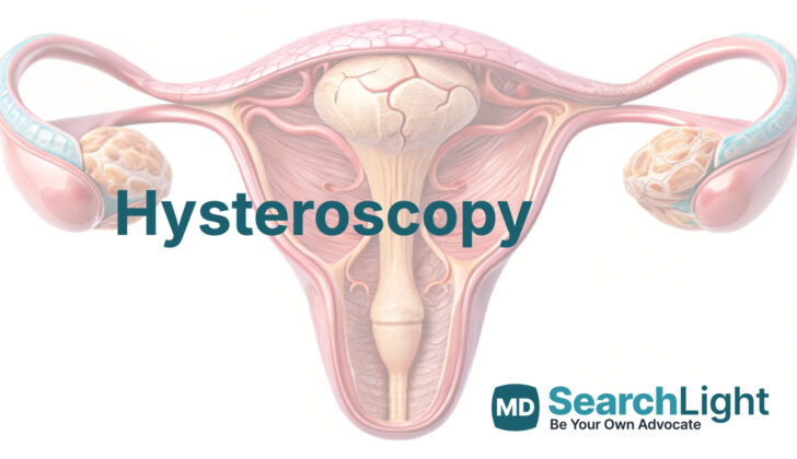
The main body of the uterus sits in the middle of the pelvis, between the bladder and rectum. It’s usually positioned slightly forward. The term “version” refers to the position of the cervix in relation to the vagina while “flexion” refers to the position of the top of the uterus (fundus) relative to the cervix. The fallopian tubes, which are the paths for eggs to travel from the ovaries to the uterus, connect to the uterus. These connections can be seen during a medical procedure called a hysteroscopy.
Why do People Need Hysteroscopy
Doctors often use a procedure called hysteroscopy to examine the uterus. There are several reasons why a doctor might recommend this procedure:
- An unusual mass or lesion in the uterus.
- Unexplained or unusual bleeding from the uterus.
- Excessive thickening of the uterus lining.
- Bleeding after menopause.
- Difficulties getting pregnant.
- Problems with the structure of the uterus present from birth (Mullerian congenital anomaly).
- Need to remove foreign bodies or materials from the uterus.
Whenever it is viable, performing the hysteroscopy in the doctor’s office is usually the preferred choice. The office procedure is often more convenient for both the patient and the doctor, and it avoids the need for general anesthesia. This in-office process generally leads to higher patient satisfaction, quicker recovery, and it’s usually more cost-effective.
However, there are situations where an in-office hysteroscopy may not be possible. For example, if there’s a large mass in the uterus, if the patient is highly anxious, if the necessary equipment or staff are not available, or if the doctor lacks the required skill or expertise for an office procedure. In these cases, the hysteroscopy may need to take place in a hospital setting.
When a Person Should Avoid Hysteroscopy
There are a few specific situations where it’s not advisable to perform hysteroscopy, a procedure that allows your doctor to look inside your uterus. These include having an active infection in the pelvic region, symptoms or actual genital herpes, and confirmed cases of cervical or endometrial cancer (cancer of the cervix or the lining of the uterus).
Moderate vaginal bleeding can potentially make it difficult to perform a hysteroscopy, but in some cases, your doctor can manage this by flushing the area with a lot of fluid. Pregnancy is another condition where hysteroscopy should usually be avoided, unless it’s necessary to remove an IUD (a small contraceptive device that sits inside the uterus) or remnants after conception.
Some underlying health issues could possibly make hysteroscopic surgery more risky. For example, a person with coronary heart disease (a condition that affects the flow of blood to the heart) or bleeding diathesis (an abnormal tendency to bleed) may not be good candidates for such a procedure.
Equipment used for Hysteroscopy
Hysteroscopes
Hysteroscopes are tools used by doctors to look inside a woman’s uterus for diagnosis and surgery. There are various types of hysteroscopes; some are flexible and some rigid. These tools can provide different views, ranging from a straight line (0 degrees) to a more side view (up to 70 degrees). They also connect to equipment that provide light and video images. To get the best views, doctors need to fill the uterus with a special fluid.
Most hysteroscopes include a built-in passageway that allows doctors to insert small surgical tools like scissors during procedures.
The STORZ Bettocchi integrated in-office hysteroscope is tiny, just 4-5 mm (or about the size of a grain of rice), and has a rounded tip, which makes it easier and less painful to insert for patient. STORZ Campo Trophyscope is even smaller and can be used for simpler procedures. However, during procedures, this may need to be switched out for a tool with a bigger diameter.
The Olympus Flexible Hysteroscope is a different type with a flexible tip that can move in a large range, and is slightly larger than the previous two at 3.1 mm.
There are also other rigid hysteroscopes that come in various sizes and viewing angles.Heavy duty tools like the resectoscope and mini-resectoscope have special capabilities like electricity, which can help the doctor cut through dense tissue more easily.
Fluid Distension Medium
A key factor in performing a hysteroscopy is to properly fill the uterus, usually with a special fluid or a gas like carbon dioxide. The reason for this is to help the doctor get a better look at the uterus. Carbon dioxide, however, is not suitable for surgical procedures because it can obscure the view if there’s any bleeding. The type of fluid used depends on the surgical tools used. For instance, if they use an electrical current loop for dissection, then the fluid should not contain electrolytes since they may carry electricity to other tissues. If a tool that uses both type of electricity (bipolar) is used, then the fluid used can contain electrolytes like normal saline.
Other Instruments
Hysteroscopy may also require additional tools such as a metal speculum, forceps, sound and cervical dilators. Local anesthesia, like 1% or 2% lidocaine, might be used to numb the area, based on doctor’s preference. A thin needle is used to administer the anesthesia. With newer methods like vaginoscopy, only the hysteroscope is needed.
Who is needed to perform Hysteroscopy?
The number of medical staff needed in a hysteroscopy (a procedure that lets your doctor look inside your uterus) will vary according to where the procedure is performed. In an operating room setting, the team will consist of highly trained professionals, including a specialist doctor (an anesthesiologist) or a certified nurse who specializes in anesthesia to help in putting you to sleep, nurses who will look after you before and after surgery, and personnel to ensure the equipment is clean and ready for the procedure. There are also people who will take care of moving you from your room to the operating area.
On the other hand, if your doctor decides to do this procedure in their office, you won’t need as many medical staff. However, it’s still important to have a professional, other than the doctor, who can monitor you throughout the procedure. It is also recommended to have a team member who is certified and equipped to handle emergencies like sudden heart problems or severe allergic reactions (anaphylaxis). You wouldn’t need a specialized doctor or nurse for anesthesia, as this procedure in an office setting is usually done with only local anesthesia (numbing a small area) or without any anesthesia.
Preparing for Hysteroscopy
Before a hysteroscopy – a procedure to look inside the uterus – certain preparations may need to occur. These are based on each patient’s unique needs and any existing health conditions that may increase risks with the procedure. Additional tests might be necessary for some patients to understand these risks better.
In women who have gone through menopause, a hysteroscopy can take place at any time. For women still in their childbearing years, it’s crucial to consider the timing of the procedure. If it’s done during a specific phase of the menstrual cycle, it could lead to false identification of uterine polyps – small growths in the lining of the uterus. This is because the uterine lining itself can appear abnormal during this stage of the cycle.
There are various opinions on whether certain drugs for opening the cervix should be used before the procedure. However, this is not something that’s typically done. Also, preventive antibiotics are not required before a hysteroscopy.
Every woman should have a detailed health evaluation, including a thorough check-up and history, before undergoing a hysteroscopy. Also, all women who still have their periods should have a pregnancy test pre-procedure.
Managing pain during an in-office hysteroscopy is still up for discussion in the medical community. Various approaches have been suggested, both drug-based and non-drug based.
And of course, like any surgical procedure, patients should always know and understand all the aspects of the procedure, including its risks and benefits, as well as any other options available to them. This is part of the informed consent process – ensuring patients have all the information they need to make the decision that’s best for them.
How is Hysteroscopy performed
Firstly, correct positioning of the body is essential for any gynecological procedure. The patient is positioned lying flat on their back with their legs spread apart; this is known as the dorsal lithotomy position. It’s crucial to make sure there is no unnecessary pressure that could potentially hurt the nerves. A two-handed examination should always be carried out before starting any gynecological procedure. It’s usually not needed to put in a catheter to drain the bladder before starting the procedure; however, a single-use catheter might be used if required.
Next, the hysteroscope, a long thin tube with a camera at the end, is prepared, the camera is adjusted and focused, and the input tract is primed. Using a method called vaginoscopy, the hysteroscope is introduced into the vagina. The vagina is then expanded slightly, and the cervix (the bottom part of the womb) and external os (the opening of the cervix) may be located by gently pushing the scope. The posterior fornix, the space behind the vagina, is usually identified easily.
When the external os is located, the hysteroscope is carefully inserted and passed through the internal os (the inner opening of the cervix) into the uterine cavity (the main part of the womb). With the traditional techniques, the cervix is visualized, grasped closer to the front with a special tool, and then dilated to the width of the hysteroscope being used. The hysteroscope is inserted while counter traction is applied with the special tool to straighten the womb.
Once inside the womb, the whole uterine cavity can be inspected. Any abnormalities can be quickly identified, and a plan made for the operation.
The operation will vary depending on the type of pathology (disease) found. For example, a growth such as a polyp or fibroid can be removed in various ways. If the patient has abnormal bleeding, hysteroscopy is often used, with structures such as polyps or fibroids often being the root cause. Moreover, issues such as trapped contraceptive devices can also be effectively removed using this technique.
There’s limited evidence suggesting that use of medication or an intrauterine device (a small plastic device placed inside the womb to prevent pregnancy) in hysteroscopy can prevent the formation of intrauterine adhesion (scar tissue in the womb), which is associated with infertility.
Most patients deal well with diagnostic and operative hysteroscopy and can go home shortly after the procedure. Vaginal spotting and discomfort are common. Rest is advised and normal activities may resume in 24 hours. A follow-up appointment should be arranged for a future review of pathology results.
Possible Complications of Hysteroscopy
In general, hysteroscopy, a procedure to inspect the inside of the uterus, is considered a safe and minimally invasive operation. However, like all medical procedures, there are possible complications.
A uterine perforation or tearing is the most common issue with hysteroscopy. This could happen at any point during the procedure but is usually more common when some parts of the uterus, known as the uterine myometrium, need to be removed. This happens in about 1% of cases. If a perforation happens and the patient is stable with no other injuries, then there’s no need for other surgeries. Long-term recovery after surgery would then be recommended, with careful monitoring of pain, bleeding, and fever.
Another potential problem is bleeding, which can happen when the innermost layer of the uterus wall is penetrated, especially during a resection. Such bleeding can be managed with electricity-caused heating (electrocautery), medicines to contract the uterus (uterotonics), or a special type of balloon catheter that helps stop the bleeding.
A fluid overload is another complication you should be aware of. This is when too much fluid gets absorbed into your blood during the procedure. It’s mostly a concern when using fluids that do not contain electrolytes, leading to a condition called hyponatremia or low sodium in the blood, resulting in swelling of the brain. Symptoms include nausea, vomiting, dizziness, shortness of breath, or headache. Safe levels of fluid absorption depend on the patient’s overall health. To avoid this condition, known as operative hysteroscopy intravascular absorption syndrome (OHIA), doctors closely monitor fluid levels during the procedure.
Another rare but serious complication is an embolism — a blockage in a blood vessel due to gas bubbles — when carbon dioxide is used in the procedure. This is a life-threatening event as it can cause heart failure. If it happens, doctors must immediately stop the procedure. Certain maneuvers and treatments can then help remove the gas bubbles from the blood circulation. To prevent this, doctors primarily use fluid-based distention media and avoid excessive use of surgical instruments that might introduce air into the uterus.
What Else Should I Know About Hysteroscopy?
Hysteroscopy is the best way for doctors to diagnose and treat problems inside the uterus. The method has several advantages and can be used for many different things, such as improving a woman’s quality of life, treating fertility problems, removing foreign objects, or identifying cancer. Hysteroscopy can be especially helpful for women who are dealing with unusual uterine bleeding, as it can significantly reduce these symptoms by removing the part of the uterus causing the issue. This method helps women avoid more invasive surgical procedures, like a hysterectomy, which is the removal of the uterus.
When compared to endometrial sampling (a method that blindly takes a tissue sample from the uterus), hysteroscopy is more accurate because it allows for targeted sampling and complete removal of uterine abnormalities like polyps. These are growths in the uterus that can potentially turn into cancer, especially in women who have gone through menopause and those with a family history of gynecological cancer.
What’s more, hysteroscopy can be performed as an outpatient procedure, which means you might not need to undergo general anesthesia or sedation. Studies have found that hysteroscopy performed in a doctor’s office is just as acceptable to patients as one done in the operating room, with similar results and less cost.


