Overview of Pregnancy Ultrasound Evaluation
Ultrasound is a tool that doctors often use to check on pregnant women and their babies. It has become a standard part of prenatal care. The use of ultrasound during pregnancy has become increasingly common. In fact, the average number of ultrasounds done during a pregnancy increased from 1.5 in the period between 1995 and 1997 to 2.7 between 2005 and 2006. And for pregnancies which are considered high-risk, doctors often use even more ultrasounds.
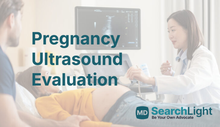
Ultrasounds have two main purposes during pregnancy. Firstly, they’re a routine part of prenatal check-ups, helping doctors monitor the development and well-being of the baby. Secondly, they are used in emergency situations, like if there’s a concern about a possible complication or if the pregnant mom gets injured.
Anatomy and Physiology of Pregnancy Ultrasound Evaluation
Ultrasound, which can be done via two methods – through the vagina (transvaginally) or through the belly (transabdominally), is a tool often used to get a picture of the female reproductive system. The transvaginal method often gives better images of the structures inside. The uterus, where babies grow during pregnancy, is positioned behind the bladder and in front of the colon (part of the digestive system). When observed using a transvaginal ultrasound, you can see three parts of the uterus, from bottom up: the cervix, body, and then the top part, known as the fundus.
Once a pregnancy begins, the uterus holds what is called the gestational sac, which is typically visible via ultrasound from around 4.5 to 5 weeks into the pregnancy. The gestational sac is usually the first thing seen in an ultrasound once pregnancy has taken place. The ovaries and Fallopian tubes, which accompany the uterus, are usually found on each side of it. A baby’s heartbeat, initially seeming like a flicker, can usually be heard for the first time about 6 weeks into the pregnancy.
The placenta, a structure that provides nourishment for the developing baby, usually becomes visible by the 10th week of pregnancy during a transabdominal ultrasound. It often looks like a rounded structure with uniform brightness and is typically observed either at the front or the back wall of the uterus. It surrounds the gestational sac and appears as a thick rim of tissue.
Why do People Need Pregnancy Ultrasound Evaluation
If a woman is pregnant and is not experiencing any unusual symptoms or discomfort, it’s generally recommended that she has an ultrasound between the 10th and 13th week of pregnancy. This first ultrasound is important for several reasons, such as confirming that the pregnancy is a normal, healthy one, figuring out the precise age of the baby, and checking if there is more than one baby.
However, if a pregnant woman is experiencing worrying symptoms like vaginal bleeding or pain in the belly or pelvic area, it’s important for her to have an immediate ultrasound. This helps doctors check for a condition called an ectopic pregnancy, where the pregnancy is developing outside the womb.
Also, if a woman who may be pregnant is involved in any type of accident or injury, whether minor or major, doctors have to assume she might be pregnant until they can test for certain. This is checked by using a pregnancy test that looks at urine or blood samples. Medical professionals use a quick ultrasound check called a FAST (Focused Assessment with Sonography in Trauma) exam as part of the process. This ultrasound lets doctors see if fluids have built up in the belly or if the woman has a pregnancy developing in her womb.
If the ultrasound scan shows that the woman is pregnant, the doctors will then check on the baby, such as by listening to its heartbeat, assessing the amount of protective fluid around the baby, noting any signs of the baby moving, and examining the placenta (the organ that supplies oxygen and nutrients to the baby).
In cases where a woman is at least halfway through her pregnancy (more than 20 weeks gestated) and experiences an injury, expert OB-GYN advice should be sought immediately. As soon as the mother’s condition is stable, she should undergo an ultrasound scan to make sure that the baby is okay as well.
When a Person Should Avoid Pregnancy Ultrasound Evaluation
There is only one absolute reason why a patient cannot have an ultrasound done through the abdomen or the vagina, which is if the patient doesn’t allow it. However, it’s not always safe or appropriate to use an ultrasound through the vagina late into a pregnancy term or for patients who are at a high risk. This method needs to be thought about carefully, especially during emergencies with pregnant patients who have vaginal bleeding and unclear medical history.
In these cases, an ultrasound through the abdomen should be done first to check the placenta (the organ that provides oxygen and nutrients to the growing baby). Following this, an ultrasound through the vagina and a thorough examination using a speculum (a tool used to open the vagina for examination) can be done if it is considered safe.
Despite these precautions, an ultrasound through the vagina is usually safe if the patient has a condition called placenta previa (where the placenta is near or covering the cervix). This is because the angle between the cervix and the ultrasound probe is usually enough to stop the probe from accidentally going into the cervix and disturbing the placenta.
Equipment used for Pregnancy Ultrasound Evaluation
To carry out a routine 2-D prenatal ultrasound, which allows us to see the baby inside the womb, we require two ultrasound probes. These probes are devices that send out sound waves to create an image of the baby. For the part of the exam that takes place through the vagina, we use a high-frequency transvaginal probe (7.5-10 MHz). And for the part of the exam that is performed through the belly, we use a curvilinear probe (1-6 MHz).
How is Pregnancy Ultrasound Evaluation performed
The techniques used for two types of pregnancy ultrasounds, transabdominal (across the abdomen) and transvaginal (inside the vagina), are generally the same for all pregnancies. However, these techniques differ when first assessing a pregnancy involving trauma or injury.
During a transabdominal ultrasound, the patient lies on their back. The healthcare professional uses a special instrument, called a curvilinear probe, which is positioned just above the pubic bone in the middle of the patient’s body. This allows them to see a clear image of the uterus, including its length, the cervix and part of the vagina. They’re looking for a particular stripe shape on the screen, which indicates the endometrial stripe, a part of the womb’s lining. Images are captured from two orientations, longitudinally (along the length) and transversely (across).
In a transvaginal ultrasound, the probe’s tactile indicator, which guides the user, points upward. The probe is inserted about 4 to 5 centimeters into the vagina in the sagittal plane (as if drawing a line from head to foot). First, they identify the bladder and the position of the uterus right next to it. This ensures the ultrasound isn’t just showing the adnexa, or surrounding areas. If there’s a high suspicion of an ectopic pregnancy (where a fertilised egg implants outside the womb), they’ll next examine the ovaries and Fallopian tubes – the most common place for an ectopic pregnancy is the ampulla of the fallopian tube.
In the case of trauma, the patient will have an abdominal ultrasound with a focus on the FAST exam, which checks for free fluid in the abdomen and around the heart. This exam uses the curvilinear probe and assesses the upper right and left quadrants of the abdomen, the area below the sternum (subxiphoid), the region just above the pubic bone (suprapubic), and pays close attention to the womb for a clear view of an intrauterine pregnancy (a pregnancy within the womb).
Possible Complications of Pregnancy Ultrasound Evaluation
Generally, ultrasound procedures are safe and carry a small risk of complications. However, undergoing a transvaginal ultrasound (an ultrasound procedure where a special probe is placed inside the vagina) might sometimes lead to side effects. These could include feelings of anxiety or discomfort, experiencing pain, or possibly some vaginal bleeding.
What Else Should I Know About Pregnancy Ultrasound Evaluation?
Ultrasounds are a go-to technique for checking on a baby’s development during pregnancy. This painless process is safe and offers fast results. It’s used in routine check-ups and emergency situations.
When doing regular prenatal care, the ultrasound’s goal is to confirm a healthy growing baby (viability), find out how old the baby is (determining gestational age), and see if there’s more than one baby (assess the number of fetuses).
To confirm a healthy baby, doctors look for a heartbeat. Usually, the heartbeat can be detected around the 6th week of the baby’s growth when it measures around 2 mm. If there’s no heartbeat, other signs in the ultrasound can help predict whether the baby will grow healthily.
Doctors use the ultrasound to study the size and growth of what’s called the gestational sac, which can be seen around the 4.5 to 5 weeks, and it’s the first sign of pregnancy. It’s measured by adding three dimensions of a specific space, adding these numbers up, and dividing by three to get a mean sac diameter (MSD). Healthily growing babies have a gestational sac that grows by a little more than 1 mm each day. If the sac measures 25 mm with no baby inside, it’s a sign of a failed pregnancy. If the sac measures between 16 and 25 mm without a baby, it could suggest a possible failed pregnancy.
Another method of checking viability is measuring the baby from head to bottom, commonly known as the crown-rump length (CRL). An absence of cardiac activity in a baby with a CRL larger than 7 mm is a sign that the baby might not be viable.
The ultrasound can also examine a little sac called the ‘yolk sac.’ When a yolk sac is not round or oval, or it’s too small (under 2 mm) or too large (over 5 mm), it could mean a higher chance of miscarriage.
Doctors also use ultrasounds to look for a condition called subchorionic hemorrhage, where there is a separation between the uterus and the membrane enveloping the baby. Larger separations are linked to higher rates of miscarriage.
Everyone pregnant should get an ultrasound between ten and thirteen weeks into their pregnancy to find out the accurate age of the baby, pick up on any noticeable physical issues the baby might have, and check if there is more than one baby.
If more than one baby is being carried, the ultrasound can see if there’s a barrier between them (amnionicity) and look at the kinds of placenta being shared or used by the babies (chorionicity). If the babies are sharing the exact same space without any barrier (called “mono-amniotic and mono-chorionic”), it could mean a higher risk for complications like entangling cords and fetal death.
During the second trimester, the ultrasound’s goal is for a thorough check-up and is typically carried out through the abdomen.


