What is Laryngotracheal Stenosis?
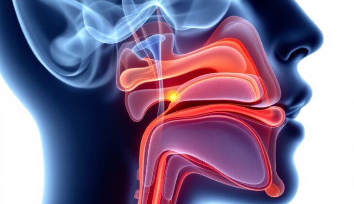
Laryngotracheal stenosis (LTS) is a medical condition that causes the upper part of the airway, between the voice box and windpipe, to become narrow. This narrowing can lead to severe problems like difficulty breathing, heart and lung issues, and even death. The affected area includes the voice box, a part known as the glottis, the area below the glottis, and the windpipe (trachea). The windpipe is shaped like a cylinder with a flexible wall at the front formed by c-shaped rings and a softer wall at the back. The windpipe splits into the right and left main bronchi (the large passageways leading into the lungs) at a spot called the carina, located roughly at the same level as the fourth bone in the upper back (T4).
The narrowing of the voice box can be due to various reasons, such as injury, complications from placing a tube in the trachea (intubation), tumors, autoimmune diseases, and infections. Some people might not experience any symptoms, while others may face difficulties related to blockage in the upper airway.
Regardless of the cause, treating LTS usually requires a team of specialists, including lung doctors, critical care doctors, ear-nose-throat doctors, and stomach doctors. Speech and language therapists and chest surgeons may also be involved. Identifying why the LTS occurred is important, as it can guide the treatment plan and inform the patient about what to expect in the future.
What Causes Laryngotracheal Stenosis?
Laryngotracheal stenosis, or the narrowing of your windpipe, can be linked to several causes. These include: accidental damage from a medical procedure specifically when a tube is placed in your throat to help you breathe (endotracheal intubation), autoimmune disorders, infections, tumors, trauma, and in some cases, the cause is unknow. Autoimmune diseases like lupus, rheumatoid arthritis, vasculitis, sarcoidosis, and scleroderma, among others, can trigger this condition.
Infections like bacterial throat infections, viral infections, and tuberculosis also contribute to this condition. Similarly, tumors, specifically squamous cell carcinoma and adenoma, are causes. Injury to the windpipe due to accidents, inhaling hot gases or radiation can also lead to laryngotracheal stenosis.
There are instances that injuries to your throat could be a part of post-intensive care syndrome, a group of health problems that remain after critical illness. The placement of breathing tubes (endotracheal tubes) during surgery often has side effects like voice changes, difficulty swallowing, and sore throat. But these side effects are more pronounced in patients who are critically ill. These tubes can injure the back of your throat, causing scar tissue to form and narrow your windpipe.
Risk Factors and Frequency for Laryngotracheal Stenosis
The understanding of laryngotracheal stenosis, a disorder which impacts the throat and windpipe, is not exact because there are several causes for this condition. Up to 30% of patients with a tracheostomy, which is a medical procedure that creates an opening in the neck for breathing, might get tracheal stenosis. Yet, this can be influenced by many factors specific to each patient, such as their existing health conditions and the reason and duration of their tracheostomy.
Signs and Symptoms of Laryngotracheal Stenosis
When a doctor suspects that a patient has laryngotracheal stenosis, it is important to review the patient’s medical history and conduct a physical examination. The doctor should inquire about any past instances of intubations, infections, autoimmune diseases, vasculitis, trauma, or surgery. The patient’s current symptoms are also vital information.
The most common symptoms of laryngotracheal stenosis include:
- Difficulty breathing (dyspnea)
- A high-pitched wheezing or whistling sound while breathing (stridor)
- Hoarseness
- Cough
In emergency situations, a doctor may not have enough time to gather all this information. In these cases, a physical examination becomes crucial. It’s important for the doctor to identify signs such as inspiratory stridor, which is a high-pitched sound caused by disrupted airflow often associated with a cough and difficulty breathing.
Different types of laryngotracheal stenosis present with various symptoms. For example:
- If the patient has extrathoracic stenosis (stenosis outside the chest), symptoms may include hoarseness, inspiratory wheezing, stridor, and a nonproductive cough.
- If the patient has intrathoracic stenosis (stenosis within the chest), symptoms may include difficulty in breathing out and discomfort when laying flat. The patient may also have trouble clearing throat secretions and may wheeze, a symptom that can be mistaken for asthma but does not respond to typical asthma treatments.
However, it should be noted that determining the exact location of the stenosis based on symptoms alone can be challenging.
Testing for Laryngotracheal Stenosis
If your doctor suspects that you have laryngotracheal stenosis, an illness where your windpipe becomes narrow or blocked, they may use a laryngoscopy or bronchoscopy to examine the area. These are procedures that use a small tube with a light on its end to check your throat and airways.
In more serious cases, where it’s difficult to directly check your throat or you’ve had an injury to your windpipe, your doctor might suggest a computed tomography (CT) scan, which is a type of X-ray that shows detailed pictures of your body. This can also help to plan for surgery. For instance, this scan may show that your windpipe is being squeezed by a large goiter, which is an enlarged thyroid gland in your neck.
Additionally, your doctor may perform a spirometry test. This simple test involves breathing into a small machine to measure how much and how quickly you can move air out of your lungs. It’s useful to check your lung function over time.
Once they’ve examined you, the doctor will classify the condition based on how severe it is and where it’s located, using one of the three classification systems. These rely on findings during surgery.
The Cotton-Myer classification is dependent on how much of the windpipe is blocked. The scale ranges from less than 50% blocked (Grade I) to completely blocked (Grade IV).
The Lano classification is based on how many different parts of the windpipe are affected (from one part to three).
Lastly, the McCaffrey classification considers how long the stenotic (or narrowed) area is, from less than 1 cm to any lesion involving the glottis (the part of the larynx containing the vocal cords).
These classifications help your doctor predict the treatment outcome and how severe the disease might become, including whether a permanent hole in the throat (tracheostomy) might be needed to help you breathe. For instance, patients graded III and IV on the Cotton-Myer scale were usually dependent on a tracheostomy, as were those with larger areas of narrowed airways in the Lano and McCaffrey classifications.
Treatment Options for Laryngotracheal Stenosis
Dealing with laryngotracheal stenosis, a condition where your airway narrows, can be complicated as it often requires multiple procedures and there’s a chance it could return. The goal of treatment is to make sure you can breathe properly, decrease the number of procedures needed, and remove the breathing tube for patients who have one. Unfortunately, there’s no standard way to approach this condition yet but different treatments have been suggested by various medical studies.
There are several treatment options available depending on what’s causing your condition and how severe and complicated it is. These options include widening your airway using an endoscope (a thin tube with a light and a camera), surgery, inserting a stent (a tube that keeps your airway open), laser therapy (using light energy to treat your airway), or using drugs that suppress your immune system.
A bronchoscopy, an examination of the airway using a flexible tube, can be used to mechanically enlarge the airway, apply laser therapy, and insert a stent to treat laryngotracheal stenosis. However, this might not be too effective in the area below your vocal cords because of its location. Procedures like dilation or laser treatments are options for narrowing due to scar tissue or growth; however, they tend to come back. Inserting a stent could cause a bigger airway injury. Using a laser should be done carefully for stenosis below the vocal cords to avoid damaging the cartilage, which houses nerves crucial to the functioning of your vocal cords. Studies have shown less than 20% success rates for laser and stenting.
Endoscopic dilation, where a device is used to widen your airway, is an effective way to preserve your voice compared to patients who have stenosis close to your vocal folds. This should be the first choice of treatment for simple stenosises, whereas more complex ones may need a team of professionals and possibly surgical evaluation.
For patients with advanced and unremovable airway-blocking cancer, a tracheal stent can be used to alleviate symptoms. This option comes with its own risks and requires a discussion about the pros and cons.
Before surgery, several assessments are performed to help the doctors decide the best treatment option. In particular, patients should be tested for MRSA, a type of bacteria that’s resistant to several antibiotics, a few weeks before possible surgery. Any positive cases are treated proactively. Patients should also have their swallowing evaluated to determine the best way to give them nutrition after the surgery.
Open surgery is an option for patients with severe stenosis, cartilage loss, or stenosis longer than 1 cm (about 0.4 inches). Usually, patients who undergo surgery need fewer repeated interventions. Open surgery may involve laryngotracheal resection (removing part of the windpipe) with reanastomosis (reattaching) or laryngotracheoplasty using native and tissue grafting.
A T-tube can be placed through the wall of the windpipe distal to the reconnection. It provides a stable airway for a long time (at least six months) until the doctors consider removing it, or in cases where removal is not possible.
Aside from these treatments, other additional treatments are currently used for laryngotracheal stenosis. Some of these include mitomycin C, a chemotherapy drug that is put directly onto the surgical incision to decrease scar tissue formation, and steroids that are given either locally or orally in pill form to treat thickened stenosis.
What else can Laryngotracheal Stenosis be?
When doctors are trying to figure out if someone has laryngotracheal stenosis (narrowing of the windpipe and voice box), they consider whether the symptoms could be caused by other conditions. It’s important to explore these possibilities to make sure the diagnosis is correct. Some of the conditions that might have similar symptoms and need to be ruled out include:
- Angioedema (swelling underneath the skin)
- Asthma
- Epiglottitis (inflammation of the tissue that covers the windpipe)
- Esophageal tumor (cancer of the tube that connects the mouth and stomach)
- Foreign body aspiration (inhaling something like a small toy or piece of food)
- Gastroesophageal reflux disease (heartburn or GERD)
- Mediastinal mass (mass in the area of the chest that separates the lungs)
- Retrosternal goiter (enlarged thyroid gland located behind the breastbone)
- Subglottic web (abnormal tissue growth below the vocal cords)
- Tracheomalacia (weakness and floppiness of the walls of the windpipe)
- Vocal cord dysfunction (changes in the voice or difficulty speaking)
What to expect with Laryngotracheal Stenosis
The outlook is generally very good for patients with idiopathic LTS (a condition where the larynx or ‘voice box’ narrows for unknown reasons) who undergo a surgical procedure to treat it. However, the outlook for patients with other causes of LTS depends on how the underlying disease that caused the LTS progresses.
Possible Complications When Diagnosed with Laryngotracheal Stenosis
Acute LTS, such as LTS appearing after tubing removal, can cause a cessation of breathing if it’s not identified and treated promptly. In idiopathic LTS (a type of LTS with unknown cause), complications can include voice changes, dependency on tracheostomy without the ability to remove the tube, and the necessity for multiple procedures.
The main objective of open surgery is to remove the tracheostomy tube. About 63 to 95 percent of patients who undergo open surgery are able to have their tracheostomy tubes successfully removed. However, the chances of remaining dependent on tracheostomy can be higher in patients with severe stenosis (as per the Myer-Cotton scale), diabetes, gastroesophageal reflux disease, diabetes, and/or a high body mass index (BMI exceeding 30).
There can be complications in open surgery, such as difficulty in swallowing arising from injury to the laryngeal nerves or the use of a stent to keep the airway open.
A study published in the Journal of Thoracic Disease by Bibas et al. found that patients with benign tracheal stenosis (such as stenosis caused by intubation and not suitable for surgical treatment) have significantly reduced quality of life. Quality of life in this context refers to the combination of physical and mental well-being of both the patient and their family members. The study included patients with a specific type of airway tube (Montgomery T-tubes), silicone stents or a tracheostomy, and the capability to complete a survey. Patients with chronic conditions such as chronic obstructive pulmonary disease, advanced heart failure, and kidney failure were not included in the study.
- Acute LTS can cause cessation of breathing.
- Idiopathic LTS can lead to voice changes, dependency on tracheostomy, and multiple procedures.
- The aim of open surgery is to remove the tracheostomy tube – 63 to 95 percent of patients achieve this.
- Severe stenosis, diabetes, gastroesophageal reflux disease, and high BMI may increase tracheostomy dependence.
- Open surgery can lead to swallowing problems.
- Patients with benign tracheal stenosis have significantly reduced quality of life according to a study.
Preventing Laryngotracheal Stenosis
Often, patients may not fully understand the need for a medical procedure called an endotracheal intubation (a tube placed in the throat to help with breathing), and its possible complications, such as Laryngotracheal stenosis (LTS), a narrowing of the airway. This is usually because the procedure is often performed in emergency situations. If patients have a history of autoimmune diseases and are at risk of developing LTS, their doctor should guide them on which signs and symptoms they should not overlook and when to urgently seek medical help. This is particularly important if they’ve previously experienced LTS.
Moreover, those suffering from LTS, especially when the cause is unknown, may find it beneficial to join online forums. Here, they can connect with other patients going through similar experiences, exchange information, share personal experiences, and offer each other emotional support.


