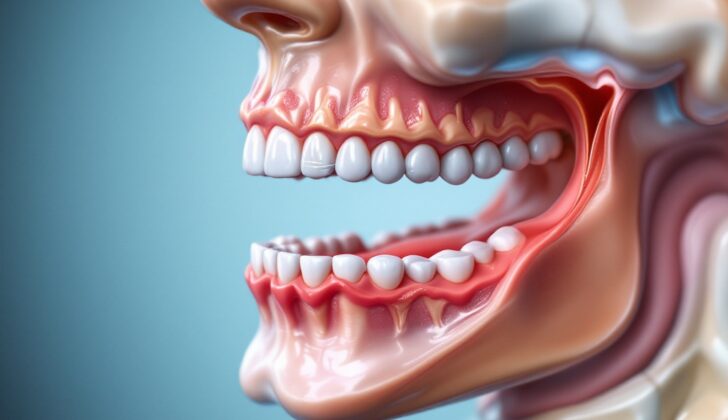
What is Submandibular Sialadenitis and Sialadenosis?
The submandibular glands are large salivary glands located in the area beneath the jaw, covered by a layer of deep tissue. They are divided into a superficial and a deep lobe by a muscle called the mylohyoid. These glands release saliva into the mouth through a duct known as Wharton’s duct, which is positioned between a smaller gland beneath the tongue and a muscle called the hyoglossus. This duct releases saliva through a tiny hole next to a fold of tissue on the bottom of your mouth.
The amount of saliva your glands produce can increase due to stimulation by the parasympathetic nervous system (which helps control bodily functions when you are at rest) and decrease due to the sympathetic nervous system (responsible for triggering the body’s response to stress). Saliva is crucial for our health as it contains higher levels of potassium, lower levels of sodium and substances that help to start breaking down food. It also contains Immunoglobulin A (IgA), which helps to maintain and protect the environment of your mouth.
Sialadenitis is a condition that causes inflammation of the salivary gland. It’s less common in the submandibular gland than in the parotid gland (a gland located near the ear). Acute sialadenitis, which occurs rapidly due to bacterial or viral infections, results in swift pain and swelling. On the other hand, chronic sialadenitis, which can be due to blockages like stones or strictures in the duct, is often characterized by recurrent or long-lasting swelling of the salivary gland without redness.
Sialdenosis is a condition where the salivary gland swells, but is not due to a tumor or inflammation. It’s associated with an increase in size of the salivary cells and a shrinking of the salivary ducts. Sialdenosis often appears as non-painful swelling that is usually seen on both sides and is symmetrical. It is often linked to metabolic conditions, which impact how the body’s cells change food into energy.
What Causes Submandibular Sialadenitis and Sialadenosis?
Submandibular Sialadenitis, or inflammation of your submandibular salivary glands, can happen due to various causes:
1. Infections:
– Bacterial: Mostly caused by a variety of different bacteria such as Staphylococcal aureus (the most common one), Hemophilus influenza, Enterobacteriaceae, to name a few. Sometimes, the infection can be anaerobic (includes bacteria that do not need oxygen to live), like Prevotella, Fusobacterium, and Peptostreptococcus.
– Viral: Mumps or HIV.
– Other types of infections: Actinomyces or Tuberculosis.
2. Physical obstructions:
– Sialolithiasis, which means there are stones in your salivary glands.
– Ductal stricture, or narrowing of a salivary duct.
– Presence of a foreign body like a fish bone, hair, or grass blade in the salivary duct.
– External compression of a salivary duct, for example, by a denture’s flanges.
3. Inflammatory causes:
– Post-radiation Sialadenitis: inflammation caused by radiation treatment.
– Contrast-induced Sialadenitis: inflammation caused by an iodine-based contrast used in medical imaging.
– Drug-induced Sialadenitis: inflammation due to certain drugs such as Clozapine, I-asparaginase, or Phenylbutazone.
– Autoimmune Sialadenitis: inflammation due to conditions where your body’s own immune system attacks the salivary glands, like Sjögren syndrome or IgG4-related disease.
– Granulomatous Sialadenitis: inflammation due to certain conditions that affect your immune system like Sarcoidosis or Xanthogranulomatous sialadenitis.
Sialadenosis, or non-inflammatory enlargement of your salivary glands, can also happen due to various causes:
1. Nutritional disorders: Bulimia nervosa or vitamin deficiency.
2. Hormonal disorders: Diabetes or Hypothyroidism.
3. Metabolic disorders: Obesity, Cirrhosis, or Malabsorption (not absorbing nutrients properly).
4. Autoimmune disorders: Sjögren disease.
5. Drug-induced: Certain drugs like Valproic acid or Thiourea may cause your salivary glands to enlarge.
Risk Factors and Frequency for Submandibular Sialadenitis and Sialadenosis
Information on how often submandibular sialadenitis occurs is not readily available. However, we do know that it makes up about 10% of all cases of sialadenitis, which is an inflammation of the salivary glands. This condition is also the reason for about 0.001 to 0.002% of all hospital admissions. It’s important to note that this condition does not favor any specific age or gender. It is commonly seen in older patients who are dehydrated. In the United States, the most common reason for swelling of the salivary gland is sialadenosis.
Signs and Symptoms of Submandibular Sialadenitis and Sialadenosis
If you’re dealing with swelling under your jaw, or submandibular swelling, doctors will conduct a detailed examination. During your appointment, they might ask:
- How long you’ve been experiencing symptoms
- If multiple glands are involved
- If the swelling is painful
- If you have a foul taste in your mouth
- How often the symptoms occur
- If certain things like meals or salivary stimulants make it worse
- If you have any symptoms of illness, like a fever, weight loss, or joint pain
- If you have dry eyes or mouth
- If you have any existing health conditions such as alcohol use, diabetes, bulimia, liver disease, or autoimmune disease
- If you’ve received radiation treatment in the past
A physical examination will include checking and feeling the gland size and texture, and seeing if it’s tender. By massaging the gland, doctors can observe the type of discharge from the duct orifice. They may also check your facial nerve and mandibular branch of the trigeminal nerve and examine your neck lymph nodes. Sometimes, a fever can be present.
Usually, the gland feels swollen, hard, and tender. In cases of infection, neck gland inflammation may be observed. Chronic or repeated salivary gland inflammation causes regular pain and swelling, often with meals and recurrent infections. By massaging the gland, doctors might notice pus-tinged saliva at the mouth of the duct. Intermittent gland swelling occurring with meal stimulation is often due to obstruction by salivary gland stones or constrictions. The blockage of saliva flow within the duct causes the gland to swell.
In the case of viral salivary gland inflammation (like mumps), you may experience acute, widespread salivary gland swelling accompanied by illness symptoms like a fever, headache, fatigue, and muscle ache.
A particular type of submandibular swelling, known as Submandibular sialdenosis, presents with painless bilateral enlargement under the jaw, possibly with mild discomfort. Around half of these cases are associated with known risk factors such as diabetes, metabolic syndrome, alcoholism, bulimia, malnutrition, and liver disease.
Testing for Submandibular Sialadenitis and Sialadenosis
To assess if someone has sialadenitis, which is inflammation of the salivary glands, doctors may use a combination of methods including asking about symptoms, conducting a physical examination, doing laboratory tests, taking X-rays, and possibly performing a biopsy, which is the removal of a small sample of tissue for testing.
1. Laboratory testing: Doctors might take a sample of the discharge from the duct (a small opening or passageway) of the salivary gland for testing. This test helps identify what bacteria have caused the infection and determine which antibiotic will best treat it. They might do this test before prescribing an antibiotic.
2. Complete blood count: This test checks for signs of infection in your blood.
3. Imaging tests: These tests can give doctors a better look at what’s going on inside your body.
– X-ray: This can help doctors to spot a salivary stone in chronic cases of sialadenitis. In fact, about 70 to 80% of salivary stones in the submandibular gland (one of the saliva glands located under your jaw) show up on an X-ray.
– Ultrasound: This test uses sound waves to create images of your salivary glands and can display a salivary stone if it’s larger than 1 mm, as well as any potential abscesses (pockets of pus).
In cases where the symptoms are particularly severe or X-rays don’t show anything, more tests might be needed:
– CT scan: This more advanced type of X-ray can also help detect salivary stones. It can also show changes in the size of the salivary gland, which can be a sign of chronic sclerosing sialadenitis, a prolonged inflammation that can cause the gland to become enlarged or shrunk.
– DSA sialography: This test uses a dye to make the salivary ducts and glands more visible on an X-ray image. Although it can help find salivary stones, duct strictures (narrowings), and loss of gland tissue integrity, it’s usually avoided during an acute inflammation.
– MRI: Doctors might need to use an MRI if they suspect the presence of a tumor.
Lastly, if doctors believe that your sialadenitis might be related to a connective tissue disorder, they might test for SSA/anti-Ro, SSB/anti-La, ANA, RF – these are markers that can signal the presence of such conditions. If the sialadenitis looks like chronic sclerosing sialadenitis, which can present similarly to a tumor, a fine-needle aspiration (FNA) cytology of the affected gland can be used. This procedure uses a thin, hollow needle to take out a small amount of cells from the gland to check for the presence of a tumor.
Treatment Options for Submandibular Sialadenitis and Sialadenosis
Acute sialadenitis, chronic sialadenitis, and sialadenosis are all conditions that affect your salivary glands. The treatment for these conditions can vary.
For acute sialadenitis, which is a sudden and severe inflammation of the salivary gland, treatment usually starts conservatively. This includes hydration, applying warm compresses, massaging the affected area, and taking medicines to alleviate pain, such as non-steroidal anti-inflammatory drugs (NSAIDs). Doctors may prescribe medicines called sialogogues that help stimulate saliva production. Some patients might also need antibiotics. Those with severe cases might require antibiotics given through an IV (a tube placed in a vein). Sometimes, medication called corticosteroids is used to reduce swelling in the soft tissue. In rare cases, the inflammation can cause an abscess (a pocket filled with pus); if this happens, a surgical procedure might be needed to drain it.
Chronic sialadenitis is a long-term inflammation of the salivary gland. Treatment is similar to acute sialadenitis and includes hydration, maintaining good oral hygiene, and pain relief. Also, broad-spectrum antibiotics, which work against a wide variety of bacteria, are sometimes used in case of infection. If the cause of chronic sialadenitis is sialolithiasis (salivary gland stones), the removal of these stones is necessary. This can be done using a procedure called interventional sialendoscopy, or through direct surgical removal. Another technique known as extracorporeal shock wave lithotripsy (EWSL), which uses sound waves to break up stones within the gland, can also be used.
If you have chronic sialadenitis that frequently recurs (more than three times in a year), or chronic sclerosing sialadenitis (an immune-mediated condition causing repeated inflammation and scarring of the salivary glands), your doctor might recommend excision (surgical removal) of the affected salivary gland.
Sialadenosis is a non-inflammatory condition in which the salivary glands enlarge without a known cause. Management of this condition involves treating whatever underlying problem is associated with it.
What else can Submandibular Sialadenitis and Sialadenosis be?
When trying to understand what’s causing an issue with the submandibular salivary glands (submandibular sialadenitis and sialadenosis), doctors consider several possibilities including:
- Infectious causes such as bacteria (like Staphylococcus aureus) or viruses (like mumps)
- Granulomatous causes like Tuberculosis, sarcoidosis, cat scratch disease, and actinomycosis
- Autoimmune causes like Sjogren disease and systemic lupus erythematosus
- Tumors, which enlist Pleomorphic adenoma, oncocytoma, ductal papilloma, adenoid cystic carcinoma, squamous cell carcinoma, and mucoepidermoid carcinoma
- Endocrine and metabolic causes such as hypothyroidism, diabetes mellitus, bulimia, cirrhosis, vitamin deficiency, and malabsorption
- Drug-related causes such as thiourea
What to expect with Submandibular Sialadenitis and Sialadenosis
Acute sialadenitis, or short term inflammation of the salivary glands, usually gets better on its own. With basic outpatient care, symptoms often disappear completely. Most of the immediate symptoms clear up within a week, though swelling may take a little longer to fully go away.
Chronic sialadenitis, or long-term inflammation of the salivary glands, can come and go multiple times.
Your prognosis, or expected medical outcome, often depends on the cause of the inflammation. If salivary stones, also known as sialoliths, are the cause and require surgery, the outlook is good. If the inflammation is due to an autoimmune condition (like Sjogren’s syndrome), symptoms often get better with medical treatment of the underlying condition. When the base cause is treated properly, sialadenosis (enlargement of the salivary glands) has an excellent prognosis.
Possible Complications When Diagnosed with Submandibular Sialadenitis and Sialadenosis
The problems that can occur with submandibular sialadenitis and sialadenosis include the following:
- Recurrence: The condition may come back after it has been treated.
- Abscess formation: The infection might spread along the neck tissue, causing a severe complication. It typically needs to be treated with incision and drainage.
- Dental decay: Reduced saliva production due to the salivary gland not functioning properly can lead to less protection against acid erosion; this paves the way for tooth decay.
Preventing Submandibular Sialadenitis and Sialadenosis
People diagnosed with sialadenitis, an inflammation of the saliva-producing glands, need to pay special attention to their oral hygiene practices such as regular brushing of teeth and staying hydrated. Once the immediate painful symptoms have lessened, gentle, repeated massaging of the gland located underneath the jawbone – the submandibular gland – can continue to be part of their routine care. It’s important to avoid medication classes known as anticholinergics and diuretics, which can interfere with saliva production.
Addressing any illnesses that might have led to developing sialadenitis, for example, Sjogren syndrome, a condition that causes dry mouth, is crucial as well. If diagnosed with submandibular sialadenosis, a non-inflammatory disorder of the saliva glands, focus should also rest on the management of any underlying conditions that could have caused it, such as hypothyroidism, diabetes, or liver disease like cirrhosis.


