What is Blepharoconjunctivitis?
Blepharoconjunctivitis is a type of eye disease that involves characteristics from two other conditions, blepharitis (which affects the eyelid rim) and conjunctivitis (which affects the area around the eye, known as the conjunctiva). It’s often connected to blepharitis and can be seen as a more advanced stage of this condition. If blepharitis is not treated early on, the inflammation can spread to the nearby conjunctiva, leading to blepharoconjunctivitis.
The condition is understood and described in different ways, for example, considering what caused it, its symptoms, which part of the eye is affected, and how it looks in standardised photographs.
In the United States, most cases are grouped using the guidelines suggested by the American Academy of Ophthalmology. They categorise cases as either anterior or posterior, depending on which part of the eye is affected. The treatment for this condition is the same as for blepharitis, and aims to ease symptoms. This involves keeping the eyelids clean and limiting exposure to things in the environment that might trigger it. Medicines like steroids, antibiotics, and antiseptics can also be applied to the eye to help manage the condition.
What Causes Blepharoconjunctivitis?
Blepharoconjunctivitis is a condition where the eyelid and the white part of the eye become inflamed. It can be tricky to determine the exact cause of this because of the close relationship between the structures of the eye. Also, the white part of the eye can quickly become involved in the inflammation.
There could be many reasons behind blepharoconjunctivitis and it often has multiple causes. It’s easier to identify these causes based on how the condition appears and progresses over time. If it develops quickly with signs of ulcers, it’s likely due to an infection, with the usual culprit being a type of bacteria called Staphylococcus. But, if it develops quickly without ulcers, it could be an allergic reaction. It’s important to remember, though, that not having ulcers does not necessarily rule out an infection.
When blepharoconjunctivitis develops over a longer period (chronically), considering the part of the eye that’s involved can help identify the cause. If it’s at the back of the eyelid, there’s a good chance that it’s due to a dysfunction in the Meibomian gland, which is a tiny gland in the eyelid that helps keep the eye lubricated. If the inflamed part is near the corner of the eye, the condition is known as angular blepharoconjunctivitis and could be triggered by a different kind of bacteria called Moraxella or in rare cases, a vitamin B6 deficiency. If the inflammation is on the front of the eyelid, this could suggest an infection, like Staphylococcus, or a condition known as seborrhea. There’s also a relationship between this inflammation and acne rosacea.
Other potential causes of blepharoconjunctivitis include parasitic infections, such as Demodex or pubic lice, and some drugs like dupilumab.
Risk Factors and Frequency for Blepharoconjunctivitis
Blepharoconjunctivitis and blepharitis are two conditions that are very similar, making it tough to separate data related to the two. Furthermore, it’s difficult to find reliable information about how common blepharitis is in the general population, because most studies look at people who go to eye clinics. In a study done in the United States, it was found that 37% to 47% of patients at eye clinics showed signs of blepharitis.
- In a study of 90 patients at an eye clinic, the average age was 50.
- In another study, a version of blepharitis called infective blepharitis was more common in females, with an average age of onset around 42.
- As for seborrheic blepharitis, another type of the disease, the average age was also around 50, and there wasn’t a noticeable difference between the sexes in terms of who it affected more.
Signs and Symptoms of Blepharoconjunctivitis
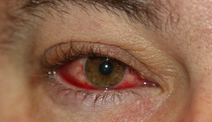
Blepharoconjunctivitis, an inflammation in the eyelids and the white part of your eyes — the conjunctiva, can lead to various symptoms and signs. People with this condition often experience discomfort in their eyes, feeling like something is stuck in their eye, and they may wake up with crusty eyelids or their eyelids being stuck shut. Their eyes might become red, and the symptoms often get worse in the morning. It’s common for both eyes to be affected, and these symptoms can fluctuate.
Upon a doctor’s examination of the eyes, several signs could potentially be noticed. The patient’s tear film — a protective layer of moisture on the eyes — might break up very quickly, leading to dry eyes. The doctor might also detect corneal erosions or ulceration, and the conjunctiva and sclera (the white part of the eye) might have a bloodshot appearance.
- Fast tear film break up
- Corneal erosions or ulceration
- Bloodshot eyes
If the inflammation starts from the front of the eyelid, the edge of the eyelid might swell up. This same edge of the eyelid could turn red, with fine red, spider vein-like lines being visible (telangiectasia). You might see crust forming at the base of your eyelashes, which looks like collarettes. Advanced stages of this condition might even cause changes to the eyelashes, such as lightening of eyelashes or depigmentation (poliosis), a change in their direction (trichiasis), or thinning of eyelashes (madarosis). There might be changes to the eyelid’s shape or a turning in or out of the eyelid.
- Swollen edge of eyelid
- Redness and fine red lines at eyelid’s edge
- Crusts at the base of eyelashes
- Changes in eyelashes color, direction, or thickness
- Changes in eyelid’s shape
If the inflammation is rooted from the back of the eyelid, the doctor might observe signs related to the meibomian glands, which produce the oil element of your tears. These glands might appear widened, and the opening might be blocked with a thick oil. In a detailed eye examination using a slit-lamp, this blockage might look like a string of pearls on the edge of your eyelid. The eyelid areas around the glands might look scarred or appear inflamed to the doctor.
- Widened meibomian glands
- Thick oil blocking the gland openings
- Scarring or visible inflammation around the glands
Testing for Blepharoconjunctivitis
Blepharoconjunctivitis, an inflammation of the eyelid and the outermost layer of the eye, is usually diagnosed based on patient’s medical history, physical symptoms and signs observed by the doctor. Tests like tear film break up time can help but aren’t always necessary. If your eye condition isn’t improving with treatment, your doctor may suggest a lid biopsy. This is a small tissue sample taken from the eyelid to exclude the possibility of cancer.
Another useful method is using fluorescein drops and a blue light to check for any corneal damage. Corneal erosions or ulcers, which are open sores on the cornea (the clear, front surface of your eye), can occur in severe, long-term cases of blepharoconjunctivitis.
Treatment Options for Blepharoconjunctivitis
Blepharoconjunctivitis is a condition affecting the eyelids and eyes, and its primary treatment involves thorough cleaning of the eyelids. This involves using a warm compress on your eyes for about 5 to 10 minutes to soften the oil inside the glands on your eyelids (called Meibomian glands). You can then gently massage your eyelid to help release the oil. It’s also helpful to clean your eyelashes with a mild shampoo to remove any dirt or debris. Keep in mind that this cleaning routine should continue even after the symptoms reduce because blepharoconjunctivitis is a long-term condition.
If you have dry eyes, over-the-counter eye drops, or ‘artificial tears’, can provide some relief. During flare-ups of the condition where your eyes or eyelids become inflamed, your doctor might prescribe eye drops containing steroids. If simply cleaning the eyelids isn’t enough to manage the condition, topical antibiotics (creams or ointments applied to the skin) might be recommended. These are especially helpful if the blepharoconjunctivitis is caused by bacteria, such as a staphylococcal infection.
If you also have seborrheic dermatitis, which is a skin condition causing flaky, itchy skin, treating this underlying condition may help manage your blepharoconjunctivitis. If you have a type of blepharoconjunctivitis called ‘posterior blepharoconjunctivitis’, your doctor might recommend an oral azithromycin, which is a type of antibiotic.
The use of supplements which contain omega-3 and omega-6 fatty acids to help dry eye symptoms is still a topic of debate among experts. A recent review of research studies suggested that Omega-3 supplements might be beneficial, but it also noted that the quality of the research on this topic is not very strong.
What else can Blepharoconjunctivitis be?
For patients whose symptoms don’t improve with treatment, it’s crucial to check if they might have cancer, primarily if the symptoms affected their eyelashes. Cancer usually impacts the lower eyelid, and if the symptoms only appear on one side, there should be a high suspicion of malignancy. People with dry eye syndrome might also experience a similar ‘gritty’ feeling, like there’s something in their eyes. However, they usually won’t have any inflammation or flaky skin along the edges of their eyelids.
What to expect with Blepharoconjunctivitis
The outlook is usually positive, and most people experience relief from their symptoms. However, because this condition is long-term or chronic, it’s important to keep the eyelids clean consistently. This would act as a maintenance therapy and help prevent sudden severe flare-ups of the condition.
Possible Complications When Diagnosed with Blepharoconjunctivitis
In long-term cases, there are a variety of potential complications that could arise. These include issues with the cornea such as sores, scarring, or the growth of new blood vessels. There may also be changes to the lid margin, causing abnormal lashes, outward or inward turning of the eyelid, or loss of eyelashes. Urgent and intensive treatment is necessary should an ulcer or perforation occur to prevent permanent damage to vision.
Complications May Include:
- Sores on the cornea
- Scarring of the cornea
- Growth of new blood vessels in the cornea
- Changes to the lid margin
- Abnormal lashes
- Outward or inward turning of the eyelid
- Loss of eyelashes
- Ulcer or perforation requiring urgent treatment
- Potential permanent vision impairment if not properly treated
Preventing Blepharoconjunctivitis
Patients should understand that their condition is long-lasting and requires steady treatment, often in the form of keeping the eyelid clean, to stop sudden flare-ups from happening. They should also learn about the signs of any worsening conditions, and get immediate medical help if they notice changes in their vision or new pain in the eye.


