What is Furrow Degeneration?
Furrow degeneration (FD) is a rare health condition affecting the eye that’s known by various names in medical writings. Most often, it’s referred to as furrow degeneration, but terms like senile furrow degeneration of cornea, corneal furrow degeneration, or age-related marginal corneal degeneration are also used. FD is identified by the thinning of the peripheral cornea – the transparent front surface of the eye, located between two other eye structures – the arcus senilis and the limbus.
Furrow degeneration hasn’t been thoroughly studied, and we don’t entirely understand its causes and development process. This could be because it typically doesn’t lead to severe problems. Unlike many other cornea-related conditions, FD does not involve blood vessel development or inflammation, which are usually involved in the disease process. Therefore, it leads to a painless thinning of the peripheral cornea, without symptoms. Even though the cornea thins out, patients won’t notice any changes in their vision because the thinning happens evenly throughout the cornea.
There have been only a few cases studied to understand how the condition develops over time. As it’s quite rare, many questions about furrow degeneration remain unanswered.
This summary will focus on how most FD patients’ physical examination is carried out and its outcomes. It will also emphasize the significance of recognizing other cornea-related conditions and distinguishing FD from them. However, it won’t delve into the specific anatomy and physiology of the cornea, and will mainly concentrate on the clinical symptoms and medical management of FD.
What Causes Furrow Degeneration?
What causes functional dyspepsia (FD), a type of indigestion, is not fully understood yet. However, we do know that getting older is a risk factor, which means that the chance of getting FD increases as you age.
Risk Factors and Frequency for Furrow Degeneration
Furrow degeneration, often referred to as FD, typically occurs in older people, usually between the ages of 60 and 70. It’s important to note that FD doesn’t favor any gender—it affects both men and women equally.

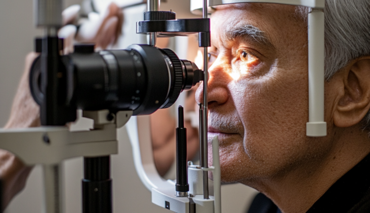
examination in a patient with Furrow Degeneration.
Signs and Symptoms of Furrow Degeneration
Patients with furrow degeneration, a condition linked to aging, usually don’t experience symptoms, changes in vision, or an increased risk of perforation. Because of this, the condition is often discovered by chance during an eye examination in older individuals. The distinguishing feature of furrow degeneration, identifiable during this examination, is a specific type of thinning of the cornea in both eyes. Despite the thinning, the surface layer of the eye remains unbroken and free of blood vessels. Except for these changes, the rest of the eye examination typically appears normal unless other conditions are present.
Testing for Furrow Degeneration
Currently, there are no regular lab tests or imaging techniques used specifically to diagnose furrow degeneration. This condition is mostly diagnosed through a hands-on examination by a doctor. For instance, one method doctors use is a tool called a slit-lamp, which help them look at your eyes and spot any thinning in the outer layer of your eyeball’s clear, front surface (known as peripheral cornea).
Sometimes, though, doctors might use computerized maps of your eye (a process called topography) to help tell the difference between furrow degeneration and other, more severe eye problems. These computerized maps use color to show different features of your eye. Both furrow degeneration and distortions caused by contact lenses show a general flattening of the outer surface of the eyeball. Yet, only in furrow degeneration will the map indicate a local steepening (or sudden peak) in the outer part of the eye.
In addition to these, there are other less common tests that can be used to confirm if the outer layer of your eyeball (cornea) is getting thinner. These tests are anterior segment optical tomography (a type of 3D imaging), videokeratoscopy (a video imaging technique), and computer-assisted corneal topography (where a computer helps make a detailed map of the surface of the cornea).
Treatment Options for Furrow Degeneration
Furrow degeneration is a condition that typically doesn’t worsen over time and often doesn’t require treatment. The main approach is to monitor the condition. However, in more severe cases, special eye drops can be used to lubricate the eye, or a procedure called ‘punctal occlusion’ can be performed. Punctal occlusion is a minor procedure that blocks the tear ducts to keep the eye’s surface moist.
What else can Furrow Degeneration be?
When a patient shows signs of furrow degeneration, it’s important for the doctor to differentiate this condition from other diseases that might affect the outer layer of the eye, also known as the cornea. These diseases pose different risks for the patient, like vision problems or increased chances of the eyeball rupturing.
Four conditions that can be mistaken for furrow degeneration are:
- Pellucid Marginal Degeneration (PMD): Like furrow degeneration, PMD doesn’t cause inflammation. Both conditions won’t show signs of scarring, blood vessel growth, or fat deposits when doctors use a special light (called a slit lamp) to examine the eye. The key difference is where the thinning part of the cornea (transparent front surface of the eye) is located. In PMD, it’s further from the center of the eye, leading to unique changes in the cornea’s shape seen during the physical examination or with a tool that maps the curve of the cornea. These changes are a beer belly or crab claw shape. For some patients, this might lead to vision problems because of uneven thinning.
- Terrien Marginal Degeneration (TMD): TMD shows up as thinning in the upper portion of the cornea and progresses over time until it affects the entire cornea. The early stage of TMD is characterized by a yellowish opaque band on the cornea with fat deposits and blood vessels growth. Most people won’t have symptoms during this stage. However, as TMD progresses, the person starts experiencing vision problems as well as increased chances of the eyeball rupturing.
- Keratoconus: In keratoconus, the cornea adopts a cone shape due to thinning. Patients usually struggle with worsening vision issues that can’t be corrected. A physical examination with a slit lamp can show this thinning, which usually occurs in the lower part of the cornea. Patients with keratoconus show specific signs that won’t appear in a furrow degeneration case.
- Keratoglobus: This condition causes the cornea to thin and bulge into a globe shape. Most people with keratoglobus suffer from nearsightedness and deformed vision, but some may experience rupturing of the eyeball. Diagnosis is based on clinical signs, but if those aren’t clear, other tools can be used.
- Dellen: Caused by local drying and shrinking of the cornea due to an interruption in the tear layer, Dellen are typically temporary, shallow, dish-like lesions on the cornea. They can be distinguished from furrow degeneration by their location and shape, and because they’re usually not an isolated condition.
In conclusion, each of these conditions can be differentiated from furrow degeneration by their unique characteristics during a physical examination and the patient’s history of symptoms.
What to expect with Furrow Degeneration
People diagnosed with furrow degeneration generally don’t experience any symptoms. In fact, one case study revealed that there was no increase in the thinning of the cornea after a year. Another case study showed that the thinning hadn’t worsened even after following up for three months.
Furrow degeneration is a non-vascular condition, meaning it doesn’t involve blood vessels. Also, it doesn’t cause any damage to the outermost layer of the eye (the epithelium). Thanks to these factors, there’s a low risk of the cornea puncturing or ‘perforating’.
People with furrow degeneration typically don’t need to be frequently monitored by their doctors. They are generally advised to stick to their yearly eye check-ups.
Possible Complications When Diagnosed with Furrow Degeneration
Given that there’s been very little research and case studies done on Furrow degeneration, there have been no reported complications related to the condition.
Preventing Furrow Degeneration
Patients are encouraged to attend yearly eye examinations to check the condition of the thin layer at the front of the eye, known as the cornea. This helps to monitor whether it’s getting any thinner. If a patient is diagnosed with Fragmentary Degeneration (FD), it’s important to know that this is a minor, non-worsening condition. This knowledge helps reduce patients’ stress and worry when they learn they have a cornea disease that’s not getting worse over time.


