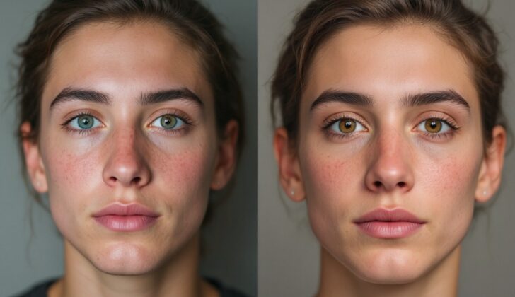
What is Hypertelorism?
Orbital hypertelorism is a condition where there is an increased space between the eye sockets or orbits. This means the eye sockets have been moved outwards. The signs of this condition can be measured and include an increased distance between the inner corners of the eyes (inner canthal distance or ICD), an increased distance between the outer corners of the eyes (outer canthal distance or OCD), and an increased distance between the pupils of the eyes (interpupillary distance or IPD). If all three distance measurements are above the 95th percentile of standard values, this confirms true orbital hypertelorism. If only the inner canthal distance is increased, it’s called telecanthus. In this context, we’ll use the term ‘hypertelorism’ to mean ‘orbital hypertelorism’. We’ll be focusing on true orbital hypertelorism, but will talk about telecanthus when we discuss how to tell it apart from other conditions.
What Causes Hypertelorism?
Hypertelorism is a condition that happens when the eyes are spaced too far apart. This can happen when there’s an issue in the development of a baby’s face between the 4th and 8th week of growth inside the womb. The part of the face that makes up the forehead and nose, known as the frontonasal prominence, normally moves and shifts as it forms. If it doesn’t develop as it should, the brain fills up that space, leading the eyes to end up further apart than they should be.
In extreme cases, if there’s a mass or growth like an encephalocele (a rare type of birth defect where parts of the brain poke out to the skin from a gap in the skull) in the frontonasal prominence, it can cause a very noticeable form of hypertelorism. Other reasons for this condition could be due to early hardening of the sphenoid bone (a bone at the base of the skull) or from craniosynostosis syndromes. The latter is where the seams in a baby’s skull close too early, affecting the normal development and movement of the eye sockets.
For example, Apert syndrome, a common type of craniosynostosis syndrome, can lead to hypertelorism. This occurs when a part of a bone in the skull called the cribriform plate of the ethmoid bone sinks down, affecting the layout of the base of the skull.
Risk Factors and Frequency for Hypertelorism
Hypertelorism is a physical symptom seen in many facial disorders, not a disorder on its own. It refers to when the eyes are spaced wider apart than normal. This can happen due to issues during early development, such as a disruption in the standard development of the frontonasal prominence – a structure in the embryo that forms the face. This can lead to a condition called median facial clefting, also known as frontonasal dysplasia. Sometimes, irregular bone development can cause hypertelorism, leading to conditions like craniosynostosis or cranial bone abnormalities.
Frontonasal dysplasia or median facial clefting is fairly uncommon, seen in about 0.17% of all presentations of cleft conditions. Cleft conditions as a whole occur about once in every 700 births. Craniosynostosis affects roughly 1 in 2,000 live births, although not all cases this condition include hypertelorism. Hypertelorism as a singular condition is quite rare, thought to occur in roughly 1 in 20,000 births.
In addition to the above, hypertelorism can show up in several genetic syndromes, including:
- Bohring-Optiz Syndrome
- Greig Cephalopolysyndactyly
- Noonan Syndrome
- Joubert Syndrome
- Trisomy 18
- 22q11 syndrome
- Neurofibromatosis type 1
- Aarskog syndrome
- Cat-Eye Syndrome
- CHARGE association
- Loeys-Dietz syndrome
- Wolf-Hirshorn Syndrome
- Morquio Syndrome
- Hurler Syndrome
Each of these syndromes is discussed separately in other articles.
Signs and Symptoms of Hypertelorism
Hypertelorism, a condition where the eyes are too far apart, is usually noticed at birth. This condition is rarely found in isolation and is often associated with other health abnormalities. Thus, a detailed physical examination is highly recommended when hypertelorism is identified. One particular study found that in cases where hypertelorism was diagnosed even before birth, about 82% had chromosomal abnormalities. Additional health problems were also noticed – issues with the nervous system, skeleton, heart, kidneys, or abdomen.
Hypertelorism can be linked to different syndromes such as Apert syndrome, Carpenter syndrome, and Crouzon syndrome. Each of these syndromes presents with a unique set of symptoms and characteristics. For instance:
- Apert syndrome is linked with facial abnormalities, vision issues, learning difficulties, excessive sweating, and a cleft palate.
- Carpenter syndrome can lead to specific skull shape, low ear setting, abnormal teeth, intellectual disability, umbilical hernia, obesity, heart disease, hip and spine deformities, and genital abnormalities.
- Crouzon syndrome can result in facial abnormalities, dental issues, hearing loss, but normal intellectual development.
In a nutshell, a thorough physical examination is required to check for additional abnormalities or distinctive features when hypertelorism is evidence in a physical examination.
Testing for Hypertelorism
If you suspect you might have hypertelorism, which is a condition where the eyes are spaced too wide apart, there are certain tests and measurements your doctor might use to diagnose this condition. The most precise way to measure this is to use special X-rays or CT scans. This technique is especially useful when planning for a surgery. However, these precise methods might not be practical in initial stages of diagnosis.
Your doctor might initially assess this condition by measuring distances between different parts of your eyes. This involves sitting in front of the healthcare professional while a small, clear ruler is placed on the bridge of your nose. Using this ruler, the distances between the inner and outer corners of your eyes can be measured and recorded. Once these measurements are done, your doctor will compare them with what is considered normal or standard measurements. You will be diagnosed with hypertelorism if your measurements are above the 95th percentile. You can easily find the normal distance values for different age groups on the internet.
When your doctor evaluates your condition, you can expect that they will also conduct a complete examination of your body looking for any abnormal physical features. This extensive examination is referred to as a dysmorphology exam. Your doctor will be looking for other signs of hypertelorism aside from the distance between the eyes. These signs may include unusual facial or body features, skin and hair conditions, and abnormalities related to the chest, abdomen, and genital area.
In adults, the severity of hypertelorism is classified based on the distance between the inner corners of the eyes. Degree categories range from the 1st to 3rd degree, with the 3rd degree being the most severe. Medical professionals also categorize the condition based on CT scan images; the orientation of the walls of the eye sockets (orbits) helps them to classify the type of hypertelorism.
Treatment Options for Hypertelorism
Surgery for hypertelorism, a condition where the eyes are set farther apart than normal, is primarily done for aesthetic reasons. This type of surgery was originally pioneered by Paul Tessier, who showed that the sockets holding the eyes can be moved without impacting a person’s sight. The main goal of these surgeries is to move the eye sockets closer together, straighten any misaligned eyes, correct the shape of the bridge of the nose, and remove any excess skin.
When considering the right time for this surgery, the age of the patient is important. It is usually conducted between the ages of 5 and 7. Before the age of 5, doing the surgery might harm the growth of the upper jaw bone and affect tooth development. Additionally, the bones may not be strong enough at this age to properly heal after surgery. While the results of the surgery can be better predicted in adults, the potential psychological impact of the appearance of hypertelorism often makes early surgery preferable. If the patient also has craniosynostosis, a condition where the bones in a baby’s skull fuse prematurely, it should be separately corrected before the age of one.
There are several types of procedures used to correct hypertelorism, like box osteotomy, facial bipartition, and U-shaped osteotomy. The choice of a particular approach depends on various factors, such as any additional health conditions the patient might have, the shape of their upper jaw, the alignment of their eye sockets, and the severity of the hypertelorism.
Box osteotomy is used when the upper jaw and bite are normal, the eye sockets are aligned correctly, and the hypertelorism is mild to moderate. On the other hand, facial bipartition is carried out when the upper jaw is narrow with front teeth being higher than molars, the eye sockets are tilted, the nasal chamber is narrow, and the hypertelorism is severe.
What else can Hypertelorism be?
The situation where someone’s eyes appear to be spaced wide apart is known as pseudo-hypertelorism. This might be seen in individuals with certain facial characteristics such as a flat nose bridge, skin folds covering the inner corner of the eye (epicanthic folds), an eye condition where both eyes point outward (exotropia), wide-set eyebrows, or small eye openings (narrow palpebral fissures).
Another term – telecanthus – is used when the inner corners of the eyes are wider apart than usual, while the outer corners remain in a normal position. This often goes hand in hand with noticeable epicanthic folds.
Several conditions are often associated with telecanthus, such as:
- Down’s syndrome
- Ehlers-Danlos syndrome
- Klinefelter syndrome
- Fetal alcohol syndrome
- Turner syndrome
- Cri du Chat syndrome
- Waardenburg syndrome
What to expect with Hypertelorism
If you have a mild case of hypertelorism, which means your eyes are slightly farther apart than usual, then you can expect a good outlook. However, when hypertelorism is part of a genetic syndrome, it could come along with other health problems.
These health problems could range from mild to severe learning difficulties. There could also be complications related to other unusual physical features that are present from birth.
Possible Complications When Diagnosed with Hypertelorism
If a surgery is chosen to correct hypertelorism (a condition where a person’s eyes are farther apart than normal), it comes with the typical risks associated with any invasive surgery. These immediate risks can include bleeding, a leak of the fluid that surrounds the brain and spinal cord (known as CSF), and infection. There are also potential delayed complications, such as the surgical correction not holding due to looseness of the mid-face soft tissue, and deficiencies in eye movement.
Possible Complications:
- Bleeding
- Leak of brain and spinal cord fluid (CSF)
- Infection
- Surgical correction not holding due to loose mid-face tissue
- Eye movement deficiencies
Preventing Hypertelorism
The structures that make up the face form within the first three months of pregnancy. This crucial development stage is when issues with the face and head (or “craniofacial anomalies”) can begin. These issues could be due to changes in the body’s blueprint (genes), or environmental factors. Dealing with these issues isn’t a one-step process – it needs a team, including eye specialists and plastic surgeons, and ongoing care.


