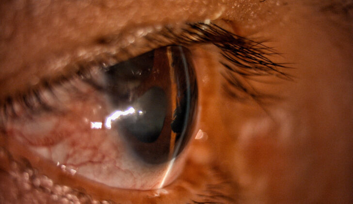
What is Iridocorneal Endothelial Syndrome?
Iridocorneal Endothelial Syndrome (ICE) is a rare eye disorder. It is caused by an overgrowth and spread of cells on the innermost layer of the cornea (the clear, dome-shaped surface at the front of your eye). These cells move to the colored part of your eye, the iris, and the angle where the iris and cornea meet. This can lead to a type of eye pressure increase called secondary angle-closure glaucoma, swollen and waterlogged cornea known as corneal edema and thinning of the iris.
This condition is usually linked to another type of glaucoma because it blocks the drain in your eye. This results in the iris sticking to the cornea or both. Patients often show changes in iris shape, a decline in how well they see, blurred vision, or a combination of these. Vision loss generally happens because of escalating glaucoma and a failing cornea. If ICE is not treated, it can lead to blindness.
Commonly, ICE affects only one eye and often happens in young adults and middle-aged women. It typically shows in three ways: Essential Iris Atrophy (EIA), Chandler Syndrome (CS), and Cogan-Reese Syndrome (CRS). EIA is marked by changes in the iris, such as full-thickness holes and weakening of the corneal endothelium, the layer of cells on the innermost part of the cornea. The most frequent type of ICE is CS, which shows less iris changes but more impaired vision, reduced iris, a displaced pupil, and a significantly swollen and dehydrated cornea. CRS usually shows with bumps on the front of the iris, a damaged corneal endothelium, and a swollen cornea.
What Causes Iridocorneal Endothelial Syndrome?
The exact causes of ICE, or iridocorneal endothelial syndrome, are still not fully understood. In our eyes, there are cells called corneal endothelial cells that typically don’t multiply in a living eye and their number decreases as we grow older. Sometimes, these cells can regain their ability to multiply and this can cause ICE. These cells play a crucial role in maintaining the amount of water in our eye and making sure our cornea, the clear front surface of our eye, remains transparent. If these cells don’t function properly, our cornea can swell and we may start having vision problems.
Scientists have proposed different theories for how ICE develops. A key one is the “neural crest theory.” According to this theory, ICE might be related to abnormal multiplication of particular cells, called neural crest cells.
Another condition, called Peters anomaly, occurs due to an abnormality during the development of the first wave of neural crest cells. This abnormality causes the lens and the cornea of the eye to stay connected, causing the cornea to become opaque. Type 1 Peters anomaly shares similarities with ICE. Yet, unlike ICE, Peters anomaly generally appears at an earlier age and is often seen in both eyes. Fuchs dystrophy, another condition, also affects both eyes but is different from ICE and Peters anomaly.
In 1978, three scientists – Cambell, Shields, and Smith – introduced their “membrane theory” to better explain ICE. They theorized that ICE initially involves the deterioration of corneal endothelial cells, which progressively leads to the formation of a membrane around the iris (the colored part of the eye). This process obstructs the trabecular meshwork, a spongy tissue located near the base of the iris, which in turn leads to changes in the iris and secondary glaucoma (a group of diseases damaging the eye’s optic nerve).
While these theories explain how ICE could progress, the exact cause is still unknown. Certain studies suggest a connection between ICE and inflammation or uveitis (a form of eye inflammation). There are also theories that a viral trigger might cause ICE. For example, a study in 1994 found DNA of the Herpes simplex virus in over 60% of ICE cases examined. When the corneal endothelial layer was removed from ICE samples, the virus’s DNA could no longer be identified, suggesting that the viral DNA primarily exists in the endothelial cell layer. Other studies suggest a link between the Epstein-Barr virus and ICE.
Risk Factors and Frequency for Iridocorneal Endothelial Syndrome
ICE, also known as Iridocorneal Endothelial syndrome, is a condition that usually affects one eye, and most commonly impacts women between the ages of 20 and 50. However, there have been instances of the condition affecting both eyes, as well as being diagnosed in men. Despite some reports linking ICE with hearing loss, there’s no solid proof confirming the connection.
Different types of ICE have been found to occur amongst various racial and ethnic groups. For example, a type called CS is most commonly found in white patients. On the other hand, CRS, another type of ICE, is reported least frequently in India, accounting for only 14.29% of ICE cases. EIA, yet another type, has the highest occurrence with 66.67% in India. Interestingly, ICE has been found in 62% of Indian male patients, which is much higher than the 17% reported in North America and Europe.
Signs and Symptoms of Iridocorneal Endothelial Syndrome
Iridocorneal endothelial syndrome (ICE) is an eye condition that changes the shape of the pupil and vision. Common symptoms include a distorted pupil due to iris deterioration and visual changes such as blurry vision, particularly when waking up, or seeing halos around lights, usually caused by a swelling of the cornea. Pain in the eye or headaches may occur if eye pressure increases significantly, potentially leading to secondary glaucoma.
The appearance of the affected eye differs based on the stage and specific type of the syndrome. Patients might not notice the deterioration of the iris, a key symptom of essential iris atrophy (EIA), without a detailed eye examination. People suffering from progressive iris atrophy (PIA) or Chandler’s syndrome may experience high eye pressure, severe glaucoma, and a swollen cornea that appears opaque due to extensive corneal dystrophy.
Doctors may find certain irregularities, given as “hammered silver” or “beaten bronze” descriptions, during a detailed eye examination using a slit-lamp, a device used to examine the eyes. They might also observe changes in the angle between the iris and cornea or unusual cupping of the optic nerve if glaucoma is present.
In the diagnosis process, apart from a detailed slit-lamp examination, it’s crucial to examine the angle of the eye with gonioscopy and measure the pressure within the eye. Specialized imaging techniques such as specular microscopy and in vivo corneal confocal microscopy (IVCM) can reveal unique findings of this disease.
Testing for Iridocorneal Endothelial Syndrome
Catching ICE (Iridocorneal Endothelial Syndrome) early is extremely important to prevent complications like corneal swelling and a type of eye pressure ailment called secondary glaucoma. Sometimes, if the swelling of the cornea is severe and blocks a clear view of the front part of the eye, a specific type of imaging called IVCM can be used.
IVCM, or In Vivo Confocal Microscopy, is a harmless imaging technique that gives a detailed view of the layers of the cornea, down to the cellular level. It can help identify abnormal corneal cells, which in patients with ICE, appear differently in size and shape, possess distinct cell boundaries, and show unusually bright nuclei (the central part of the cell).
Another imaging tool used to check the cornea is specular microscopy. This helps to identify abnormally large and round cells in the corneal epithelium, a telltale sign of ICE. The distinctive feature of these cells is something called a “light-dark reversal” pattern. This means the cells appear darker as opposed to the usual light pattern observed in normal cells.
Gonioscopy is another necessary procedure that looks at the iridocorneal angle (the angle where the iris (the colored part of the eye) and cornea meet). In cases of ICE, broad-based synechiae (adhesions) are often found in this area.
If corneal swelling makes it difficult for gonioscopy to be conducted, another imaging technique known as ultrasound biomicroscopy can be used. This technique can clearly show the iridocorneal angle and help identify any abnormalities. Combining gonioscopy with ultrasound biomicroscopy provides a comprehensive evaluation of the site and extent of these eye abnormalities.
Patients with ICE, particularly those who subsequently develop secondary glaucoma, should undergo specific eye tests. These tests include visual field testing with Humphrey’s method, measuring the thickness of the cornea, checking the pressure in the eye with a method called Goldmann applanation tonometry, and examining the optic nerve.
Moreover, imaging like optical coherence tomography of the optic nerve head and retinal nerve fiber layer should be done. Optical coherence tomography is a technique that uses light waves to create a detailed map of the nerve fibers in the retina (the light-sensitive tissue lining the back of the eye). This helps monitor changes to the optic nerve over time.
Treatment Options for Iridocorneal Endothelial Syndrome
Treatment for ICE, or iridocorneal endothelial syndrome, focuses on addressing corneal damage and preventing irreversible loss of vision due to glaucoma. Cosmetic or practical surgical procedures may also be conducted to manage changes to the iris.
Endothelial keratoplasty is a surgical technique that is often preferred for treating ICE-related corneal swelling. There are two forms of this procedure, Descemet stripping automated endothelial keratoplasty (DSAEK) and Descemet membrane endothelial keratoplasty (DMEK). DMEK produces better results and a quicker recovery time compared to DSAEK. However, DSAEK is the preferred choice for patients with significant changes to their iris. If other surgical approaches aren’t successful, an artificial cornea (keratoprosthesis) may be considered.
In people with ICE, the trabecular meshwork (the tissue responsible for draining the fluid in the eye) doesn’t function properly, which leads to secondary glaucoma. So, medications that decrease the production of eye fluid tend to be more effective than those that increase its outflow. This form of secondary glaucoma usually involves eye drops that include beta blockers, carbonic anhydrase inhibitors, and alpha agonists. Prostaglandins, another type of eye drops, have been linked to negative reactions in the eye for patients with ICE, therefore they should be avoided.
Managing glaucoma resulting from ICE can be difficult. If eye pressure isn’t controlled with medication, surgery might be necessary. Options include goniotomy (a procedure to remove the blockage caused by the ICE membrane), trabeculectomy (creating a new drainage path for eye fluid), glaucoma drainage implants, or cyclodestructive procedures (destroying part of the eye’s fluid-producing tissue).
The success rates for these surgical procedures can vary. Trabeculectomy results are typically less successful in ICE patients compared to patients with other types of glaucoma. The timely scheduling of the procedure early in the progression of ICE can increase success rates. The use of antimetabolites (drugs that inhibit cell growth) or glaucoma drainage implants can help to reduce eye pressure post-surgery. However, if the implant is blocked by the membrane, laser treatments may be utilized to correct the issue.
In cases where surgical interventions fail to control the eye pressure and result in a painful blind eye, cyclodestructive procedures that destroy part of the eye’s fluid-producing tissue can be used.
Finally, treatments such as femtosecond-assisted keratopigmentation (KTP) or implanting iris prosthesis devices (artificial iris implants) can help address common problems in ICE patients, such as double vision and light sensitivity, while also improving cosmetic appearance.
What else can Iridocorneal Endothelial Syndrome be?
If a young woman experiences one-sided corneal swelling, vision issues, iris irregularities, or glaucoma, she might have a condition called iridocorneal endothelial syndrome (ICE). This syndrome can seem like other diseases, so doctors should get a thorough medical history and conduct a detailed physical exam to pinpoint the problem.
The factors that may suggest conditions other than ICE, especially a variation called Cogan-Reese Syndrome (CRS), include Tapioca melanoma and diffuse malignant melanoma of the iris. Tapioca melanoma has similar iris nodules to CRS, but these are usually less pigmented than the darker nodules in CRS. Plus, CRS’s typical features like iris atrophy or changes in eye pressure aren’t present in Tapioca melanoma. Diffuse malignant melanoma of the iris doesn’t typically show changes in the eye’s pupils or pressure, which are more indicative of CRS.
The conditions similar to another ICE variant – Chandler’s Syndrome (CS), include posterior polymorphous corneal dystrophy (PPCD) and Fuchs endothelial dystrophy. PPCD is an inherited disorder that typically affects both eyes, just like ICE. Patients with PPCD and ICE both have changes that increase their chances of getting glaucoma. But, in PPCD patients, their Descemet membrane (a layer of the cornea) is thicker and might show cloudy patches, while it’s usually normal in CS. Fuchs endothelial dystrophy also affects both eyes but doesn’t show iris changes that are common in ICE. Its identifying feature is having less shiny nuclei in cells.
The third ICE variant – Essential iris atrophy (EIA) has some conditions similar to it: Axenfeld-Rieger syndrome (ARS), aniridia, and iridoschisis. ARS is an inherited disorder mostly seen at birth with potential associated issues such as abnormal teeth. ARS usually affects both eyes unlike ICE, which is mostly one-sided. Both ARS and ICE display a certain pattern of a single layer of cells extending over the cornea, angle between the iris and cornea, and iris. But, since ARS is present from birth, it is assumed that the abnormal iris-cornea layer is due to cells not developing properly. A clear difference between ARS and ICE is the thicker corneal edge with iris strands in ARS.
Aniridia usually affects both eyes and is often linked with other conditions such as underdeveloped optic nerve. An important factor setting it apart from late-stage EIA is the age of onset – it typically shows up within the first six weeks of life, often with associated sensory defects like hearing loss and diminished smell sensitivity. Iridoschisis, on the other hand, involves the layers of the iris gradually separating, usually affecting both eyes, and tends to happen in older patients.
What to expect with Iridocorneal Endothelial Syndrome
The future health condition (or prognosis) of a patient with ICE (Iridocorneal Endothelial Syndrome), a rare eye disorder, depends on how advanced the illness is at the moment of diagnosis, as well as whether there are any additional health issues. Procedures to correct issues with the cornea (the clear outer layer of the eye) may not be able to completely remove the abnormal cells and, as a result, may not fully prevent the condition from getting worse or from causing secondary glaucoma (a condition that can cause loss of vision if left untreated).
A small research study in 2018 examined eight eyes in seven patients with both ICE and another eye disorder called PPCD (Posterior Polymorphous Corneal Dystrophy). This study found a significant improvement in all eyes’ best-corrected vision (vision achieved when using glasses or contact lenses) at both six months and two years after a procedure called DMEK (Descemet Membrane Endothelial Keratoplasty).
A larger study in 2020 looked at 86 patients who got their first cornea transplants because of ICE. This study examined the survival rates of the transplanted cornea (how long the new cornea lasted without problems) over five years in patients who got either two types of cornea transplant surgery – penetrating keratoplasty (PKP) or endothelial keratoplasty (EK). The results showed that there wasn’t a significant difference in how long the new corneas lasted after PKP or EK with rates of 64.3% and 66.8% respectively.
Possible Complications When Diagnosed with Iridocorneal Endothelial Syndrome
The complications of ICE syndrome can include abnormalities in the iris, swelling of the cornea, cornea damage, and an increased risk of secondary glaucoma, a condition that causes high pressure inside the eye and can lead to vision damage. Nearly half of all patients with ICE syndrome develop glaucoma, especially those with the EIA variant of the syndrome. However, swelling and damage to the cornea are more likely in patients with the CS subtype.
The severity of the disease can also affect how doctors approach eye surgeries, such as cataract removal. For example, in cases where the disease has severely affected the cornea, patients might get a combined treatment that includes DSAEK surgery to treat the cornea and cataract extraction for removal of cataract.
Common Complications:
- Iris abnormalities
- Swelling of the cornea
- Cornea damage
- Increased risk of secondary glaucoma
- Possible changes to treatment approach for eye surgeries, like cataract removal
Preventing Iridocorneal Endothelial Syndrome
ICE, which stands for Iridocorneal Endothelial syndrome, is quite a rare eye condition. If you are a woman aged between 20 and 50, and you notice symptoms like swelling in one eye (known as ‘unilateral corneal edema’), changes in the color or shape of your iris (the colored part of your eye), alterations in your vision, or signs of increased pressure in your eye that could indicate glaucoma, it’s important to get your eyes checked for this condition.
A careful examination can help detect ICE early. Making a prompt diagnosis can prevent the condition from worsening. It can help avoid damage to the cornea (the clear front part of the eye) due to swelling, and also prevent secondary glaucoma, which is a serious condition that can occur when the pressure in the eye increases because certain bodily fluids can’t exit the eye properly.
Being informed about ICE and its different forms, and getting diagnosed early, can help prevent this disease from advancing, thereby saving your vision from potential threats.


