What is Lipemia Retinalis?
Lipemia Retinalis (LR) is a change in your eye’s appearance that happens when there are too many fats, specifically triglycerides, in your blood. Looking through a special device called an “ophthalmoscope,” doctors can see a creamy-white color in your eye’s blood vessels. In mild cases, these changes only affect the vessels at the edge of your eye; however, in advanced cases, these changes also involve the vessels right at the center of the eye. In extreme cases, the back of the eye, or the “fundus,” may even take on a salmon color. Despite these changes, LR does not impact your ability to see.
What’s important about LR, however, is that it can signal other severe but easy-to-treat conditions that affect your body’s ability to process fats. This article provides an overview of the causes, frequency, how it develops, tissue changes, methods to evaluate, management, and prognosis of LR. Understanding this condition can help ensure it is detected and treated promptly to prevent any further health complications.
What Causes Lipemia Retinalis?
Lipemia retinalis (LR) is a condition that was first detailed by Heyl in 1880, and later graded in 1970. LR is tied to the transportation of lipids (fats) in our blood. There are five different kinds of these lipid transporters, known as lipoproteins: chylomicrons, very-low-density lipoproteins (VLDL), intermediate-density lipoproteins (IDL), low-density lipoproteins (LDL), and high-density lipoproteins (HDL). Depending on their size, some lipoproteins scatter light and can’t be seen, while others, like chylomicrons, are larger and contribute to LR.
Most of the fat we eat is transported from our intestine to the rest of our body via chylomicrons. If the body has trouble processing these chylomicrons, their high concentration of fat can lead to LR. When blood has a high level of chylomicrons, it can give the eye a creamy-white appearance due to the light bouncing off the chylomicrons in the retina’s blood vessels.
Elevated levels of triglyceride fats (hypertriglyceridemia or HTG) can be inherited or result from other diseases. These high levels in our blood can cause HTG which can result in serious conditions such as pancreatitis (inflammation of the pancreas). When too many chylomicrons are in the blood, the blood appears cloudy and milky. This could be confused with fat embolism, a condition where large, colorless fat globules exist in the blood and can block vessels, leading to eye problems.
Chylomicronemia happens when the blood’s triglyceride fat level is above 1000 mg/dl. It is defined by having one or more of the following symptoms: eruptive xanthoma (skin condition with yellowish, waxy deposition), lipemia retinalis, and abdominal pain or pancreatitis. Some experts believe a triglyceride level above 2000 mg/dl is a more accurate lower boundary for defining chylomicronemia. Patients can show a number of symptoms, like recent memory loss in 85% of patients.
One of the main reasons for LR is an increased level of triglyceride fats in our blood. It can be inherited due to the presence of certain inhibitors or deficiencies in our bodies or even due to an abnormality in our genes. Alternatively, it can be secondary to other diseases or external factors like poor diet, particular types of medication, hormonal imbalances, or underlying systemic diseases.
Risk Factors and Frequency for Lipemia Retinalis
Lipemia retinalis is a condition that is not fully understood, including how frequently it occurs and what causes it. However, we do have information on some of the risk factors associated with this condition. For example, a lack of a substance called lipoprotein lipase can result in a condition known as familial HTG, which involves high levels of a fat/protein combo called chylomicrons in the blood. This lipoprotein lipase deficiency is quite rare, affecting less than 1 in 1 million individuals.
This issue can also be a symptom of a condition called familial combined hyperlipidemia, which affects between 1% and 2% of people on a global scale. Furthermore, reports from the National Health and Nutrition Examination Survey used data from 2001 and 2006 to estimate that around 1.7% of the entire U.S. population, or 3.4 million people in America, have severe HTG. This condition involves having large amounts of fat in the blood, ranging from 500 to 2,000 mg/dl.
- Most individuals with severe HTG are men (75.3%).
- The majority of HTG patients are non-Hispanic whites (70.1%).
- Most patients with this condition are aged between 40-59 years (58.5%).
- About 14% of HTG cases are associated with diabetes.
- Hypertension is associated with around 31.3% of HTG cases.
Signs and Symptoms of Lipemia Retinalis
Lipemia retinalis, a condition where the blood vessels in the eyes turn creamy or milky, is often detected during a routine eye test or as a part of investigation for lipid-related disorders. It has also been found in infants during screening for retinopathy of prematurity (ROP), a potentially blinding eye disorder. Interestingly, there have been reports where stomach pain and digestive issues were the first signs of high fat levels in an infant’s blood, before lipemia retinalis was detected. Family history of high lipid levels, due to various genetic or enzyme-related disorders, might be present. It is necessary to exclude possible secondary causes of high triglyceride levels through a detailed medical history and physical examination.
An examined person will have various systems of their body checked including the cardiovascular system, skin, abdomen, and eyes. For the cardiovascular system, this includes the examination of heart sounds and peripheral pulses. There will also be a check for any unusual sounds from the vessels supplying the kidneys and neck. Skin features might include xanthomas, which are small, yellowish bumps with a red base, usually found on the buttocks and elbows due to a high amount of triglycerides in the skin cells. Physicians will assess the abdomen for enlargement of the liver and spleen and to exclude pancreatic diseases. The eye’s examination is pivotal and includes a check of the area around the eye sockets, the front part of the eye, and the back part.
Observations during the eye exam might include:
- Xanthelasma or yellowish deposits of fat on the eyelids
- Tuberous palpebral xanthomas or lumps on the eyelids
- Rarely seen changes in the front part of the eye, such as yellow deposits on the white part of the eye, a ring around the cornea in older or younger individuals, lipid deposits in the cornea, yellowish-white patches on the coloured part of the eye, and fat collection in the transparent front part of the eye
A detailed examination of the back part of the eye is necessary for diagnosing and classifying lipemia retinalis. Other uncommon findings in cases of high lipid levels might include yellow deposits in the retina or the layer beneath it and adult-onset Coats disease, a rare eye disorder that leads to vision loss.
Testing for Lipemia Retinalis
To diagnose high triglyceride levels, doctors perform blood tests after you’ve fasted. When your blood sample stands in a tube, a creamy white layer may form on top if your triglyceride levels are high. This is because particles called chylomicrons, which carry triglycerides in your blood, make your blood look cloudy.
In some cases, your doctor might also order a genetic test to check for specific changes in your genes. These changes could be causing your high triglyceride levels.
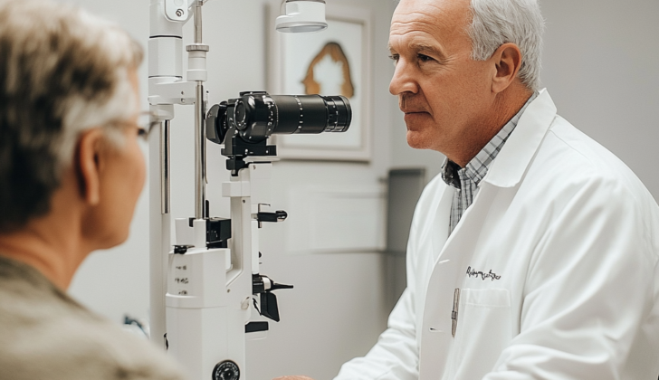
A procedure called a fundoscopy, which involves looking at the back of your eye, helps diagnose lipid-rich retinopathy – a condition related to high triglyceride levels that affects the eyes. Fundus photography, taking pictures of the back of your eyes, lets your doctor keep track of any changes in your condition.
Another test your doctor may perform is an optical coherence tomography (OCT) scan of your eye. This scan can show if chylomicrons, the particles that carry triglycerides, are in your retinal blood vessels.
Doctors can also use an electroretinogram (ERG) to measure the electrical responses of various cells in your eyes, both before and after treatment.
There are other types of eye tests, like fluorescein and indocyanine angiography, but these usually do not show any noteworthy findings when it comes to lipid-rich retinopathy.
Treatment Options for Lipemia Retinalis
Lipemia retinalis is a condition that can sometimes disappear within hours. However, quickly correcting high levels of fats in the blood can lead to a better outcome. There are several ways to achieve this.
A strict low-fat diet can be effective, especially in infants. They should not be given breast milk supplements. This method helps reduce the levels of fats in the blood and prevent lipemia retinalis. Adjusting the fat levels in your diet can reverse any abnormal findings within a week.
Lipemia retinalis does not require direct treatment. However, it’s an important sign of a potentially dangerous metabolic disorder that can be easily treated. The treatment focuses on reducing blood fat levels to below 500 mg/dl. There are various medications available that can lower fats and cholesterol levels. These drugs include fibrates and n-3 polyunsaturated fatty acids, also known as fish oil. Both can reduce fats in your blood by around 50%.
Keep in mind that individuals with liver or kidney issues should not take fibrates. Huge amounts of n-3 polyunsaturated fatty acids can cause gastrointestinal issues. Nicotinic acid is another option that can decrease fat levels by 15-35%. If you have both lipemia retinalis and diabetes, it’s crucial to have strict control over your blood sugar levels.
There’s no specific surgery for lipemia retinalis. However, an exchange transfusion can be used in severe cases to reduce blood fat levels quickly. This process involves replacing your blood with donor’s blood. Ileal bypass surgery can also show improvements in all blood fat parameters, but it’s usually reserved for severe, treatment-resistant cases.
What else can Lipemia Retinalis be?
When diagnosing lipemia retinalis, doctors must consider other conditions that may present with similar symptoms, including:
- Leukemia
- Severe diabetic eye disease leading to hardened vessels
- Blockages in the retina’s blood vessels due to kidney failure or hyperparathyroidism
- Blockage in a branch of a retinal artery or vein
- High blood pressure causing damage to the retina (hypertensive retinopathy)
- A type of birthmark in the eye lining (choroidal hemangioma) that causes the back of the eye to appear red or salmon-colored
In the case of leukemia, retinal veins appear reddish-pale while arteries are pale yellow. However, during lipemia, both arteries and veins present the same color and can only be differentiated by their size, with veins being larger.
For blockages from kidney failure or hyperparathyroidism, an irregularity in kidney function results, and a higher parathyroid hormone level could serve as a pointer to an accurate diagnosis.
On an initial examination, retinal vasculitis may be considered, but this condition typically affects only segments of the vessels and is associated with an influx of inflammatory cells around the blood vessels and signs of inflammation inside the eye.
What to expect with Lipemia Retinalis
Changes in the eye’s retina due to a condition called lipemia retinalis tend to get better once the amount of fat (triglycerides) in the blood returns to normal. Generally, one’s ability to see (visual acuity) is not greatly impacted but tends to improve after the high amounts of triglycerides in the blood (hypertriglyceridemia or HTG) are corrected.
Photopic and scotopic responses, which are measurements of the eyes’ responses to light in bright and dim conditions respectively, show improvement after HTG correction. This is observed through a test called an electroretinogram, which measures the electrical responses of various cell types in the retina, including the light-sensitive cells called rods and cones.
People with HTG are at a heightened risk for heart-related diseases like coronary artery disease, heart attack (myocardial infarction), and stroke. They also have an increased risk for certain eye conditions, such as blockages in the retinal artery or vein and a lack of blood flow to the retina (retinal ischemia). As such, these patients should continue to see their doctor or eye care professional for regular check-ups and monitoring.
Possible Complications When Diagnosed with Lipemia Retinalis
Complications from having high levels of fats in the bloodstream, a condition known as hyperlipidemia, can have damaging effects on various parts of the body. For instance, this condition can lead to diseases of the heart, high blood pressure, heart attack, and stroke. In some people, hyperlipidemia presents as Chylomicronemia, which is characterized by symptoms such as acute pancreatitis, abdominal pain, enlarged liver and spleen, difficulty breathing, and recent memory loss.
Eye-related complications due to hyperlipidemia are less common but can occur. These might include blockage of the blood vessels in the retina, lack of blood supply to the retina, growth of new blood vessels and bleeding into the jelly-like substance that fills the back of the eye.
Common Complications:
- Cardiovascular diseases
- High blood pressure
- Heart attack
- Stroke
- Acute pancreatitis
- Abdominal pain
- Enlarged liver and spleen
- Difficulty breathing
- Recent memory loss
- Blockage of the blood vessels in the retina
- Lack of blood supply to the retina
- Growth of new blood vessels in the eye
- Bleeding into the jelly-like substance in the back of the eye


