What is Ocular Cellulitis (Eye Infection)?
Orbital cellulitis is an infection that causes swelling in the eye socket, behind a thin barrier known as the orbital septum. This specifically affects the fat around the eye and the muscles that move the eye but doesn’t affect the eye itself. On the other hand, there’s another type of infection called preseptal, or periorbital, cellulitis. This type of infection occurs in front of the orbital septum, mainly in the eyelids.
While periorbital cellulitis occurs more often, especially in children, it’s crucial to be able to tell it apart from orbital cellulitis. The complications from an orbital cellulitis infection can be much more severe and could potentially be life-threatening.
What Causes Ocular Cellulitis (Eye Infection)?
The main causes of orbital cellulitis, a type of eye infection, are bacteria known as Staphylococcus and Streptococcus, specifically Staphylococcus aureus and group A Streptococcus. There’s also growing concern over more cases of a resistant strain of bacteria called MRSA (methicillin-resistant Staphylococcus aureus).
In the past, bacteria named Haemophilus influenzae and Streptococcus pneumoniae were significant in causing these types of infections, but thanks to vaccines, these cases have significantly reduced. However, worldwide, it should be noted that Haemophilus influenzae type B still causes about 20% of orbital cellulitis cases which result in an abscess under the bone covering.
Another type of bacteria, non-spore forming anaerobic bacteria, are also linked to orbital cellulitis, but less frequently than the aforementioned organisms. In the United States, the most common associated bacteria of this type are Fusobacterium and Peptostreptococcal species.
For people with weakened immune systems, fungal infections can be a concern. Here, Mucormycosis and Aspergillus species are the most common causes of orbital cellulitis. We must watch out for situations such as Mucormycosis causing a rapidly progressing sinusitis infection in diabetic patients in Ketoacidosis, and Aspergillus infecting the orbit in patients with suppressed immune systems due to HIV or severe neutropenia. However, compared to bacterial orbital cellulitis, fungal orbital cellulitis usually progresses more slowly and over a longer period.
Risk Factors and Frequency for Ocular Cellulitis (Eye Infection)
Orbital cellulitis, an infection of the eye area, happens more often in young children than in older children or adults. This is because it’s usually linked to bacterial rhinosinusitis, an inflammation of the nasal passages, or an upper respiratory infection, which are common in young kids. Some studies indicate that between 86% and 98% of all orbital cellulitis cases are related to rhinosinusitis, with cases relating to the ethmoid sinus, located near the eye, being particularly frequent.
Another reason orbital cellulitis is more common in children is its peak occurrence in winter, a time when rhinosinusitis is also more common.
- Other possible causes of orbital cellulitis include complications after eye surgery or use of anesthesia near the eye,
- physical injury or foreign objects in the eye,
- severe infection of the tear ducts (dacryocystitis), or
- spread of infections from the teeth, face or middle ear to the eye area.
Signs and Symptoms of Ocular Cellulitis (Eye Infection)
Orbital cellulitis is a severe eye infection. It’s crucial to tell it apart from preseptal cellulitis. Historical clues and physical symptoms can typically do this.
Patients with orbital cellulitis often have a past record of sinusitis or upper respiratory tract infection. They may also have recently undergone ophthalmological surgery or have past sinus or dental diseases. It’s important to check these things as they have been strongly linked to orbital cellulitis.
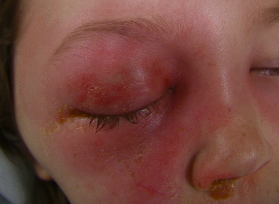
Symptoms usually start with sudden swelling or redness of the eyelid. This rapidly progresses to eye pain (especially when moving the eye), reduced vision, and eye redness. Along with eye discomfort, patients often experience fever, general weakness, and lack of appetite. It’s also essential to know if there’s a family history of autoimmune diseases or cancer to rule out other possible causes for the symptoms. These conditions can also cause unilateral proptosis, a symptom that can mimic orbital cellulitis.
- History of sinusitis or upper respiratory tract infection
- Recent ophthalmological surgery
- Past sinus or dental diseases
- Sudden eyelid swelling
- Eye pain with movement
- Reduced vision
- Eye redness
- Fever
- General weakness
- Lack of appetite
In a physical examination, common symptoms include linear swelling of the eyelid accompanied by warmth and eyelid tenderness. These symptoms are often augmented by vision loss, eyeball bulging (proptosis), puffiness around the eye (chemosis), and paralysis of eye muscle movements (ophthalmoplegia). However, eyelid swelling can sometimes make physical examination difficult. Collaborative examinations with an eye specialist (ophthalmologist) can help, especially when looking for fundoscopic findings (eye examination performed with an ophthalmoscope). Alarmingly, or alarmingly? signs can include swelling of the optic disc with orbital nerve edema, enlargement of disc vessels, or the presence of retina artery blockages, all of which can indicate the spread of the infection to the brain.
Testing for Ocular Cellulitis (Eye Infection)
When a patient is suspected to have orbital cellulitis, a thorough and careful evaluation is necessary. This involves looking out for symptoms like fatigue and checking vital signs. The most important step, however, is an extensive physical examination.
The doctor may also request a complete blood count or CBC. This can show an increase in white blood cells, particularly a type known as “neutrophils”, which suggests the body is trying to fight an infection. However, a CBC is not essential for diagnosis. While some suggest getting blood cultures to look for bacteria in the blood, this hasn’t proven to be especially helpful. The best way to identify the bacteria causing the infection is through surgical aspiration of an abscess located beneath the outer layer of the bones (subperiosteal) or inside the eye socket (orbital), but this is only necessary if surgery is required. Starting treatment does not require a routine aspiration or biopsy.
Imaging can provide more information, though some disagree on its use. Some suggest a CT scan should be a routine part of the first evaluation for patients suspected of having orbital cellulitis. This allows doctors to get a better look at the eye socket and adjacent sinus chambers. It’s particularly useful for detecting an abscess under the bone coat, which could require surgical intervention. If a CT scan is done, using an intravenously-administered contrast dye could help to tell the difference between a real abscess and tissue inflammation. If the CT scan isn’t done initially, it’s recommended for patients who don’t improve or get worse within 48 hours of starting antibiotic treatment via IV.
Magnetic resonance imaging (MRI) is a good alternative that exposes the patient to less radiation than a CT scan. This is especially beneficial for pediatric patients, though MRIs do provide a clearer picture of the eye socket tissues and might be a better choice for adults. Because an MRI takes longer, children might need to be sedated, which requires coordination with an anesthesiologist.
Finally, it’s important to call on the services of an ophthalmologist in any case of orbital cellulitis. The initial evaluation should include a comprehensive examination of the eye, involving visual function tests, testing of the pupil’s light reflex, and ophthalmoscopy to look for optic nerve swelling and twisted veins, especially when there’s significant swelling and fluid accumulation in the eyelid.
Treatment Options for Ocular Cellulitis (Eye Infection)
When a patient is diagnosed with orbital cellulitis, it’s crucial they start receiving antibiotics promptly due to the severe complications that can occur with this condition. The antibiotics used are typically intravenous (IV) and designed to target the most common bacteria causing the condition, namely Staphylococcus and Streptococcus and some other types of bacteria called gram-negative bacilli. If the infection extends to the brain, antibiotics against anaerobic bacteria are added. It’s important to consider the local prevalence of bacteria resistant to antibiotics (like MRSA) when deciding on the antibiotics.
Generally, the treatment is a combination of different IV antibiotics. In patients with healthy kidney function, the prescribed treatment commonly includes Vancomycin with another antibiotic like ceftriaxone, cefotaxime, ampicillin-sulbactam, or piperacillin-tazobactam. If the infection is suspected to have reached the brain, a combination of vancomycin and either ampicillin-sulbactam or piperacillin-tazobactam should be given – in this case, no additional treatment is needed. But if ceftriaxone or cefotaxime is added to vancomycin, metronidazole should also be given for additional anaerobic coverage.
If the patient also has a sinusitis, the use of an oxymetazoline nasal spray can alleviate nasal congestion and saline nasal wash can aid in sinus drainage, both assisting in recovery.
For patients allergic to certain antibiotics like penicillin or cephalosporin, the treatment may consist of vancomycin combined with either ciprofloxacin or levofloxacin. However, antibiotics from the fluoroquinolone group (ciprofloxacin and levofloxacin) may not be very effective in preventing the spread of the infection to the brain, as they don’t get into the central nervous system well.
After a noticeable improvement in the patient’s condition, usually within 48-72 hours of starting IV antibiotics, the patient can be transitioned to oral antibiotics and continue their treatment at home. Antibiotics like clindamycin or trimethoprim-sulphamethoxazole (that cover MRSA), combined with an antibiotic effective against Streptococcus and anaerobic bacteria are typically recommended. The entire duration of antibiotic therapy usually lasts 14 days but can be extended to 21 days in complicated situations.
Surgical intervention is usually reserved for special cases such as the presence of large abscesses under the lining of the bone (subperiosteal) or within the eye socket (orbital). Surgery may be needed if the patient’s visual function changes or if there’s no improvement in symptoms like fever, swelling, eye bulging (proptosis), or pain, prompting further imaging studies to check for abscesses. Subperiosteal abscesses bigger than 10mm, orbital abscesses, or extensions of the infection into the brain may need to be assessed and possibly drained by the surgical team.
What else can Ocular Cellulitis (Eye Infection) be?
If a person doesn’t show common symptoms of orbital cellulitis, such as swollen eyelid, bulging or pushed-out eye, pain when moving the eye, fever, or generally looking unwell, then doctors may need to look into other possible causes of a protruding eye. These could be tumors, inflammation, autoimmune diseases, or issues with the bones around the eye, among other things.
The most frequently occuring tumors that could be causing the eye to bulge are rhabdomyosarcoma and retinoblastoma, which are cancerous tumors. Neuroblastoma, another type of cancer, is the most likely to spread to the eye or its surrounding areas. Melanoma, a type of skin cancer, is also known to potentially spread to these areas.
Some autoimmune diseases could be causing the eye to bulge. These include orbital pseudotumor (also known as idiopathic orbital inflammatory disease), Tolosa-Hunt syndrome, and Exophthalmos that happens due to Graves’ disease. Doctors can usually rule out these conditions based on a patient’s medical history, lab tests, and imaging.
Imaging and examination can also help to rule out things like foreign objects, blood-filled cysts, bone cysts caused by an aneurysm, tumors like ossifying fibromas, and pseudoaneurysms of the bones around the eye.
What to expect with Ocular Cellulitis (Eye Infection)
In general, the outlook for orbital cellulitis – an infection of the eye tissues – is usually positive as long as it’s diagnosed promptly and treated immediately. Most patients start showing improvement within the first 24 to 48 hours of starting IV antibiotics, especially in straightforward cases.
Although uncommon, complications can occur if treatment is not promptly administered. These events can be quite serious.
Most cases get better with IV antibiotics and are typically completely resolved with continued oral antibiotics taken at home. However, if there’s a large build-up of pus (known as a subperiosteal abscess that’s greater than 10 mm), or if an abscess forms in the eyeball, or if the nerves or blood vessels are compromised, surgery may be needed.
Possible Complications When Diagnosed with Ocular Cellulitis (Eye Infection)
Orbital cellulitis, an infection in the tissue surrounding the eye, can lead to a range of complications. The most common issue is the formation of abscesses, specifically under the dense, fibrous membrane that lines the eye socket (known as subperiosteal abscesses). These typically occur on the inner side and base of the eye socket, as these areas tend to be thinner. Although it’s rare, abscesses can also develop inside the eye socket itself and can result in a persistent fever even after two days of strong, intravenous antibiotics.
A significant drop in vision quality or a loss of color vision can indicate possible danger due to swelling of the optic disc – this can occur from blockage of the small arteries in the retina due to infected blood clots. Similarly, if the pupil doesn’t respond normally to light, it may indicate that the optic nerve is affected.
Severe complications can arise if the infection spreads to the brain. These complications can be cavernous sinus thrombosis (a blood clot in the brain’s drainage system), meningitis (an infection of brain’s protective membrane), or an abscess within the brain. Particularly alarming is the sudden onset of the inability to move the eye laterally in a patient suffering from acute orbital cellulitis who is not getting better with antibiotics – this may suggest the development of a cavernous sinus thrombosis. This is because the nerve that controls lateral eye movements (the sixth cranial nerve) is located along the wall of the brain’s venous sinus (a cavity that drains deoxygenated blood).
Possible complications involve:
- Formation of abscesses
- Persistent fever
- Decrease in vision
- Loss of color vision
- Abnormal pupil response to light
- Cavernous sinus thrombosis (a serious brain condition)
- Meningitis (a brain infection)
- Brain abscess
- Paralysis of lateral eye movements


