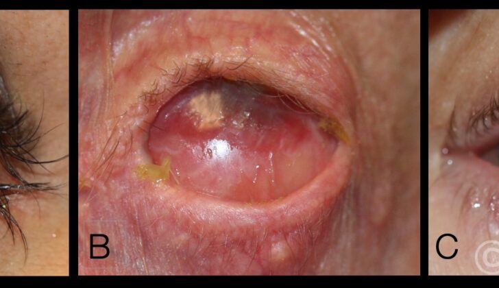
What is Ocular Pemphigoid?
Ocular cicatricial pemphigoid (OCP) is a type of disease that affects the clear, thin tissue covering the white part of the eye and the inside of the eyelids, technically known as mucous membrane pemphigoid (MMP). This condition is characterized by a long-term, on-and-off inflammation in both eyes. People with this autoimmune disease – where the body’s immune system wrongly attacks healthy tissue – will eventually face eye scarring, and if the disease doesn’t respond to treatment or if left untreated, it can lead to clouding of the clear front surface of the eye (corneal opacification) and permanent loss of vision.
In addition to the eyes, MMP can also affect the skin and the moist lining of the mouth, nose, food pipe, genitals, and rear end, causing sores, blisters and narrowings. Around 60% to 70% of people affected by MMP are those with ocular cicatricial pemphigoid.
What Causes Ocular Pemphigoid?
Ocular cicatricial pemphigoid is caused by an overactive immune response, known as a type II hypersensitivity response. This happens when someone who is genetically predisposed gets exposed to a certain environmental factor or trigger. In such cases, their body produces proteins, called antibodies, that attach to specific markers, or antigens, on the surface of the lining of the eye, called the conjunctiva.
This binding process attracts and activates more proteins and substances that cause inflammation, known collectively as complement proteins and inflammatory cytokines. The outcome is inflammation, which can cause scarring. HLA-DR4 and HLA-DQ7 are two genetic markers that have been associated with people with ocular cicatricial pemphigoid, hinting that genetic factors may play a role in who gets the disease.
Risk Factors and Frequency for Ocular Pemphigoid
Ocular cicatricial pemphigoid is a rare eye disease that mostly affects females more than males at a ratio of 2:1. It usually starts around the age of 60 or above, and it can affect people of any race. The chances of getting this disease are quite low, with an estimated 1 in 10,000 to 50,000 people being diagnosed.
Signs and Symptoms of Ocular Pemphigoid
Ocular cicatricial pemphigoid is a medical condition affecting the eye. It starts as a long-term inflammation in both eyes that comes and goes. At first, it can be mistaken and misdiagnosed due to the subtle and generic early signs and symptoms. Patients may experience eye redness, tearing, a burning sensation, sensitivity to light, and a feeling like there’s something in the eye – all of which can look a lot like dry eye syndrome and other eye disorders. In most cases, there’s little to no eye discharge, helping to rule out infections
As the disease progresses, specific signs that point to ocular cicatricial pemphigoid start to show, such as the joining or adhesion of certain parts of the eye.
Initially, the part of the eye called the conjunctiva shows signs such as excessive redness and swelling, dry eye syndrome, and an eye condition due to the loss of certain cells. As the disease moves to the middle stage, there’s scarring under the conjunctiva and a particular part of the eye starts to shorten. At this point, most patients get diagnosed. In the later stage, further changes occur leading to a condition where the skin gets harder and smoother in the corners of the eye.
The disease also affects the cornea (the clear front surface of the eye). Early signs here include small patterns of wearing away of the top layer of the cornea, cornea inflammation, surface flaws, edge-tissue infiltration, ulcers, and blood vessels growing into the cornea. If the condition worsens, the cells at the edge of the cornea that help keep it clear may fail, leading to hardening and the conjunctiva spreading over it. Late in the disease, corneal scarring can occur.
In addition, the eyelids also suffer from changes. Signs include inflammation of the eyelid edges, misdirected lashes, and lid-turning due to under-skin scarring and hardening of the eyelid edge. In very severe cases of ocular cicatricial pemphigoid, patients might even develop a condition where the upper and lower eyelids fuse together.
Testing for Ocular Pemphigoid
To track the progression and monitor ocular cicatricial pemphigoid, a condition that causes inflammation and scarring in the eye, doctors use several evaluation and grading systems. Two systems often used are the Foster staging system and the Mondino and Brown system.
The Foster staging system is based on the visible changes on the eye:
- Stage 1 – includes scarring and fibrosis, or the thickening and scarring of connective tissue, underneath the clear mucous membrane lining the front of the eye.
- Stage 2 – includes fornix shortening, which is when the upper part of the eye socket where the eyelid and eyeball meet becomes smaller.
- Stage 3 – occurs when a symblepharon form, that is, an adhesion or joining together of the eyelid to the eyeball.
- Stage 4 – involves the formation of ankyloblepharon, a condition where the upper and lower eyelids are partially or completely fused together, and keratinization of the ocular surface, which means the eye surface becomes hardened and thick.
Meanwhile, the Mondino and Brown system measures the disease’s progression based on how much shallow, or loss, in the lower part of the eye socket:
- Stage 1 – 0% to 25% loss
- Stage 2 – 25% to 50% loss
- Stage 3 – 50% to 75% loss
- Stage 4 – 75% to 100% loss
By using these systems, doctors can accurately track the disease’s progression and plan the appropriate treatment.
Treatment Options for Ocular Pemphigoid
Ocular Cicatricial Pemphigoid (OCP) is a condition that can cause inflammation and scar tissue in the eyes, and strategies to manage it generally focus on controlling this inflammation and stopping the scar tissue from getting worse. This might be achieved through the use of local treatments, medications throughout the body, small procedures, and more significant surgeries when needed.
A common first step is treating dry eye, a syndrome that often accompanies OCP. This can help improve eye comfort, clear vision, and prevent damage to the cornea, the clear front surface of the eye. This typically involves using artificial tears and lubricating ointments frequently. Various types of eye drops may also be used to reduce inflammation and treat symptoms of dry eye. However, prolonged use of some eye drops known as steroids may lead to undesirable side effects such as high eye pressure and cataracts. Cataract surgery may carry an increased risk for patients with OCP since it could lead to infection and abnormal healing in the areas of the surgery.
Eye drops made from the patient’s own blood serum, also known as “autologous serum drops”, can be beneficial for individuals with severe dry eye. These drops can help promote the healing of the eye surface. Additionally, scleral lenses, a type of gas permeable contact lens, can provide moisture to the ocular surface and help with healing. These lenses can also correct irregular astigmatism, a type of blurred vision caused by corneal damage from the disease. However, these lenses come with risks, so regular follow-up care is essential.
The eyelids also need to be treated, typically for a condition called blepharitis, or inflammation of the eyelids. If left untreated, blepharitis may lead to serious complications. Methods to alleviate these symptoms may include lid hygiene practices, warm compresses, antibiotic ointments, and sometimes oral antibiotics.
While these local treatment methods can help manage symptoms, preventing the progression of OCP often requires systemic — or body-wide — treatment. Several medications, including Dapsone and other sulfonamide antibiotics, can be prescribed for this purpose, though they have to be carefully managed due to the potential side effects. More potent medications might be necessary for individuals with more severe disease or if there is no response to the first-line medicines.
Surgical interventions, such as minor procedures to remove eyelashes or repair the inward turning of the eyelid, might also be necessary. More significant surgical interventions, like corneal transplants or artificial corneas, might be required in advanced stages of the disease. However, surgeries should only be considered when the disease is in a passive phase, as surgeries may otherwise lead to uncontrolled inflammation and worsening of scarring.
What else can Ocular Pemphigoid be?
When a doctor is trying to diagnose a case of chronic conjunctivitis, which is characterized by uneven inflammation in both eyes and scarred conjunctiva (the clear layer covering the front of the eye), a number of different conditions need to be considered. These include:
- Stevens-Johnson syndrome
- Toxic epidermal necrolysis
- Trachoma
- Graft-versus-host disease
- Dry eye syndrome
- History of adenoviral conjunctivitis
- Chemical burn
- Medicamentosa – side effects from topical glaucoma medications and anti-viral medications for herpetic eye disease
- Atopic keratoconjunctivitis
- Radiation exposure
- Systemic lupus erythematosus
- Sjogren syndrome
A progressive symblepharon, which is an adhesion between the eyelid and eyeball, is a prominent feature of a specific conjunctivitis condition called ocular cicatricial pemphigoid (OCP). There are instances where the other conditions listed above can also present this symptom, but in those cases, the symblepharon usually forms and then remains stable. Only in rare cases like neoplasia (abnormal growths), lichen planus (a skin disease), and paraneoplastic pemphigus (an autoimmune disorder), there might be a progression in the scarring or adhesion.
What to expect with Ocular Pemphigoid
In simple terms, patients diagnosed with MMP, a type of skin disease, often have different health outcomes depending upon which parts of the body are affected. For instance, those who have eye-related issues due to MMP tend to have a harder time compared to those only affected in the skin and/or inside the mouth.
Good news is, with proper medical care, the disease progression related to the eyes can be halted in around 90% of patients; however, there’s still a chance (between 20% to 30%) of disease coming back, but this can vary greatly.
An interesting fact about MMP affecting eyes (also known as ocular cicatricial pemphigoid) is that it can sometimes lead to “white inflammation”. This essentially means that the disease progresses underneath the lining of the eye (subepithelial) leading to abnormal scar tissue formation (fibrosis and cicatrization), but there would be no visible red flags. In this situation, the patient doesn’t feel any discomfort and on checking, the eye area (conjunctiva) will seem perfectly normal despite the disease actually progressing. This can be confirmed by a sample of eye tissue showing signs of inflammation. Due to these hidden symptoms, managing this condition can be somewhat challenging.
Research shows that between 25% to 30% of patients can end up with vision loss, typically due to a clouding of the eye’s clear front surface (corneal opacification) that happens because of the disease. The key thing to remember is ocular cicatricial pemphigoid is a lifelong condition requiring regular doctor visits and continuous care, even when the disease symptoms seem to have disappeared (remission).


