What is Periorbital Cellulitis (Infection Around Eye)?
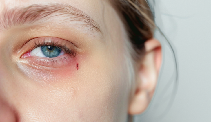
Periorbital cellulitis, also known as preseptal cellulitis, is an infection in the skin and soft tissues around the eye, in front of a structure called the orbital septum. Despite its rare serious complications, it can look very similar to the more severe condition called orbital cellulitis, an infection behind the orbital septum. Periorbital cellulitis mainly affects children and is mainly caused by injuries or sinus infections. It often results in swelling and fluid build-up in one eyelid.
However, orbital cellulitis symptoms can be similar but also include eye indications like bulging eyes, eye pain, blurred vision, and limited eye movement. It’s crucial to distinguish between the two conditions because their treatments differ. Diagnosing these conditions involves a medical examination and a CT scan of the orbits (eye sockets) and sinuses.
When it comes to periorbital cellulitis, the treatment focuses on antibiotics that fight against the most common bacteria involved in these cases, Staphylococcus aureus, and the Streptococcus species. In most cases, proper antibiotic treatment leads to the resolution of periorbital cellulitis within five to seven days.
What Causes Periorbital Cellulitis (Infection Around Eye)?
Periorbital cellulitis, a type of eye infection, often happens because of a nearby infection spreading like sinus infections or an infection occurring after an injury. To understand how this infection spreads, it’s helpful to know about the structures surrounding the eyes. The eye socket, also known as the orbit, is lined with a tough tissue called the periosteum and is surrounded by air-filled spaces known as sinuses. The different types of sinuses include the frontal sinus above the eye, the ethmoid sinus on the inner side, and the maxillary sinus below the eye. The most common type of sinus infection leading to the periorbital or orbital cellulitis is acute ethmoiditis.
This infection from the ethmoid sinus spreads quickly because the only thing separating it from the eye socket is a thin layer, called the lamina papyracea. This thin layer has holes and gaps that allow the passage of nerves and blood vessels, making it easy for the infection to spread from the sinuses to the areas around and inside the eye socket.
The orbital septum, a thin skin-like structure attaching to the periosteum, determines whether the infection is around the eye, known as periorbital, or inside the eye socket, known as orbital. Generally, infections around the eye don’t cause severe problems but can sometimes progress into an infection inside the eye socket. Misdiagnosis of an infection inside the eye socket as an infection around the eye is common, leading to the wrong treatment. The infection can also spread via the veins in the face, which don’t have valves, allowing a free flow of blood and thus the spread of infection into and beyond the eye socket. The veins above the eye and the eye’s blood vessels directly drain into a space in the brain called the cavernous sinus, increasing the risk of infection spreading inside the brain. As the infection spreads and goes untreated, it can become severe and lead to complications including blindness, and brain-related problems.
The Chandler classification is a system used to group severity levels of eye and eye socket infections to help provide the right treatment. Although not a measure of disease progression, it’s useful for diagnosis, especially due to similarities in how different conditions can present. The classification groups are: 1) Group 1 pre-septal cellulitis (infection around the eye), 2) Group 2 orbital cellulitis (infection inside the eye socket), 3) Group 3 sub-periosteal abscess (pus-filled pocket below the periosteum), 4) Group 4 orbital abscess (pus-filled pocket inside the eye socket), and 5) Group 5 cavernous sinus thrombosis (blood clot in the cavernous sinus, a brain structure).
Risk Factors and Frequency for Periorbital Cellulitis (Infection Around Eye)
Periorbital cellulitis is a condition that can affect people of any age, but it tends to be particularly prevalent among children. It occurs more frequently than orbital cellulitis. Some research suggests that if periorbital or orbital cellulitis leads to complications in the brain, the mortality rate can range from 5% to 25%.
Signs and Symptoms of Periorbital Cellulitis (Infection Around Eye)
Periorbital cellulitis is a condition that can usually be assessed based on the patient’s medical history and a physical examination. It often follows a sinus or respiratory infection, trauma, local infection or insect bites. Symptoms include eye complaints, fever, and swelling around the eye area.
This condition is difficult to distinguish from another called orbital cellulitis. They both show up with similar features, like redness and swelling around the eyes. However, periorbital cellulitis typically only affects one side and doesn’t involve deep eye tissues or muscles, so the patient’s vision or eye movements are usually normal.
On the other hand, orbital cellulitis is more serious as it affects areas behind the eye, causing symptoms such as bulging eyes, limited eye movement and potential vision loss, in addition to redness and swelling. Though fever is usually absent with periorbital cellulitis, some cases can present with fever, teary eyes, and discharge.
Periorbital cellulitis can also be confused with conditions like allergic reactions, trauma without cellulitis, orbital tumors, eye deformities, or eye infections. If not treated properly, it can lead to severe conditions including orbital cellulitis or even brain sinus thrombosis, which is a blood clot in the brain.
The Chandler classification system lists these complications in five groups:
- Group 1 – Preseptal cellulitis, which affects the eyelid and surrounding structures
- Group 2 – Orbital cellulitis, a bacterial infection in the eye causing swelling and potential vision loss
- Group 3 – Subperiosteal abscess, a pus collection between the eye wall and surrounding structures, potentially causing a displaced eye
- Group 4 – Orbital abscess, a separate pus collection in the eye tissue leading to serious vision impairment
- Group 5 – Cavernous sinus thrombosis, a blood clot in the brain that can affect the other eye and potentially lead to meningitis in severe cases
Although these groups aren’t always connected, they share similar symptoms and should be considered when evaluating a patient with eye complaints.
Testing for Periorbital Cellulitis (Infection Around Eye)
Periorbital cellulitis, an infection of the eyelid and skin around the eye, is usually diagnosed based on symptoms and a doctor’s physical evaluation. Sometimes, it can be hard to tell it apart from a more severe infection called orbital cellulitis. A CT scan- an imaging technique that provides detailed pictures of the inside of the body- can help differentiate between the two. This scan also allows doctors to check how far the infection has spread.
If there is still uncertainty, patients are generally treated as though they have orbital cellulitis just to be safe. When doctors think there might be an abscess (a pocket of infection), they’ll use a CT scan of the head to check if the infection has reached the brain. A CT scan might also be done if the patient’s eyelid is very swollen, they have a fever, a high white blood cell count, or if there’s no improvement after 24 hours of taking the right antibiotics.
In a CT scan of periorbital cellulitis, expect to see swelling of the eyelid, but no pushing forward of the eye, no spread of infection to the fat around the eye, and no involvement of the muscles used in eye movements. In a study, about 41% of people with periorbital cellulitis showed signs of sinusitis (inflammation of the sinuses) on their CT scan.
Blood tests aren’t typically part of the diagnosis process for periorbital cellulitis. They can be hard to perform on patients with this condition and usually don’t provide new information.
Treatment Options for Periorbital Cellulitis (Infection Around Eye)
The way we treat periorbital cellulitis, an infection of the skin and tissue around the eyes, depends on how severe the condition is and the age of the patient. The most common treatment is antibiotics, and these are chosen to fight against certain bacteria, such as S. aureus, the Streptococcus species, and anaerobes.
If a patient is over a year old and only has mild symptoms, they can be treated at home with oral antibiotics. But babies less than a year old, or older patients with severe symptoms, need to be admitted to hospital for treatment.
The types of antibiotics we use have changed. In the past, amoxicillin-clavulanic acid, cefpodoxime or cefdinir were common choices. But the rise of MRSA, a type of bacteria that’s resistant to many antibiotics, has made us rethink our approach. To protect against MRSA, we might recommend treatments like Trimethoprim-sulfamethoxazole (TMP-SMX), Clindamycin, or Doxycycline. However, TMP-SMX and Doxycycline don’t work against group A Streptococcus bacteria and we don’t usually give doxycycline to children under eight. That’s why the current recommendation is to use Clindamycin or TMP-SMX along with Amoxicillin-clavulanic acid, Cefpodoxime or Cefdinir. In some cases, where a patient has not been immunized against H.influenzae, we might recommend an antibiotic from the beta-lactam family. Typically, the patient will stay on antibiotics for five to seven days, unless the infection lasts longer.
Usually, antibiotics work quickly and completely. If someone doesn’t feel better after one or two days of treatment at home, they should be admitted to the hospital. There, they might receive a wider range of antibiotics, have a CT scan, or be considered for surgery to drain the infection. Steroids aren’t usually given, because there’s no evidence to suggest they would prevent the cellulitis from coming back or developing complications.
Surgery is necessary for patients in the severe stages of the condition, according to the Chandler classification system. If there is any doubt about whether a patient has periorbital or more serious orbital cellulitis, even after a CT scan, it’s best to treat the situation as if it’s orbital cellulitis.
What else can Periorbital Cellulitis (Infection Around Eye) be?
These are some conditions that doctors may examine if someone has specific eye or facial symptoms:
- Swelling, typically around the eyes or lips (Angioedema)
- Insect bites
- A blood clot in cavernous sinuses located behind the eyes (Cavernous sinus thrombosis)
- A lump in the eyelid caused by a swollen oil gland (Chalazion)
- Infections of the eyelash follicle and oil gland (Internal and External Hordeola)
- A type of fungal infection that typically affects the sinuses or the lungs (Mucormycosis)
- A kidney disorder causing excessive protein in urine and low protein levels in the blood (Nephrotic Syndrome)
- An infection that causes inflammation and can spread around the eye (Orbital Cellulitis)
- A rare complication of sinusitis where an abscess forms on the forehead (Pott’s Puffy Tumor)
- A severe skin infection that develops rapidly around the eyes (Periorbital Necrotizing Fasciitis)
It’s important for physicians to look into these possibilities carefully and carry out relevant tests to pinpoint the actual cause of the symptoms.


