What is Punctal Stenosis?
Punctal stenosis is a condition where the external opening of the lacrimal canaliculus, or tear duct, becomes narrow or blocked. It’s different from punctal agenesis, a condition you’re born with. Sometimes, punctal stenosis can be linked to a blockage in a shared canal in the tear duct system. Most people with this condition experience excessive tearing. It’s quite common, affecting anywhere from 8% to 54.3% of the population.
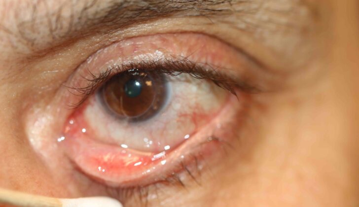
For a little context, the tear duct openings, known as puncta, are found on the inner corner of your eyelid edge. There’s one at the top and another at the bottom, and the top one is slightly toward the middle compared to the lower one. When you close your eyes, they meet. These openings allow tears to enter your tear duct system which includes the lacrimal duct, the lacrimal sac and the nasolacrimal duct. The puncta sit on small, bump-like structures that are surrounded by muscle fibers and a fibrous ring. The typical size of each puncta ranges from approximately 0.2-0.5mm. Punctal stenosis is diagnosed when the puncta is smaller than 0.3mm, or when a 26G cannula (a thin tube used in medical procedures) can’t enter the puncta without making it bigger.
Depending on the shape of the external puncta, there are four types of punctal stenosis:
- Membranous (makes up 31% of cases)
- Slit type (makes up 13% of cases)
- Horseshoe (makes up 31% of cases)
- Pinpoint (makes up 32% of cases)
What Causes Punctal Stenosis?
Punctal stenosis, a narrowing of part of the eye that helps drain tears, can be caused by many factors. The most common trigger is chronic blepharitis, a long-term inflammation of the eyelids, which causes about 45% of the cases. Sometimes, the exact cause isn’t known (making up 21% of the cases), while in other instances, punctal stenosis can be caused by ectropion (23%), a condition where the eyelid turns outwards, or by certain medications (5%).
Certain infections of the eyelids, like herpes simplex and trachoma, add to the risk of developing punctal stenosis. Inflammatory conditions such as benign mucous membrane pemphigoid disease and Stevens-Johnson syndrome can cause punctal stenosis as well.
The use of certain eye drop medications, used to treat glaucoma like timolol, latanoprost, betaxolol, dipivefrin hydrochloride, echothiophate iodide, and pilocarpine, can also increase the risk for punctal stenosis. Certain chemotherapy drugs used systemically (which means they travel throughout the body), specifically 5-fluorouracil and docetaxel, are also known to increase the risk for this condition.
Risk Factors and Frequency for Punctal Stenosis
Punctal stenosis, a condition where the tear ducts in the eyes narrow or become blocked, becomes more common as people get older. A study found that over half (54.3%) of patients visiting a general outpatient clinic had this condition. However, it’s important to remember that not everyone diagnosed with punctal stenosis will have symptoms. The average age of patients with this condition is around 69.4 years, and they can be anywhere between 39 and 90 years old. The condition does not seem to favor any particular gender or racial group.
Signs and Symptoms of Punctal Stenosis
Punctal stenosis is a condition causing excessive tear production and discomfort in the eyes, often resulting in red, watery eyes. It’s usually linked to chronic inflammation and may cause patients to have mixed seborrheic and ulcerative blepharitis, leading to uncomfortable symptoms.
A type of examination known as a biomicroscopic examination is used to evaluate the condition and categorize punctal stenosis into different grades, which show its severity. The grading system involves assessing the eyelid margin, punctal orifice (the tear duct opening), and tear meniscus (the thin layer of tears covering the surface of the eye).
- Grade 0: No punctum or tear duct opening can be identified
- Grade 1: Papilla (part of the tear duct) is covered with a membrane or fibrosis (scar tissue)
- Grade 2: Smaller than normal, but recognizable punctum
- Grade 3: Normal, easily recognized
- Grade 4: < 2mm in size
- Grade 5: > 2mm in size, larger than usual punctum
To confirm the diagnosis, probing and irrigation (flushing out the tear ducts) are conducted. Obstruction in the tear duct system is marked by a soft resistance during probing. The lower lacrimal (tear duct) system is assessed with the help of normal saline and a lacrimal cannula. In case of any obstruction, regurgitation of the saline solution through the punctum will be noticed.
While assessing a patient for punctal stenosis, it is important to note the resting position of the lower punctum as it is not normally visible under the biomicroscope. In normal cases, the punctum points backwards in the medial tear lake. If the lower punctum is pointing upwards or outwards, it could signal lower eyelid laxity and ectropion, even if the lower eyelid appears normal on examination. If left uncorrected, these conditions can lead to a gradual stenosis or narrowing of the punctum.
Testing for Punctal Stenosis
The process of identifying a condition generally involves taking a look at your medical history, conducting a dye disappearance test, and carrying out a ‘slit-lamp examination’. This diagnosis doesn’t usually require any laboratory testing.
The dye disappearance test is first on the checklist, and it’s done before dilation. A particular solution, called a 2% fluorescein, is inserted. After a window of 3 to 5 minutes, doctors will look at the tear meniscus, which is the little bit of tear fluid that sits at the edge of your eye – this is done to see if any dye still remains. If there’s punctal stenosis, a condition which involves the narrowing of the tear duct openings on your eyelids (called puncta), this remaining dye will still be noticeable even after 5 minutes.
The next step is punctal dilation. What does this mean? Well, the puncta in your eyes will be expanded using a tool called a Nettleship dilater. To get it ready, this dilater is lubricated with ointment.
Following this, two other procedures are performed – canalicular probing and nasolacrimal duct irrigation. Both are carried out under topical anesthesia (a local numbing agent). To dilate the punctum, a punctal finder is used. Then, doctors insert a canalicular probe. Depending on the ‘stop’ you notice – a soft stop signals canalicular obstruction, while a hard stop symbolizes a patent canalicular system, which is a clear pathway for tears to drain.
Irrigation is the next step; this is where a 5ml normal saline solution is used for rinsing. If all goes well, the fluid will flow freely down into your nose or nasopharynx, without any reflux or backflow.
After dilation, the disappearance of the fluorescein dye is examined. If the tear flow goes back to normal after dilation, the dye would disappear in under 3 minutes.
Treatment Options for Punctal Stenosis
Punctal stenosis is a condition where the opening that drains tears from your eyes, known as the “punctum”, becomes narrow. There are several ways to treat it. The main goal of these treatments is to widen this opening and improve tear drainage, while still keeping the tear pump working properly. However, doctors have not yet agreed on the best treatment technique.
Here are some of the treatment options:
Placing a perforated punctal plug: This involves inserting a special plug into the punctum, leaving it there for around two months. According to one study, this method improved symptoms such as excessive tearing in 87% of patients. However, more research is needed to understand the long-term success of this procedure.
Punctoplasty: This is a process that widens the punctum using a dilator. Although it may work initially, it typically does not provide a permanent solution. There are several types of this procedure, such as:
One-snip punctoplasty: This involves making one vertical cut along the tear duct with a special knife to open it up. However, this method often fails because the wound closes back up over time.
Three-snip punctoplasty: This method involves making three separate incisions in the shape of a triangle at the back of the punctum. According to studies, this method has a high success rate – up to 86% for reducing excessive tearing. However, some patients continue to have tearing issues despite this procedure.
A variation of the three-snip punctoplasty called the rectangular three-snip punctoplasty is used to help maintain the shape and function of the punctum and tear duct system. This modification has improved the treatment’s success rate to 94%.
Stent cannulation: A small tube or stent is inserted into the punctum after it has been dilated. One study found this method relieved symptoms in 82% of patients.
Wedge punctoplasty: In this technique, a small wedge-shaped portion of the punctum and tear duct is removed. This has shown promise with 95% of patients having an open punctum and 92% experiencing relief from symptoms.
Punch punctoplasty: Similar to wedge punctoplasty, a punch is used to remove a segment of the punctum. This method had a 94% success rate.
Use of mitomycin C: This is a medication that can be used before or after other procedures. It has been shown to effectively help treat punctal stenosis.
Balloon dilation: In this procedure, a small balloon is used to widen the narrow opening. Study results are mixed, however, and this technique is generally more effective in treating other conditions, such as nasolacrimal duct obstruction, which can affect tear drainage.
What else can Punctal Stenosis be?
Epiphora, also known as excessive tearing, can happen due to several causes. They include:
- Dry eye and reflex tearing
- Canalicular obstruction (blockage in tear drainage canal)
- Nasolacrimal obstruction (blockage in the tear drain pipe near the nose)
- Congenital glaucoma (a rare type of glaucoma present at birth)
- Ectropion without punctal stenosis (lower eyelid turning outwards without narrowing of tear duct opening)
What to expect with Punctal Stenosis
In most cases, patients have a good outlook. However, a small number of these patients might experience a return of symptoms even after surgery.
Possible Complications When Diagnosed with Punctal Stenosis
Punctal stenosis often presents two main difficulties, namely canalicular fibrosis (a hardening or thickening process affecting 46% of cases) and restenosis (the re-narrowing of your eye’s tear duct). To deal with restenosis, doctors typically employ the rectangular three-snip punctoplasty method. On the other hand, to manage canalicular fibrosis, a procedure called mini-Monoka punctocanaliculoplasty (MMPC), which is usually preceded by a widening of the punctum (the eye’s tear duct), is used.
Key Problems and Solutions:
- Canalicular Fibrosis – Managed through Mini-Monoka Punctocanaliculoplasty (MMPC).
- Restenosis – Rectangular three-snip punctoplasty technique is effective for this.
Preventing Punctal Stenosis
Punctal stenosis, a condition where the small openings (puncta) in the eyelid that drain tears become narrow, often leads to excessive tearing. Many people with this condition may notice their vision gets blurry due to the increased tear film covering their eyes. If you are experiencing this, it’s advisable to have your eyesight checked. It’s also suggested that you avoid driving until your vision is clear, and to inform the authorities who issue your driving license about your condition.


