What is Retinopathy of Prematurity?
Retinopathy of prematurity (ROP) is a disease that affects the blood vessels of the eye’s retina in infants who were born early and need oxygen treatment. This oxygen therapy can lead to unhealthy growth of vessels in the developing part of the eye which can seriously harm the eye, causing possible retina detachment and abnormal creases in the eye’s center, known as macular folds.
In order to prevent ROP, a leading cause of blindness in children, screening guidelines are in place to identify the disease early. Screenings are recommended based on how soon a baby was born and their birth weight, though other factors can increase the likelihood and seriousness of the disease. Early treatment using cold therapy, laser light treatment, and medicine to control blood vessel growth has helped improve vision in patients. However, early recognition through screening is essential for successful treatment.
Preventing ROP requires a team approach, starting even before the baby is born and continuing throughout childhood.
What Causes Retinopathy of Prematurity?
While still in the womb, the light-sensitive tissue lining the back of the eye (the retina) exists in a state of low oxygen or hypoxia. Higher levels of a substance called vascular endothelial growth factor (VEGF), helps create new blood vessels in the retina, a process known as angiogenesis.
The normal blood vessel development in the retina occurs in two stages: vasculogenesis, which is from the 14th week to the 21st week of pregnancy, and angiogenesis, starting at the 22nd week and continuing until the retina is fully equipped with blood vessels after the baby is born.
Since the nasal and temporal (sides) parts of the retina develop late in pregnancy, between 32 and 40 weeks respectively, babies born prematurely have incomplete blood vessel growth in these areas. The low oxygen condition that was promoting vessel growth is replaced with a state of high oxygen (hyperoxia), as many premature infants are exposed to extra, supplemental oxygen in addition to the normal amount in the air.
Risk Factors and Frequency for Retinopathy of Prematurity
The Early Treatment for Retinopathy of Prematurity study identified several key risk factors for Retinopathy of Prematurity (ROP), a disease that can affect the eyes of premature babies. This condition was prevalent in 68% of infants weighing less than 1251g. In 2010, ROP affected 184,700 infants globally, out of which 14.9 million were preterm. ROP led to blindness or severe visual impairment in 20,000 infants, while 12,300 developed mild to moderate visual impairment.
- The two most impactful risk factors are the baby’s gestational age (how far along the pregnancy was at the time of birth) and birth weight. Research involving over 4000 infants revealed that every 100g increase in birth weight and each additional week in gestational age reduce the chances of developing severe ROP by 27% and 19% respectively.
- The amount of oxygen an infant receives also plays a critical role. The combination of supplemental oxygen with the oxygen naturally present in the air results in higher oxygen levels in the body, leading to retinal ischemia and overgrowth of retinal vessels, causing ROP. The more oxygen delivered, the higher the risk of ROP. For instance, giving oxygen levels that result in a PO2 (a measure of oxygen in the blood) of ≥80mmHg for every 12 hours, doubles the risk of ROP. The duration of oxygen therapy is also a significant factor for severe ROP.
- Other potential risk factors include maternal high blood pressure during pregnancy, diabetes, medication use, maternal age and smoking, conception via fertility treatments, birth outside a study center hospital, and multiple births (like twins or triplets).
Signs and Symptoms of Retinopathy of Prematurity
Screening for a condition called ROP is incredibly important for babies who are born prematurely or are of low birth weight. According to guidelines from major health organizations like the American Academy of Ophthalmology, the American Academy of Pediatrics, and the American Association for Pediatric Ophthalmology and Strabismus, any infant born that is ≤30 weeks old or weighs ≤1500 grams at birth should be screened for ROP. In some cases, even larger babies could benefit from this screening. Typically, each infant needs about 3.4 follow-up exams.
Testing for Retinopathy of Prematurity
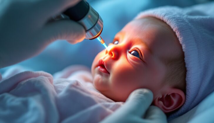
All babies born before 30 weeks of pregnancy or weighing less than 1500g (about 3.3 lbs) should have an eye exam to assess the size and shape of the back of their eyeball, which is known as the fundus. This type of exam should begin when the baby is 4 weeks old or around 30-31 weeks adjusted-age (based on the due date) and can be performed both if the baby is still in the hospital or outside.
Before the exam, eye drops containing 2.5% phenylephrine hydrochloride and 1% cyclopentolate or tropicamide are used to widen (or dilate) the pupils. These drops are given twice, 15 minutes apart. It’s important to be careful and prevent these drops from being absorbed into the body. In severe cases of Retinopathy of Prematurity (ROP, a potentially blinding disease), the pupils may not dilate properly. As infants may temporarily stop breathing or have slow heartbeats during the exam, a nurse should always be present.
A tool known as the Postnatal Growth and Retinopathy of Prematurity Screening Criteria (G-ROP) has been developed to avoid unnecessary eye exams while ensuring all cases of ROP are caught. This tool also considers how much weight the baby is gaining. It has shown excellent results in a study of almost 4000 babies, where it accurately identified all cases of ROP and reduced the number of eye exams by 30%.
Diagnosing ROP involves classifying the disease based on its location (zone) and severity (stage). The location is the part of the retina (the light-sensitive layer at the back of the eye) where the disease is most advanced. The retina is divided into three zones that are centered around the optic disc, which is where the optic nerve (which carries vision signals to the brain) enters the eye.
There are 5 stages of ROP severity. Stage 1, the least severe, is identified by a thin line distinguishing the blood vessel-filled part of the retina from the area without blood vessels. Stage 2 is marked by a raised ridge where this line is. In stage 3, abnormal blood vessels grow from this ridge into the jelly-like substance (vitreous) that fills the eye. Stage 4 involves part of the retina detaching, while stage 5 involves total detachment of the retina. If there is unusual dilation or twisting of the blood vessels in the back of the eye, the condition is classified as “Plus disease”, which indicates severe ROP.
Threshold ROP is a particular severity level of the disease, which is diagnosed when there are at least 5 continuous or 8 total hours in a day of Stage 3 ROP in zone 1 or 2, along with Plus disease.
Treatment Options for Retinopathy of Prematurity
Retinopathy of Prematurity (ROP), a potentially blinding eye disorder, doesn’t always need treatment as soon as it’s detected. The decision to treat depends on the severity and type of ROP. Type I ROP, which is a more severe form, usually needs treatment. In contrast, Type II ROP, which is less severe, can typically be monitored without immediate treatment.
If treatment is necessary, the most common approach is a surgical one. Initially, cryotherapy was used; this process involves freezing the parts of the eye affected by the disorder. However, this method required general anesthesia, was time-consuming, and had other possible complications.
Due to those drawbacks, laser treatment (photocoagulation) has become the go-to for treating ROP. This approach has led to fewer complications and has been a significant improvement over cryotherapy.
Another possible treatment uses anti-VEGF agents. These drugs work against a protein called VEGF which plays a role in ROP’s progression. One particular study tested the effects of anti-VEGF agent bevacizumab versus laser treatment with significant results. There were fewer cases of ROP recurrence in the group treated with bevacizumab compared to the laser-treated group. However, there are some concerns about long-term side effects, including late reactivation of the disorder, even years after birth.
Anti-VEGF treatment does have benefits, especially as VEGF can be crucial for healthy development in infants. However, a study in 2019 found higher mortality and poorer cognitive outcomes in infants treated with bevacizumab than with other treatments.
If ROP progresses to the point where the retina detaches, the risk for poor vision increases significantly. At these advanced stages, surgery becomes necessary. The most common procedures at this point are scleral buckling or lens sparing vitrectomy. For the most severe cases, other methods such as lensectomy with vitrectomy or open sky vitrectomy are used. Success is typically measured by whether the retina is reattached after surgery, and the improvement in the child’s vision, which can take time and may vary.
What else can Retinopathy of Prematurity be?
Some conditions that may present similar symptoms to what you’re experiencing include:
- Familial exudative vitreoretinopathy
- Persistent fetal vasculature
- Incontinentia pigmenti
What to expect with Retinopathy of Prematurity
Advances in medicine have made treatments more effective but even then, some patients undergoing treatment may still experience reduced vision quality. One of the main aims of eye treatment is to prevent unfavorable outcomes that could affect the nerve layer at the back of the eye. These outcomes, determined by a study known as the ETROP study, may include harmful folds, detachment of the nerve layer, the development of tissue or mass behind the iris, or the need for eye surgery.
A follow-up to this study found that after two years, adverse outcomes had dropped to 9.1% in patients with high-risk eyes that were treated early.
Further study, a six-year follow-up, determined that 34.6% of children had visual clarity of 20/40 or better, meaning they could see clearly at 20 feet what should normally be seen at 40 feet. Around 40.3% had acuity greater than 20/40 and less than 20/200, indicating their vision was not as clear. And unfortunately, 23.7% had vision worse than 20/200, which includes just light perception and blindness.
Possible Complications When Diagnosed with Retinopathy of Prematurity
The most common issue resulting from Retinopathy of Prematurity (ROP) is retinal detachment, which often leads to poor vision. Macular folds, folds in the central area of the retina, are another common complication. As a child grows, issues with their visual clarity can persist, with shortsightedness being the most commonly seen long-term effect. Other possible complications include glaucoma, lazy eye, cataracts, and misalignment of the eyes.
A child with poor vision due to Retinopathy of Prematurity is more likely to face a range of developmental, educational, and social challenges. An extended study of children, who were born weighing less than 1251 grams and had severe ROP, showed that by ages 5 and 8, children who had significant visual problems were considerably more likely to have developmental disabilities, epilepsy, special educational needs, and were often performing below their grade level at school.
Key Health Concerns:
- Retinal detachment, leading to severe vision loss
- Macular folds
- Persistent vision problems leading into adulthood, with shortsightedness being common
- Other possible complications: glaucoma, lazy eye, cataracts, misalignment of the eyes
- Developmental, educational, and social challenges in cases of severe vision loss
- Increased chances of developmental disabilities, epilepsy, and special educational needs
- Lagging academic performance
Recovery from Retinopathy of Prematurity
After surgery, premature babies often need assisted breathing through devices like ventilators, feeding through an intravenous line, and antibiotics. They are also closely monitored using pulse oximetry (a tool to measure oxygen levels in the blood), neurological screenings, ultrasound screenings, and checks for conditions like bronchopulmonary dysplasia (a lung condition) and patent ductus arteriosus (a heart problem that can affect blood flow).
As part of a long-term treatment for a vision disorder called amblyopia, glasses might be used, or the stronger eye may be patched for 6 to 12 hours a day. Another option is using a drug-based approach, in which a medication like atropine is used to intentionally blur the vision in the stronger eye. This encourages the weaker eye to work harder and get stronger.
Preventing Retinopathy of Prematurity
Providing oxygen to babies is very important in preventing Retinopathy of Prematurity (ROP), which is an eye disease that occurs in premature babies. In the past, medical experts believed that maintaining an oxygen saturation (SpO2; amount of oxygen in the blood) at a lower target could decrease the chances of ROP without increasing the risk of death.
In 2001, a study seemed to support this theory. However, to better determine the optimal level of oxygen in the blood for premature babies, a global network of newborn specialists called the Neonatal Oxygenation Prospective Meta-analysis (NeOProM) was formed. These professionals conducted five studies comparing a lower oxygen level (85% to 89%) with the risk of death or major disability in infants born before 28 weeks.
The results, however, showed that having too little oxygen increases the risk of death. They suggested that the ideal oxygen target should be between 90% to 94%.
Given these results, specialists agreed that keeping the oxygen level lower is not the best solution, and began to explore other approaches. These include better nutrition, managing infections and pain, maintaining the right temperature, and supporting the baby’s development. Globally, it has been noticed that addressing ROP more successfully is linked to financial investment in these areas. These best practices were outlined in 2019 by Darlow and colleagues, and continue to guide neonatal care today.


