What is Uveitis?
Generally, uveitis is a medical term used to describe the inflammation in the uvea, a part of the eye that includes the iris, ciliary body, and the choroid. However, inflammation can affect any part of the eye not just the uvea. There are four types of uveitis; anterior, intermediate, posterior, and panuveitis, named according to the main part of the eye that’s inflamed.
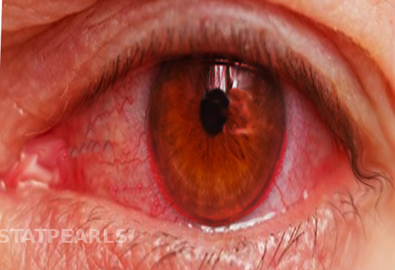
The symptoms and effects of uveitis can vary widely, from pain and redness in the eye (also known as conjunctival injection), to total loss of vision. Anterior uveitis mainly affects the front part of the eye, while intermediate uveitis involves inflammation of the vitreous cavity and pars plana, terms used to describe parts of the eye. Posterior uveitis impacts the retina and choroid, which are at the back part of the eye. Inflammation in panuveitis affects all parts of the eye.
The different types of uveitis based on where they occur in the eye are:
- Anterior
- Intermediate
- Posterior
- Panuveitis
What Causes Uveitis?
Uveitis, a condition causing inflammation in the eye, is often unexplained but can be linked to injuries, inflammation, and infections. Sometimes, patients might also have other symptoms or illnesses that suggest the issue extends beyond the eye. Roughly 48 to 70% of uveitis cases happen for unknown reasons.
There are several health conditions that often coincide with a form of uveitis called anterior uveitis, including HLA-B27-associated conditions, juvenile arthritis, inflammatory bowel disease, sarcoidosis (a disease that leads to inflammation in various body organs), Behcet’s disease, or tubulo-interstitial nephritis (an inflammation of the kidneys). Sarcoidosis, tubulo-interstitial nephritis, and multiple sclerosis (a disease causing nerve damage in the brain and spinal cord) are typically linked with another form of uveitis known as intermediate uveitis. Similarly, posterior uveitis (a rarer form of uveitis) could be associated with conditions such as Vogt-Koyanagi-Harada syndrome, leukemia (a type of blood cancer), lupus, Behcet’s disease, and multiple sclerosis.
Behcet’s disease can also cause inflammation across your whole body, and might result in pan-uveitis, which is an inflammation of all layers of the uvea in the eye.
Additionally, infections are believed to cause about 20% of all cases of uveitis, but the exact infection causing it can differ depending on where you live. These infections could be viral (such as HSV, VZV, CMV), bacterial (like endophthalmitis, syphilis, tuberculosis), or caused by parasites or worms (such as toxoplasmosis, Lyme Disease, toxocara, Bartonella sp. or other uncommon infections).
Risk Factors and Frequency for Uveitis
Uveitis is a condition that can strike anyone, regardless of age and where they live. A study conducted in 2006 and 2007 revealed that the occurrence of uveitis was 24.9 cases for every 100,000 people. During these same years, there were 57.5 and 58 instances of uveitis per 100,000 people respectively. The occurrence of uveitis was the same for both men and women, but more women were found with the condition. The most common type of uveitis is “anterior uveitis”, which makes up about 50% of all cases, while “posterior uveitis” is less common. If uveitis is left untreated and the inflammation continues, it could be to blame for roughly 10% of blindness cases in the United States.
- Uveitis can affect anyone, regardless of their age or where they’re from.
- In 2006 and 2007, the occurrence of uveitis was 24.9 cases for every 100,000 people.
- The amount of people found to have uveitis during these years was 57.5 and 58 per 100,000 people respectively.
- Both men and women had the same chance of getting uveitis, but more women had the condition.
- The most common form of uveitis is anterior uveitis, which is 50% of all cases. Posterior uveitis is the least common.
- If uveitis and the resulting inflammation are not treated, it could cause about 10% of the cases of blindness in the United States.
Signs and Symptoms of Uveitis
Diagnosing conditions like uveitis requires a comprehensive past medical, family, and ophthalmic (eye-related) history. It’s also helpful to review all body systems as some systemic diseases can affect the eyes. Depending on which part of the eye is inflamed, symptoms can vary. Acute anterior uveitis, which affects the front part of the eye, can cause pain, blurred vision, sensitivity to light, and redness around the cornea. The pupil may be smaller or irregularly shaped compared to the healthy eye due to changes in the lens caused by inflammation. Intermediate uveitis, typically doesn’t cause pain, but can lead to blurred vision and floaters. Posterior uveitis affects the back of the eye and often causes vision loss. Panuveitis, where multiple areas of the eye are inflamed at once, can cause a combination of all these symptoms.
An eye exam can include tests for visual acuity, an in-depth examination using a special microscope called a slit lamp, measurement of inner eye pressures, and an exam with dilated pupils. If a patient forgets their glasses or is thought to have uncorrected vision problems, a simple method using a pinhole in a cup can be used for a quick visual acuity test. Specific signs during the slit lamp exam can include diagnosing redness around the cornea, presence of white blood cells in the front chamber of the eye, and changes in the clarity of the fluid within the chamber. Any unexpected findings might signify a serious condition that requires ophthalmologist evaluation.
Other symptoms like changes in the lens, growth of blood vessels on the iris surface, and collections of calcium on the cornea may indicate a longstanding inflammation or previous event, which can help in diagnosing the disease. Inflammation cells can be seen in the vitreous (the jelly-like substance inside the eye) in cases of intermediate uveitis, and can even form clumps known as “snowballs”. Posterior uveitis is characterized by changes in the retina and choroid, the back parts of the eye, and symptoms depend on the nature of these changes.
The most common symptoms and signs for different types of uveitis are:
- Anterior uveitis: Red, painful eye and changes in the front chamber of the eye
- Intermediate uveitis: Increased presence of floaters, decreased vision and changes in the vitreous and back of the eye
- Posterior uveitis: Decreased vision, changes in field of view, and changes in the retina and choroid
- Panuveitis: Red, painful eye, severely reduced vision, floaters and changes in multiple parts of the eye
Testing for Uveitis
If you have an eye condition known as anterior uveitis, your doctor might not need to do any lab tests during the first episode. However, if it comes back, or if you have other types of uveitis such as intermediate, posterior, or panuveitis, more tests might be needed. This is because these types of uveitis can be linked to other diseases that affect the whole body, and they increase the risk of permanent visual damage.
Further tests might be necessary to rule out any underlying autoimmune or infectious diseases if recurrent anterior uveitis is experienced.
For anterior uveitis, a range of tests might be considered by your doctor. These might include HLA-B27 typing, FTA/RPR test, serum ACE/lysozyme test, a chest x-ray, and the quantiferon gold test. If you have intermediate uveitis, which tests are best will depend on other symptoms and risk factors. However, they are likely to include FTA/RPR, a chest X-ray, ACE/lysozyme, and quant gold. In some cases, your doctor might consider brain imaging if there’s a chance you might have multiple sclerosis.
A disease called syphilis, which can affect all parts of the body, is often referred to as the “great imposter” because its symptoms can resemble many other diseases. It should be taken into account in all cases of uveitis. Special medical textbooks provide a detailed list of possible diseases that need to be considered.
In rare cases (less than 0.5% of cases at specialised medical centres), uveitis can be caused by the medications a patient is taking. One example includes the antiviral drug, cidofovir, which can cause anterior uveitis. Recent cancer drugs, called checkpoint inhibitors, can also cause different types of uveitis.
Your doctor will aim to rule out syphilis, sarcoidosis (a disease that causes inflammation in different parts of the body), and tuberculosis in every case of uveitis. They are likely to recommend lab tests such as FTA AND RPR, ACE/lysozyme test, chest X-ray, and a quantiferon gold or PPD test.
Treatment Options for Uveitis
The main goal of uveitis treatment is to reduce inflammation and pain. This is typically done using steroids and medicated eye drops. Additional treatments may be necessary if other conditions are present. For example, if the uveitis is caused by herpes, antiviral medication is required. If it’s a result of a parasite, an antibiotic called bactrim might be used.
In case of chronic, non-infectious uveitis or when it’s linked with diseases causing inflammation throughout the body, more potent medications may be needed. These include drugs that lessen your body’s immune response. Some examples are methotrexate, azathioprine, mycophenolate, cyclosporine, adalimumab, and infliximab. However, these drugs have potential side effects and should only be prescribed by a healthcare provider trained in their use.
Anterior uveitis, the most common form of the condition, is usually treated with topical steroids and cycloplegics (medicated eye drops). Steroids are used to reduce inflammation, but they should be used mindfully to avoid inflammation bouncing back when the dosage is reduced. Their use can also increase intraocular pressure (IOP), which can harm your eyesight. This is something your doctor will monitor closely.
Cycloplegics, like atropine or cyclogyl, are used to minimize pain. They work by preventing muscle spasms in the eye and can keep the iris (colored part of the eye) from sticking to the lens of your eye. These medications are typically applied twice a day until inflammation has reduced significantly.
If uveitis affects the middle or back of the eye or the entire eye (intermediate, posterior, and panuveitis), the treatment becomes more complicated and will be directed by eye specialists known as ophthalmologists. If possible, a uveitis specialist will guide your care.
What else can Uveitis be?
There can be several reasons why your eye is red, painful, and has variations in vision, which are commonly seen by doctors in primary care or emergency rooms. When a patient comes in with these symptoms, doctors are trained to think about few different potential causes since many share similar symptoms. Doctors will use specific tools and skills to diagnose the condition, which may include testing your vision clarity, checking eye pressure, using a special microscope called a slit lamp, or using a special dye for examination.
One potential cause of a red, painful eye could be Conjunctivitis, also known as pink eye. This often comes with sensitivity to light and possibly a discharge from the eye. The type of discharge can help diagnose the cause: bacterial conjunctivitis presents with a thick, mucus-like discharge, while viral or allergic conjunctivitis usually cause a thinner, watery discharge. Eye swelling and inflamed tissue on the inside of your eyelid are clues that can help a doctor suspect conjunctivitis.
Another possible condition is Keratitis, a serious inflammation of the cornea (the clear, round dome covering the eye’s iris and pupil). This often includes eye pain, light sensitivity, and injection around the cornea. Those who wear contacts frequently face this issue. Keratitis can make your cornea appear cloudy, which is not typical for uveitis, another eye condition. In keratitis, the pupil is usually normal, while in uveitis it might be constricted.
Acute angle closure glaucoma is a condition that can cause a red, painful eye and a change in vision. Patients may experience pain on one side of the head, feel nauseous and even vomit. The affected eye’s pupil is usually mid-sized and reacts slower to light. The pressure in the eye will also be significantly increased.
Corneal abrasion can also cause severe eye pain, sensitivity to light, and tearing. Generally, the patient will have a history of some injury to the eye. This condition is known to respond well to local anesthesia, which is not typical for uveitis.
Finally, panuveitis could cause severe inflammation resulting in a red, painful eye. This condition requires immediate attention from an eye specialist. If eye inflammation is accompanied by alarmingly high intraocular pressure, it could be a sign of acute retinal necrosis or endophthalmitis, both of which are ocular emergencies that require immediate treatment.
Most common reasons for a red, painful eye:
- Anterior uveitis
- Keratitis
- Corneal abrasion
- Acute angle closure glaucoma
- Conjunctivitis
- Scleritis
What to expect with Uveitis
In most scenarios, the outlook for uveitis, an inflammation of the middle layer of the eye, is generally positive provided it is detected early and treated correctly. While a significant number of patients may develop complications related to their eyes, the right treatment and potentially, surgery, can significantly lessen the likelihood of permanent vision loss.
Determining the root cause of uveitis is also crucial, as some specific illnesses that can lead to uveitis are associated with significant sickness and even death. So, its always important to treat any underlying condition effectively.
Possible Complications When Diagnosed with Uveitis
Before starting any treatment, it’s important to correctly determine what the issue is and set up a plan for regularly monitoring it. In cases of anterior uveitis, a condition affecting the eye, the go-to treatment is usually topical corticosteroids. However, these can lead to increased pressure within the eye. As a result, patients need to have regular follow-ups with their eye doctor both to see if the condition is improving and to keep an eye on any pressure changes.
If the inflammation in the eye is not treated adequately or for long enough, it might cause harm to the eye and even lead to permanent vision loss. There are many potential complications, including cataracts, the sticking together of parts of the iris and lens, the formation of an abnormal membrane behind the retina, swelling in the back part of the eye, formation of abnormal deposits in the cornea, low eye pressure, development of glaucoma, and swelling of the optic nerve head.
It’s important to remember that each compartment of the eye has different forms of inflammation that should not be treated with medications either directly in the eye, around the eye, or taken orally before ruling out causes from an infection. This is because these treatments can potentially make the condition worsen and impact the vision further.
It is also suggested that if a person is suspected to have inflammation extending beyond the front portion of the eye, they should seek a specialized eye doctor for further evaluation and treatment.
Common Eye Issues and Complications:
- Increase in intraocular pressure
- Permanent vision loss due to unaddressed inflammation
- Various complications including cataracts
- Treatments can potentially worsen conditions
- Parts of the iris and lens sticking together
- Abnormal membrane behind retina
- Swelling in the back part of the eye
- Abnormal deposits in cornea
- Low eye pressure
- Development of glaucoma
- Swelling of the optic nerve head
Preventing Uveitis
It’s very important to explain to the patient about their condition and highlight the need to follow the treatment plan and attend follow-up appointments. Patients should be told about possible problems that could happen, including losing all of their vision. They should be clearly told when they need to seek immediate help, like if their pain gets worse or their vision changes, if they start seeing floating spots, or if they become sensitive to light.


