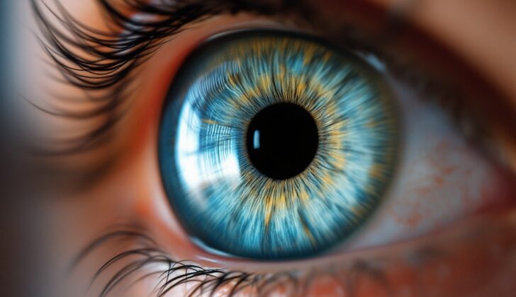
What is Megalocornea?
Megalocornea, also called by other names like anterior megalophthalmos, X-linked megalocornea, or macrocornea, is a rare birth defect that affects both eyes, where the clear front surface of the eye (the cornea) is larger than normal (greater than 12.5 to 13 mm in diameter) at birth. This doesn’t progress as the child grows but the child’s eyes have a deeper than normal anterior chamber (the space in the eye in front of the iris) but with normal eye pressure. The cornea in such cases often tends to be thinner than usual.
This defect is a part of disorders related to the development of the front part of the eye known as anterior segment dysgenesis. It has links with other conditions like Axenfeld-Rieger syndrome, Peters anomaly, primary congenital glaucoma, aniridia (absence of the iris), congenital hereditary endothelial dystrophy (a type of corneal dystrophy), and sclerocornea (part or all of the cornea becomes opaque and resembles the white part of the eye).
Two ways in which Megalocornea can present itself are when it occurs on its own with no other visible issues in the eyes or any other body system – this is referred to as primary megalocornea; or as part of other eye and body problems.
The focus in this context will be on primary congenital megalocornea and any possible simultaneous conditions and syndromes.
This condition was first identified in 1869, though only in the last ten years have we understood the genes that cause this disease. It is generally inherited through the X-chromosome, although other patterns of inheritance are also observed. The condition generally doesn’t have symptoms in childhood but might cause blurred vision due to irregularly shaped corneas (astigmatism).
In adulthood, those affected may experience early development of age-related eye cloudiness (cataract), usually between 30 and 50 years of age, glaucoma (a condition that damages your eye’s optic nerve), arcus juvenilis (a grey or white ring in the cornea), lens dislocation, and a mosaic pattern of different colors in the cornea. Other related conditions can include iris atrophy (loss of iris tissue), coloboma (missing tissue in or around the eye), abnormalities in the fibers that hold the lens in place, being able to see light through the iris, lens displacement, retinal detachment (where the retina falls off at the back of the eye), and different corneal size in the two eyes.
What Causes Megalocornea?
The cornea is the clear layer at the front of the eye and is crucial for vision. When it becomes enlarged, several different diseases or conditions could be at fault. It’s also possible that genetics and inheritance play a role in this. And sometimes, it’s not clear whether the person also has other genetic abnormalities, or if it’s just this one problem with the cornea.
One specific condition related to an enlarged cornea has been studied extensively, known as X-linked megalocornea. This condition is named so because it is linked to the X chromosome that we inherit from our parents. In 1991, scientists discovered that the gene affected in this condition is located at a specific region on the X chromosome, known as Xq13-q25. However, because that region is quite large, it wasn’t until 2012 that they pinpointed the exact gene responsible, called the Chordin-like 1 (CHRDL1) gene. Various changes or mutations in this gene have been linked to the typical features of X-linked megalocornea.
Aside from the X chromosome, other chromosomes can also play a role in causing an enlarged cornea. For example, disruptions in the 16p region of an autosomal chromosome (a type of chromosome that isn’t involved in determining sex) could lead to an enlarged cornea, even when no other birth defects or acquired conditions are present.
Risk Factors and Frequency for Megalocornea
Megalocornea, a condition often referred to as “rare” in medical reports, doesn’t really have specific numbers regarding how often it occurs. There are no studies that provide a count of how many people have this condition. However, other health issues that are linked with megalocornea may have their own facts and figures.
Signs and Symptoms of Megalocornea
Kids with a condition called megalocornea might not complain about their vision. Sometimes, doctors even miss the diagnosis during regular eye check-ups. Megalocornea is marked by a deeper-than-normal front part of the eye, but the length of the eye is normal overall. Because of this, the colored part of the eye (iris) and the lens are located further back than usual.
Those with the condition usually have wide, open angles between the iris and cornea. Their eye’s drainage system, the trabecular meshwork, might have a lot of pigment too. In some situations, the shape of the cornea might be different, allowing doctors to see these angle structures without special tools. In a specific type of megalocornea linked to the X chromosome, the cornea might appear globular, a feature that is unique to this type.
- Deep front part of the eye
- Normal eye length
- Iris and lens are located further back than usual
- Wide, open angles between iris and cornea
- Pigmented trabecular meshwork
- Possible different shape of the cornea
- Globular cornea in X-linked megalocornea
If megalocornea patients develop complications such as cataracts or retinal detachment, they would experience symptoms related to these issues such as blurry vision, glare, seeing flashes or floaters, and more.
Testing for Megalocornea
Congenital megalocornea, a condition where the front part of the eye (the cornea) is abnormally large, may not cause any symptoms for some people. Therefore, it might not be diagnosed until complications occur. Although CHRDL1, a gene associated with this condition, plays a role in retina development (the part of the eye that helps us see), individuals with CHRDL1 related megalocornea usually have normal eyesight and optic discs (the area in the back of the eye where the optic nerve connects).
Some tests, like the electroretinograms (a test that measures the electrical responses of different cells in the eyes), have shown dysfunction in the cone system (cells responsible for color and detailed vision) in some patients. Also, visual evoked potentials, which measure the electrical signals in the brain related to eye sight, may show some delays due to reduced white matter myelination (a process that covers nerve cells to ensure fast signal transmission). Despite this, patients’ cognitive abilities (thinking, learning, understanding) remain normal, or even slightly above average.
Therefore, diagnosing this condition can be difficult, especially in patients whose corneas are only slightly enlarged.
Given the many discovered mutations associated with this condition, and the likelihood that more exist, genetic testing across the entire coding region in the X chromosome (the part of our DNA that’s linked to this condition) is the most definitive way to confirm a diagnosis and provide the best care. Should other conditions or syndromes be suspected, additional genetic or diagnostic tests may be required.
Treatment Options for Megalocornea
Megalocornea is a condition where the corneas, which are the clear, dome-shaped surface of the eyes, are bigger than usual. Unfortunately, there is no cure or treatment for the enlarged corneas caused by this condition. This is due to the anatomy of the eye and the intricate nature of the issue.
Patients may experience complications which can make it challenging to manage. One example is cataract surgery, a common procedure for people with megalocornea. If the patient has a larger-than-usual ciliary ring and capsular bag (the parts of the eye where the lens sits), but a normal-sized lens, it may complicate the surgery. Using ultrasound technology can help measure the size of the capsular bag and assist in the process. However, these surgeries, despite their complexity, have been successfully performed, showing there’s hope for individuals with the condition. Techniques in cataract surgery have also improved over the years, with the use of a method called ‘phacoemulsification’ being quite effective.
Choosing the right power for the artificial lens to replace the natural one in cataract surgery can pose a challenge. This is because in normal eyes, some established formulas are used to calculate the power, but these don’t work well with the large structures associated with megalocornea. Some formulas may produce better results than others, and are encouraged to be used, as they account for the deeper anterior chamber (front part of the eye) and larger corneal curvature found in people with this condition. Still, it is tricky to land on the ideal power, and follow ups are necessary to monitor the success of choosing the power of the lens.
Different complications may arise due to larger structures in the eye requiring additional procedures. Standard artificial lenses may not sit correctly, and other methods like suturing to the iris, scleral fixation (anchoring the lens to the white part of the eye), or using a specific type of lens that is designed to be attached to the iris, may be needed. Some issues include potential wound leakage and low endothelial cell counts, which are the cells that line the inside of the cornea. Procedures like making incisions in the sclera (the white part of the eye) and stitching the wound may limit the impact on the cornea and potentially prevent low eye pressure after surgery.
While it’s clear the approach to surgery should be tailored to the specific needs of the patient, generally, the use of a particular type of artificial lens attached to the iris is recommended. This is seen as the best option to avoid further procedures and to ensure the best possible long-term visual outcomes for patients with megalocornea.
What else can Megalocornea be?
In infants with enlarged corneas (known as megalocornea), it can be hard to distinguish this from baby glaucoma. There are features that could help in making a diagnosis:
- An infant with megalocornea typically has normal eye pressure, a family history of the condition, and an intact inner layer of the cornea (Descemet’s membrane)
- On the other hand, babies with glaucoma usually have high eye pressure and cracks in Descemet’s membrane, they may also have a swollen cornea
- Signs specific to primary megalocornea such as pigment dispersion and iris transillumination are typically not seen in infantile glaucoma
- Other conditions causing eye enlargement like keratoglobus and megalophthalmos also need to be considered
There are also different syndromes or health problems that can be linked to megalocornea:
- Neuhäuser syndrome, also known as megalocornea-mental retardation syndrome, could show up as learning difficulties, seizures, low muscle tone and unique facial features
- Frank-Ter Haar syndrome affects multiple areas including the eyes, heart, and bones. It is commonly linked with deformities in the skeleton, face, and various birth defects of the heart
- Buphthalmus, or enlargement of the entire eyeball, can also occur alongside megalocornea. This is usually accompanied by glaucoma and a lack of family history with the disease
- Crouzon syndrome can lead to changes in the shape of the skull. It may lead to bulging eyes or shallow eye sockets
- Marfan syndrome a disease caused by a mutation in fibrillin, has been reportedly linked with megalocornea
- Other conditions that have features similar to Marfan syndrome might also have a connection to megalocornea
- Albinism, in some instances has been associated with megalocornea in addition to other health issues
- Ritscher-Schinzel syndrome presents with various eye, heart, and face anomalies. Megalocornea has been reported as the most common eye problem here
- Wolfram-like syndrome is a disease that typically presents with eye atrophy, diabetes, and gradual hearing loss from birth. Megalocornea could be one of the eye problems that come with it
- Lamellar ichthyosis is usually seen as a skin disorder, but there have been cases of it being associated with megalocornea
- Osteogenesis imperfecta, a collagen-based connective tissue disease, is commonly linked with ‘brittle’ bones and may have blue sclera. There has been one case where megalocornea was also related
What to expect with Megalocornea
The outlook for a patient with megalocornea, a condition where the cornea is larger than normal, largely depends on if it is linked to another disease or syndrome that comes with additional health problems. If someone has primary megalocornea, meaning they only have this condition and it’s not connected to anything else, the outlook is typically quite good. This is because existing conditions can often be addressed successfully with glasses, medication, or surgery.
Possible Complications When Diagnosed with Megalocornea
Megalocornea, a condition where the cornea is larger than usual, typically doesn’t cause issues in children, and they can usually see just fine. However, because their cornea is so large, it could cause a condition called astigmatism, which may make their vision blurry. There is a treatment for this called Photorefractive keratectomy which has been recommended.
On the other hand, the condition could also lead to cataracts forming earlier than usual. This is a complex eye surgery due to changes in the eye’s anatomy. Even so, the chances of success are high, and patients usually recover well.
Other eye issues that may develop include glaucoma, arcus juvenilis, lens subluxation, and mosaic corneal dystrophy.
Possible Complications:
- Astigmatism (due to enlarged cornea)
- Presenile cataract formation
- Glaucoma
- Arcus juvenilis
- Lens subluxation
- Mosaic corneal dystrophy
Preventing Megalocornea
Megalocornea at birth, also known as congenital megalocornea, can sometimes be tied to different genetic issues. It’s helpful for both the patient and, in the case of babies and young children, their parents, to be fully informed. Since most conditions that result in megalocornea are rare, specific tests – including tests for genetic abnormalities – may be needed to pinpoint the exact issue. But, to avoid unnecessary stress and costs, doctors should avoid extra tests if they’re not helpful. It’s also important that patients and their families are told about the possibility of further eye problems that might come up later in life. In many situations, it may be beneficial to receive genetic counseling to better understand the condition and its potential impacts.


