What is Osteogenesis Imperfecta (brittle bone disease)?
Osteogenesis imperfecta (OI), often referred to as brittle bone disease, is a genetic condition that affects the body’s connective tissues due to problems producing or processing a protein called type I collagen. This protein plays a vital role in strengthening our bones. That’s why individuals with OI are more prone to bone fractures and have lower bone density.
Additional symptoms of OI can include a blueish tint to the whites of the eyes (blue sclerae), issues with tooth development (dentinogenesis imperfecta), shortness in height, and hearing loss in adulthood. Some patients may experience heart valve issues and a condition where the part of the body that connects the heart and the aorta (the main blood vessel) becomes larger (aortic root dilation).
Less severe symptoms can also include increased flexibility (generalized laxity), frequent bruising, developing hernias, and excessive sweating. The range of OI’s symptoms can vary greatly from person to person. Some people have mild symptoms and may not even realize they have the condition, while others may have severe forms showing signs at birth with delicate ribs, a fragile skull, and long bone fractures that can sadly result in increased death rates shortly after birth.
What Causes Osteogenesis Imperfecta (brittle bone disease)?
Osteogenesis imperfecta, also known as OI, is a rare disease that is usually inherited. It often comes from changes in the genes known as COL1A1 and COL1A2. Research has recently found more gene changes that might be linked to OI.
The International Society of Skeletal Dysplasias has a list of types of OI based on how it is inherited and which genes are involved. Here are these types:
The first one is the Nondeforming OI (Type I). Inheritance of this type is generally AD which means it’s commonly inherited from one affected parent. The genes involved are COL1A1, COL1A2 and PLS3.
Perinatal (Type II) can be inherited in either an AD (one affected parent) or AR (both parents are carriers) mode. The genes involved are COL1A1, COL1A2, CRTAP, LEPRE1, PPIB, and BMP1.
For Progressively deforming (Type III), it can also be inherited via an AD or AR mode. It involves more genes; these are COL1A1, COL1A2, CRTAP, LEPRE1, PPIB, FKBP10, SERPINH1, SERINF1, and WNT1.
Moderate (Type IV) is similar to the prior, either inherited via an AD or AR mode. The genes which may be impacted are COL1A1, COL1A2, CRTAP, FKBP10, SP7, SERPINF1, WNT1, and TMEM38B.
Calcification of interosseous membrane or hypertrophic callus (Type V) typically follows an AD inheritance pattern. This OI type six involves a gene called IFITM5.
Worth to remember that AD means autosomal dominant inheritance where you can get the disease from one affected parent, while AR means autosomal recessive inheritance where both parents must be carriers for a child to be affected.
Risk Factors and Frequency for Osteogenesis Imperfecta (brittle bone disease)
Osteogenesis imperfecta, a rare condition, affects about 1 in 15,000 to 20,000 babies. Around the world, the condition’s first type shows up in about 2.35 to 4.7 out of every 100,000 people. The second type of the disease is found in about 1 to 1.4 out of every 100,000 babies. For the third and fourth types, there are fewer cases compared to the first type, but the exact numbers are not known.
- Reports show that in various types of Osteogenesis Imperfecta:
- About 19% of cases are type “congenita A”.
- Approximately 31% of cases are type “congenita B”.
- About 25% of cases are type “tarda A”.
- And the remaining 25% of cases are type “tarda B”.
Signs and Symptoms of Osteogenesis Imperfecta (brittle bone disease)
Osteogenesis imperfecta, also known as brittle bone disease, has different types, each with different symptoms and ways they can be inherited. Two experts, named Sillence and Shapiro, have created systems to classify these types based on their clinical and genetic features.
Let’s have a look at the Sillence Classification:
- Type I: This type runs in families and half the amount of a particular protein called collagen is produced in the body. Other symptoms include weak bones which are prone to breaks, blue-tinted whites of the eyes, and mild stunting of growth.
- Type II: Originally thought to have been passed down from both parents, it is found to be due to spontaneous mutation meaning the chances of the disease in future pregnancies are about 7%. Symptoms include extreme bone fragility and delayed skull ossification, leading to perinatal death.
- Type III: Whether passed down from one or both parents, it is associated with qualitative and quantitative alterations in type I collagen. Symptoms include blue sclera in infancy which changes to normal colour in adolescence, and severe bone fragility and deformities.
- Type IV: Another type that runs in families, with symptoms ranging from moderate to severe and includes changes in part I collagen. Other signs involve normal sclera, bone fragility, and deformity of the long bones
- Type V: Genetics involve a mutation in the IFITM5 gene and displays mild to moderate severity. Specific features include a calcification of the membrane between the bones, and thicker than normal skin near the end of long bones.
- Type VI: This mutation includes the SERPINF1 gene and presents with moderate to severe skeletal manifestations, normal sclera, and a lack of teeth involvement.
- Type VII, VIII, IX: These types all share a similar defect in protein formation, and are inherited from both parents. Type VII often presents with severity in upper arm and thigh bone shortening and coxa vara. Type VIII is associated with arm and thigh bone shortening and from moderate to lethal severity. Type IX represents a severe form of autosomal recessive.
- Type X and XI: Both types involve a defect in collagen chaperones and are inherited from both parents. Type X features severe bone dysplasia and other symptoms. Type XI is characterized by bone dysplasia, laxity in the ligaments, abnormal lateral curvature of the spine, and a flattening of the vertebrae.
The Sillence Classification, although useful, has some limitations, particularly related to the variability of severity within different types of the disease.
In contrast, the Shapiro’s modification of the Looser classification system provides practical use for predicting survival and mobility potential in individuals suffering from osteogenesis imperfecta.
- Congenita A: Not survivable, as fractures occur in the womb or at birth, identifiable by distinct changes visible in x-ray.
- Congenita B: Survivable; like Congenita A, fractures also occur within the womb or at birth, but x-ray shows differently shaped bones.
- Tarda A: Fractures occur before patients start to walk, and the onset of fractures does not predict their ability to walk in the future.
- Tarda B: The first fracture occurs after the age when the patient starts to walk. Most patients are typically able to walk normally.
These classifications are crucial in understanding this disease’s various complexities and provide ways to manage and treat the different types.
Testing for Osteogenesis Imperfecta (brittle bone disease)
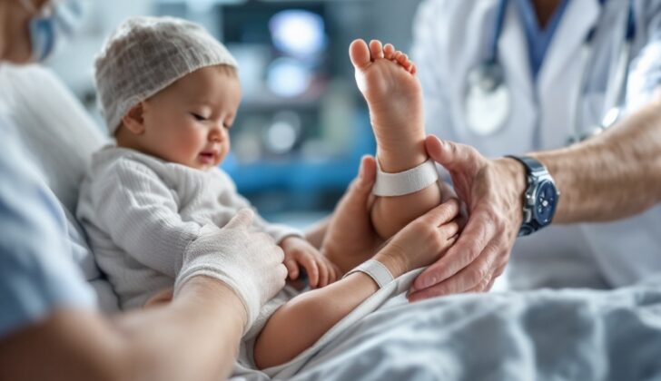
When someone arrives at the hospital with multiple fractures, it is key to first make sure these injuries were not inflicted on purpose; in medical terminology, this is referred to as “non-accidental trauma”.
The diagnosis for the underlying condition causing the fractures is often based on several factors: the patient’s clinical history (symptoms they report), their family history (any related conditions in the family), their bone mineral density (how solid their bones are) tested on the lumbar vertebra (the lower part of the spine), bone biochemistry (a closer look at the chemical composition of the bone), and radiographic features (images from X-rays or other forms of medical imaging).
For most types of a condition known as osteogenesis imperfecta (OI), or brittle bone disease, the main symptom is brittle or easily fractured bones. The specific symptoms for each type of OI were outlined by experts Van Dijk and Sillence.
Commonly, those with OI exhibit four key symptoms:
* Decreased bone mass, making bones more fragile
* Blue-tinted whites of the eyes (sclera)
* Dentinogenesis imperfecta, a condition affecting tooth dentin (the layer just below the enamel)
* Hearing loss
Additional features may include loose ligaments, increased flexibility of joints, short stature, and easy bruising.
Fractures are a major aspect of OI. Generally, if fractures occur earlier in a person’s life, their outlook is less favorable. While fractures often heal at a normal rate, sometimes there is unusually abundant healing tissue (hypertrophic callus) during fracture healing, which can look like bone cancer. Deformities can also occur as a result of fractures. Depending on the type of OI, facial features and other physical manifestations can be varied.
In terms of laboratory testing, there is currently no commercial diagnostic test for OI due to the wide variety of genetic mutations that can cause the condition. Laboratory values are typically normal, however, alkaline phosphatase (ALP), a protein involved in bone mineralization, may be slightly elevated.
Imaging tests like X-rays and CT scans can help provide a clearer picture of the condition. They can reveal features like thinning cortical bone (the hard outer layer of bone), abnormal bone structure, and lesions at the site of fractures.
Prenatal ultrasound can be vital for identifying OI in unborn babies, and may show a number of signs including bone fractures, angulated and shortened long bones (bones of arms and legs), and excessive amniotic fluid (polyhydramnios).
CT and MRI scans can also be used to detect certain features associated with OI, including otosclerosis (a hearing condition) and wormian bones (extra bone pieces in the skull).
To confirm a diagnosis of OI in uncertain cases, fibroblast culturing is used analyze type I collagen, the main skin protein affected in OI. Biopsy, including collagen analysis of a punch biopsy (small sample taken from skin) or an iliac crest biopsy (bone sample taken from the uppermost wing of the pelvic bone), can also confirm diagnosis. The biopsy samples show a decrease in the thickness and volume of bone, along with increased bone remodeling (breakdown and building of bone).
Treatment Options for Osteogenesis Imperfecta (brittle bone disease)
The management of a disease can vary depending on a patient’s age, how severe the disease is, and the patient’s overall health. Here’s a general overview:
For mild cases of the disease, patients may be asked to limit certain activities and avoid sports where physical contact may lead to fractures. Any fractures that occur are treated accordingly.
For moderate to severe cases, the treatment may include rehabilitation, orthopedic interventions, and addressing any acute fractures or curvature of the spine.
If the disease is severe, a procedure using something called an intramedullary rod could be used. This rod is implanted in the bone to help correct severe bone deformities.
There are numerous ways the disease can be medically managed. In the past, hormones, supplements such as calcium and vitamins C and D, and other methods have been tried. However, these have had mixed or unsuccessful results.
Bisphosphonates, a type of medication designed to strengthen bones, have shown helpful effects such as decreased fracture risk, improved bone mineral density, and better mobility.
Gene therapy and cell transplantation, which aim to correct the faulty genes causing the disease, are currently not widely available. However, they are showing promising results in experimental studies in animals.
Other medicines, like anabolic bone therapy, have shown potential in small studies, but are not officially approved for use with this disease at this point.
Orthopedic management is a key part of treatment and aims to improve a patient’s ability to function, prevent and correct deformities, and keep track of complications. This can involve orthotic treatments like braces, walking aids or wheelchairs, managing fractures and bone deformities, and if necessary, using intramedullary rods for children who frequently fracture their long bones.
Managing spinal deformities like basilar invagination (a condition where the upper part of the spine moves up into the skull), kyphoscoliosis (a deformity of the spine causing it to curve and twist), and spinal fractures is also an essential part of the treatment approach.
What else can Osteogenesis Imperfecta (brittle bone disease) be?
When a child presents symptoms of certain bone disorders, doctors need to rule out a few possible conditions. These could be:
- Congenital hypophosphatasia (a rare genetic disorder affecting bone and teeth development)
- Achondroplasia (a type of short-limbed dwarfism)
- Pyknodysostosis (a rare genetic condition resulting in bone abnormalities and short stature)
- Initial stages of leukemia, which can cause overall weakness of the bones (osteopenia)
- Idiopathic juvenile osteoporosis (unexplained bone loss in children)
- Child abuse or battered child syndrome (where unexplained fractures may indicate physical abuse)
What to expect with Osteogenesis Imperfecta (brittle bone disease)
The outlook can vary greatly from person to person due to the diverse nature of the disease. As stated earlier, one classification system, called Shapiro, can be a useful tool to predict the outcome of the disease. The age when fractures in the long bones (like arms and legs) first occur also plays a key role in predicting whether a person will be able to walk in the future.
Life expectancy: The severity and location of fractures, along with the general appearance of the skeleton in x-ray images, turn out to be the most important factors in determining life expectancy.
A study by Engelbert and his team showed that children who could sit and stand without assistance by 12 years of age were ultimately capable of walking. Furthermore, they found that children who were able to sit or stand independently at 12 months old are likely to be able to walk in the future.
Possible Complications When Diagnosed with Osteogenesis Imperfecta (brittle bone disease)
Osteogenesis imperfecta, a condition that weakens bones, can present with various complications. Here are a few examples:
- Rare occurrence of very large areas of hardened tissue. This is usually managed by careful, palliative (symptom relieving) radiotherapy – although this treatment does carry risk of secondary cancer – and medication called bisphosphonates.
- A type of bone cancer called osteogenic sarcoma.
- Basilar invagination, which is a condition that causes the bottom of the skull to push into the spinal canal. This can impact your cranial nerves, cause direct pressure on the brain stem, and change the flow of cerebrospinal fluid, or the fluid around your brain and spine.
- Malignant hyperthermia, which is a severe reaction to certain drugs used during general anesthesia. Both the surgeon and the anesthesiologist should be aware of this possibility during anesthesia.
Preventing Osteogenesis Imperfecta (brittle bone disease)
If your child has a genetic disorder, it’s important to understand what to expect regarding their condition, including their survival rate, potential deformities, disabilities, and ability to walk. In future pregnancies, you may need to undergo genetic counseling and prenatal screening (like ultrasounds) to check if your unborn baby has also inherited this condition. Despite any physical disabilities, it’s crucial to remember that your child can still have normal intelligence and social skills. Parents should also be careful to prevent falls in their children, as this could lead to more bone breaks.
One specific genetic disorder that doctors can detect before birth through an ultrasound is called Osteogenesis Imperfecta (OI) Sillence type II. This can be identified as early as 16 weeks into pregnancy. Other types of OI (types I, III, and IV) can also be detected depending on how severe they are.
If you’ve previously had a child affected by OI type II, there’s a 2% to 7% chance that your next child could also have it. In such cases, doctors can use DNA analysis of samples from the placenta (called chorionic villus samples), which are obtained through ultrasound to make a diagnosis before the baby is born.


