What is Schizencephaly?
Schizencephaly is a rare birth defect of the brain where an abnormal cleft, filled with a brain-and-spinal cord fluid known as cerebrospinal fluid (CSF), forms. This abnormal slit extends from the surface of the brain to the surface of the brain’s fluid-filled spaces called the lateral ventricles. This cleft is lined with a misplaced type of brain tissue called heterotopic gray matter.
The first mention of Schizencephaly was by a person named Wilmarth in 1887. The name of this condition comes from a Greek term “schizen” meaning ‘to divide’. This name was introduced in 1946 by Yakovlev and Wadsworth, who provided more detail on the condition based on their studies. They classified Schizencephaly into two types:
* Type I (closed-lip): This is where the cleft has joined together and does not allow the CSF to pass through.
* Type II (open-lip): In this type, the cleft is open, allowing the CSF to move between the ventricular cavity (the space in brain where CSF is produced) and the subarachnoid space (a space between layers surrounding the brain).
It’s important to note that Schizencephaly can happen on one or both sides of the brain. It occurs in about 1.48 out of every 100,000 live births.
In recent years, the way we understand and classify Schizencephaly has changed slightly. Now, it is believed that having a cleft filled with CSF is not a compulsory characteristic to diagnose the condition. Thus, it’s now classified into three types:
* Type 1 (trans-mantle): This does not have a cleft filled with CSF when looked at through an MRI scan, but does contain a column of abnormal gray matter.
* Type 2 (closed-lip): Includes a cleft that holds CSF, but the lining lips of abnormal gray matter touch each other.
* Type 3 (open-lip): Here, there is a cleft containing CSF, but the lining lips of abnormal gray matter aren’t touching each other.
What Causes Schizencephaly?
The exact cause of schizencephaly, a rare birth defect in the brain, isn’t fully understood by scientists yet.
A few potential causes that have been suggested include the following:
1. Prenatal exposure to harmful substances or viral infections
2. Genetic factors
3. Stroke while still in the womb
4. Younger mothers
There are certain environmental factors that could potentially play a role in the development of schizencephaly as well:
1. Harmful substances including alcohol, warfarin medication, or cocaine
2. Viral infections, with particular attention to the cytomegalovirus (CMV), and more recently, the Zika virus
3. Unsuccessful abortion attempts
4. Lack of oxygen during the eighth week of pregnancy
5. Procedures during pregnancy like amniocentesis (which involves taking a small amount of amniotic fluid) or chorionic villus biopsy (testing of placental tissue)
6. Physical trauma experienced by the mother
A type of bleeding in the baby’s brain that is caused by a defect in a protein called type IV collagen has also been suggested as a possible cause for schizencephaly.
Also, the presence of mutations in certain genes have been reported as being potentially responsible for causing schizencephaly. So far, the primary genes identified in this context are these:
1. The COL4A1 gene
2. EMX2-germline gene
3. The SHH gene
4. The SIX3 gene
Risk Factors and Frequency for Schizencephaly
Schizencephaly is a rare condition that affects the brain, occurring between 0.54 and 1.54 times per 100,000 live births. The estimated rate is about 1.48 per 100,000 births. This condition usually occurs out of the blue and isn’t specific to a particular gender. It’s been noted that it happens more often in children who are adopted or abandoned, which suggests that it might be related to negative conditions in the womb. Furthermore, there’s a higher occurrence of schizencephaly in babies born to younger mothers.
- Schizencephaly is a rare brain condition, seen in 0.54 to 1.54 out of every 100,000 live births.
- Its estimated prevalence is 1.48 per 100,000 births.
- It typically appears without any familial links.
- There is no known preference for either gender.
- This condition is more commonly observed in adopted or abandoned children, suggesting possible negative conditions during pregnancy.
- It happens more frequently in babies from younger mothers.
Signs and Symptoms of Schizencephaly
Schizencephaly is a condition that can present with a wide range of symptoms. Some people with this condition have normal thinking abilities but start experiencing seizures as adults. More often, individuals with schizencephaly present with motor deficits such as partial paralysis and developmental delay, although the severity can range from mild to significant.
The specific symptoms a person might present with actually depend on the type of schizencephaly they have:
- Type I, also referenced as “closed-lip” schizencephaly, usually has a more mild impact. In some cases, symptoms might only become apparent in adulthood. Common symptoms include seizures and minor motor deficits.
- Type II, or “open lip” schizencephaly, typically has a more severe course of symptoms. People with this type of schizencephaly often have epilepsy that may not respond well to treatment, intellectual disabilities, and various levels of paralysis. If the condition is favoring one side of the body, this can result in hemiparesis. If it affects both sides of the body, it can lead to quadriparesis, which is paralysis of all four limbs.
People with schizencephaly can also present differently depending on whether the condition is one-sided or affects both sides:
- Unilateral, or one-sided, schizencephaly might present with hemiparesis on the opposite side of the body and uneven muscle tone.
- Bilateral, or two-sided, schizencephaly can result in seizures, developmental delay, all-four-limbs paralysis, and severe intellectual deficits.
On rare occasions, schizencephaly may only become evident in adulthood, potentially presenting with psychiatric symptoms such as paranoia, irritability, and impulsive spending behavior.
Testing for Schizencephaly
The best way to get an image of schizencephaly, which is a rare brain malformation, is by performing a magnetic resonance imaging (MRI) scan. This scan can detect a fluid-filled linear cleft in the brain, which is a key characteristic of schizencephaly. For those not familiar with the term, a cleft is an opening or split.
There are two types of schizencephaly. Type 1 can be seen on an MRI scan as a small projection from the brain’s surface. Recent findings also suggest that the size and symmetry of certain areas in the brainstem might be related to the hand function in people with schizencephaly.
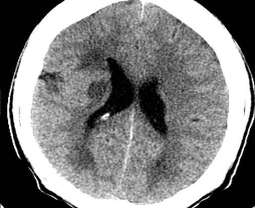
A Computed Tomography (CT) scan could be used as well, but it doesn’t provide as clear a picture of the gray matter – an important part of the brain. This makes it harder for CT scan to differentiate between schizencephaly and other brain conditions that also involve excess fluid build-up, such as arachnoid cyst, porencephaly, and hydranencephaly. However, if a cleft in the cerebral hemispheres is visible by two-dimensional ultrasonography (a type of ultrasound), schizencephaly might be suspected even before birth.
An electroencephalogram (EEG) – a test that measures electrical activity in the brain – might also be used. This test can pinpoint the exact location in the brain where abnormal electrical signals are occurring. This could be within the cleft itself, near the cleft, or even in the opposite hemisphere of the brain (called a mirror focus).
Schizencephaly is often associated with other conditions such as septo-optic dysplasia (which affects the development of the eye and the brain), optic nerve hypoplasia (underdevelopment of the optic nerve), absence of septum pellucidum (a thin membrane separating the front part of the brain), brain cortex malformations, and the appearance of arachnoid cysts.
Treatment Options for Schizencephaly
Schizencephaly treatment varies based on things like the specific symptoms a person has and how severe their condition is.
The typical treatment plan generally includes medication to prevent seizures and physical therapy. If the condition is complicated by a buildup of fluid in the brain, also known as hydrocephalus, a surgical procedure can be performed to place a shunt, or tube, that can help redirect the fluid to another part of the body where it can be absorbed.
In terms of managing the condition, the primary focus is providing supportive care. This can include rehabilitation for issues like muscle or movement problems, learning difficulties, and managing seizures. In situations where there’s excessive pressure inside the skull, also known as increased intracranial pressure, or in cases with hydrocephalus, surgery could be considered.
What else can Schizencephaly be?
The main condition that doctors need to distinguish from arachnoid cysts is schizencephaly. This distinction is typically made through imaging studies, which consider some key factors:
- Presence of heterotopic gray matter that lines the margins of the cleft
- The mass effect, which may take form in displacement of the longitudinal fissure, ventricular compression, or local obliteration of brain sulci and fissures
- Thinning and bulging of adjacent cranial bone
With schizencephaly, the heterotropic gray matter is missing, and there’s no mass effect or bone deformity. For an arachnoid cyst, these features may be present.
There are also other conditions doctors need to consider, including:
- Acquired cysts, related to trauma, surgery, hemorrhage, or postictal events. These typically show areas of gliosis around the lesions and are identified based on medical history.
- Hydrocephalus
- Holoprosencephaly
- Focal cortical dysplasia, which may show a cleft on the cortex that doesn’t extend to the ventricles.
- Grey matter heterotopia, seen as a linear cleft, but with periventricular gray matter typically bulging into the ventricle.
- Porencephaly, which extends from the cortex to ventricles but is lined by gliotic white matter. Some medical experts refer to schizencephaly as ‘true porencephaly.’
What to expect with Schizencephaly
The outlook for a patient varies depending on the size and type of the gaps or separation in brain structure they have.
* Type I speaks to a more minor condition. This type may show no signs or symptoms or only become noticeable in adults. When it does, it’s often through epileptic seizures and small motor deficits (slight difficulty with voluntary movements).
* Type II is more serious and can be recognized by severe signs such as epilepsy that is often difficult to treat, intellectual disability, varying levels of paralysis, half-body weakness if the separation is on one side of the brain (unilateral schizencephaly), and weakness in all four limbs if it’s on both sides (bilateral schizencephaly).
Type II gaps are more frequently linked with worse epilepsy outcomes, and a larger separation can lead to early onset epilepsy compared to a smaller one. Therefore, the type and size of the schizencephaly (the medical term for the gaps or separations in the brain) should be considered when treating epilepsy. Patients should be closely monitored with electroencephalography (a test used to detect electrical activity in the brain).
Possible Complications When Diagnosed with Schizencephaly
Schizencephaly is a condition affecting the central nervous system that often results in epilepsy. While many people with this condition are able to control their seizures, some may struggle with intense seizures that they cannot control. These severe, uncontrolled seizures can sometimes lead to sudden, unexpected death (called SUDEP). It’s even possible for someone to start having seizures for the first time when they become an adult.
People with schizencephaly often have large spaces filled with fluid in their brains. This can lead to an increase in pressure inside the skull, which can cause serious issues like the brain shifting out of its normal position (herniation). These individuals might require surgery that involves placing a tube (or shunt) to drain the fluid, but this procedure can have complications. Some potential risks associated with this surgery include:
- Bleeding
- An accumulation of cerebrospinal fluid under the outer membrane of the brain (subdural hygroma)
- The collection of pus in a cavity (empyema)
- Excessive accumulation of cerebrospinal fluid in the brain (hydrocephalus)
- Infections such as meningitis
If a shunt is used, there may be additional risks like heart infection (endocarditis) and kidney damage related to the shunt.
Preventing Schizencephaly
Parents of kids with schizencephaly need to understand the potential complications and be on the lookout for certain signs. It’s essential that parents know what steps to take if their child starts having seizures, as well as what signs could indicate increased pressure inside the skull (known as intracranial hypertension). With this knowledge, parents can ensure their child receives the right care when needed.


