What is Spinal Dysraphism and Myelomeningocele?
Spinal dysraphism refers to birth defects that cause abnormal formation of the spine and/or spinal cord. It’s caused by unusual development of specific types of tissues during fetal growth. This condition is categorized into two main types based on the visibility of the abnormality: aperta (open) if the defect can be seen, and occulta (closed) if it’s not visible on the skin.
Some common examples include conditions like meningocele, myelomeningocele, lipomeningocele, and others. Spinal dysraphism can also be associated with certain skin conditions such as port-wine stains, hemangiomas (clusters of blood vessels), hypertrichosis (excessive hair growth), and others.
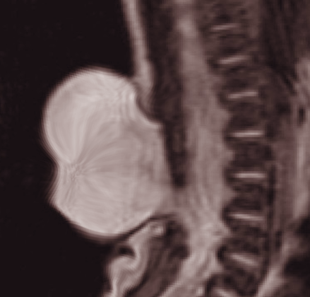
One specific type of spinal dysraphism is myelomeningocele, where the spinal cord and its surrounding tissues herniate, or bulge out, through a birth defect in the middle part of the spine. This falls under the ‘aperta’ category and is also referred to as an open neural tube defect. This is a serious condition and can often lead to life-threatening complications. Myelomeningocele is the most common form of spinal dysraphism, accounting for about 80% of the cases.
What Causes Spinal Dysraphism and Myelomeningocele?
Spinal dysraphism is a condition where parts of the spinal cord don’t form correctly. Multiple factors contribute to its development. In cases of a specific type of spinal dysraphism called myelomeningocele, taking folic acid during pregnancy has been shown to significantly reduce the chances of a child being born with the condition. Other possible causes include genetic factors and racial backgrounds. It’s more common for babies to be born with spinal dysraphism to mothers who have previously given birth to children without the condition. However, once a child is born with the condition, there’s a 1 in 20 chance of it happening in subsequent pregnancies. Some babies with spinal dysraphism can also have chromosomal anomalies, such as trisomies, duplications, deletions, or single-gene mutations. There’s some evidence that pre-existing diabetes in the mother increases the risk of nervous system defects in the baby, including spinal dysraphism.
There’s no single factor known to cause all types of spinal dysraphism. Nutritional deficiencies are the most common risk factor and are thought to be responsible for up to 50% of all cases. Alongside a deficiency in folic acid, a lack of zinc is also linked to the condition. Conversely, an excess of certain nutrients, including vitamin A, can increase the risk of spinal dysraphism. Certain substances found in food and drink, such as nitrates which are present in canned meat and some water sources, and a fungal by-product called cytochalasin, are known to cause spinal dysraphism. Some medications, including the seizure medicine sodium valproate, can increase the chance of a myelomeningocele developing in a baby when taken during pregnancy by 1 to 2 percent.
Risk Factors and Frequency for Spinal Dysraphism and Myelomeningocele
Spinal dysraphism, or defects in the spine, occur in 0.5 to 8 out of 1,000 newborns. This rate can vary widely depending on location, with developing countries often seeing more cases. Thanks to improved nutrition and increased awareness about the importance of folic acid during pregnancy, the incidence has gone down over the years. The rate is lower in the United States, with 0.3 to 0.4 cases per 1,000 live births because of initiatives like mandatory folic acid addition in cereals and better prenatal education. Myelomeningocele, a specific type of spinal dysraphism, is seen in 1 out of 1200 to 1400 births. Spinal dysraphism affects all ethnic groups equally, but occurs slightly more in females than in males.
The condition can impact various regions of the spine, but it most commonly affects the lumbar and sacral sections. On the other hand, the cervical spine (the uppermost part of the spine) is affected the least. Cervicothoracic spinal dysraphism, which involves the cervical and upper thoracic spine, is fairly uncommon.
- 0 to 5% affects the cervical spine (Uppermost part of the spine).
- 5% to 10% affects the thoracic spine (Mid-section of the spine).
- 20% to 30% affects the thoracolumbar spine (Where the mid-section and lower portion of the spine meet).
- 20 to 30% affects the lumbar spine (Lower part of the spine).
- 30 to 50% affects the lumbosacral spine (Where the lower spine and pelvis connect).
- 5 to 15% affects the sacral spine (The bottom of the spine, connected to the pelvis).
Signs and Symptoms of Spinal Dysraphism and Myelomeningocele
Open spinal dysraphism or myelomeningocele are usually diagnosed while a baby is still in the womb or at birth. There are different symptoms for open and hidden (occult) spinal dysraphism. Undiagnosed cases of open spinal dysraphism are discovered when a mass is identified in the newborn’s back with varying degrees of nerves poking out through gaps in bone and skin. The surrounding skin can easily breakdown, leading to cerebrospinal fluid leakage and exposure of the spinal cord, which can cause infections like meningitis.
Kids with spina bifida cystica can show signs like sluggishness, poor feeding habits, irritability, stridor (noisy breathing), ocular motor incoordination (difficulty controlling eye movements), and developmental delays. Older kids might show changes in cognition or behavior, weakening muscles, increased muscle stiffness, changes in bowel or bladder function, issues with lower cranial nerves, back pain, and worsening deformities in the spine or lower extremities. Hidden spinal dysraphism might not be detected early. Some patients only have skin signs like a clump of hair, a birthmark, or a sinus that leads to more tests and diagnosis. Infections like meningitis can prompt further investigations in patients with the dermal sinus. Another important sign in hidden cases is tethered cord syndrome.
- Symptoms of tethered cord syndrome include back pain
- Shooting leg pain
- Urinary dysfunction
- Fecal incontinence
These symptoms can appear in childhood or adulthood. During a physical exam, healthcare providers will check for motor and sensory dysfunctions and focus on certain neuro-segmental levels based on muscle strength of specific nerve roots. Motor weakness can be uneven and may not match the sensory level. People with myelomeningocele are classified according to the affected spinal segment. In tethered cord syndrome, there can be different skin findings during the physical examination. Generally, there’s a skin lesion in the central part of the lower back. Sometimes, there’s just a dimple in the skin in the midline above the buttocks. The neurological examination may reveal motor weakness, sensory loss in the lower body, or decreased anal tone.
Testing for Spinal Dysraphism and Myelomeningocele
If there are concerns of spinal dysraphism, which is a congenital defect in the spine, certain tests are needed for an accurate diagnosis. Catching the disease early is especially important in its hidden, or occult, type to prevent permanent disabilities.
One of these tests is called amniocentesis, which is typically done between 16 to 18 weeks of pregnancy. It involves collecting and testing a small amount of the fluid around the baby in the womb, known as the amniotic fluid. If there is a higher-than-normal amount of a substance called alpha-fetoprotein (AFP) in this fluid, it might suggest that the baby has a defect in the neural tube, which forms the spine and the brain.
Once amniocentesis shows higher AFP levels, an ultrasound test on the fetus could be done to confirm the diagnosis. Through ultrasound, the doctor can check for indications such as the baby’s head’s shape, size of the area involving spinal cord, or abnormalities in brain size and structure. These test results also help decide on what kind of treatments would be the most suitable.
In some cases, soft tissue masses can be checked using ultrasound due to its effectiveness in diagnosing abnormal changes in the spinal cord in children, particularly those with a neurogenic bladder – a condition where a person lacks bladder control due to a brain, spinal cord or nerve problem.
An X-ray of the lower spine is often the first screening tool to be used. It helps the doctors visualize the bones in the spine and identify the location and extent of the abnormality in the spine.
In addition to these, a CT scan of the brain and spine might be required to look for additional complications like hydrocephalus – a condition that can occur in spinal dysraphism where the fluid in the brain increases, causing brain swelling.
However, for the most complete and accurate insights into the spine and brain, an MRI is considered the most reliable tool. It’s particularly good at capturing soft tissue images, making it the best choice to identify malformations of the spinal cord, and specific associated conditions.
Certain physical features might call for these imaging tests. For example, children with certain marks, and those with marks located a certain distance from the rectum, or of a certain size, require imaging due to higher risks of hidden spinal dysraphism.
Tests evaluating mental abilities and functions might also be necessary in patients who have additional brain fluid or show issues with speech, cognition, or academic skills.
Treatment Options for Spinal Dysraphism and Myelomeningocele
If a baby is born with myelomeningocele, a form of spina bifida where the spinal cord and its coverings protrude from an opening in the spine, the first course of action is to provide the baby with intravenous antibiotics. Surgeons then close the opening in the baby’s spine within 24 to 48 hours of birth. This is done to prevent infections and complications down the line. If the baby develops a condition called hydrocephalus, where excess fluid builds up in the brain, a ventriculoperitoneal shunt (a device that helps drain the fluid) might be placed in the brain.
If the baby has other serious health issues at birth, such as severe difficulty breathing or other severe congenital physical anomalies, the immediate focus would be to stabilize the baby’s health.
The surgery to close the opening in the spine is conducted under general anesthesia. Surgeons begin by separating and stitching the exposed part of the spinal cord. After that, they separate the dura, the outermost layer of the spinal cord, and close it. Then they stitch the skin back together.
After surgery, it’s critical to take good care of the baby to ensure the surgery’s success. This includes giving the baby antibiotics to prevent infection, providing painkillers for comfort, ensuring the baby gets proper nutrition, and taking special care of the surgical wound to prevent infections. The baby should be placed prone, or face-down, to avoid aspiration (breathing in fluids into the lungs) and to prevent the surgical wound from reopening. The baby should also be monitored and treated for any urinary problems.
Fetal surgery to close the spinal opening before the baby’s birth is also performed in some specialized centers. This could reduce the risk of the baby developing hydrocephalus later, however it might increase the risk of complications during pregnancy and premature birth. Meningocele repair, a surgery to close the spinal opening, is usually elective, meaning it’s planned in advance, as the skin over the defect is typically intact. After this surgery, the baby will require life-long follow-ups and care from a multidisciplinary medical team.
Occult spina bifida, a less severe form of spina bifida where the spinal cord does not protrude from the spine, usually doesn’t require surgery unless the person develops symptoms. Surgery is done when symptoms become new or progressively worse. The surgery attempts to release the tension on the spinal cord by removing factors that anchor it in place. Most people with myelomeningocele show radiological evidence of a tethered spinal cord but do not need surgery unless symptoms progress or become worse.
Many children with hydrocephalus require revisions of their shunt procedures throughout their lifetime. If they have a common type of malformation seen in myelomeningocele called Chiari II, where part of the brain is pushed downward into the spinal canal, they might require decompression surgery at the base of the skull. If they already have a ventricular shunt, it’s always checked to ensure it’s working properly before performing a decompression surgery. The development of a fluid-filled cavity (syrinx) in the spinal cord may point to a tethered cord, and appropriate treatment should be performed.
What else can Spinal Dysraphism and Myelomeningocele be?
These are some conditions that may present with similar symptoms or might require similar attention:
- Sacrococcygeal teratomas
- Benign teratomas
- Subcutaneous lipomas
- Lymphangiomas
- Cystic teratomas
- Spinal epidural abscess
- Spinal cord masses
- Pilonidal cyst
- Inclusion dermoid
- Caudal regression syndrome
What to expect with Spinal Dysraphism and Myelomeningocele
The future health outcomes of a condition known as spinal dysraphism can vary greatly, as it largely relies on certain factors. These include the degree of neurological damage, any present birth defects, the speed of treatment, and the quality of care provided. Generally, if the spinal dysraphism is located lower down and is less severe, there tends to be a better outcome compared to more serious cases or those where there is fluid build-up in the brain (hydrocephalus).
Patients with milder and lower lesions may even retain the ability to walk. The majority of patients with a type of spinal dysraphism called myelomeningocele have normal intelligence, but about 60% may struggle with some learning difficulties. Those with lesions nearer to their head usually have significant fluid build-up in the brain and often face academic challenges.
Most children with myelomeningocele will need lifelong treatments targeting their damaged spinal cord and nerves. Their health is usually monitored closely through clinic visits every six months during their childhood, then annually once they reach adulthood.
The lifespan of patients with spinal dysraphism is largely influenced by the size of the lesion. Individuals with larger, more complex lesions tend to have shorter lifespans. Surprisingly enough, it is estimated that 40% to 50% of babies with severe defects pass away during infancy. However, patients with smaller lesions located closer to their head and without accompanying fluid buildup in their brain tend to live longer.
One of the leading causes of death among these patients is kidney failure. Despite these challenges, improvements in healthcare have greatly increased their life expectancy. However, most patients often still rely heavily on their parents and carers as adults. Presently, the majority of individuals with myelomeningocele have a near-normal lifespan, if they are fortunate enough not to encounter additional health complications.
Possible Complications When Diagnosed with Spinal Dysraphism and Myelomeningocele
Bladder Problems: In most people with a spinal condition called myelomeningocele, bladder control issues are common. To prevent infections, bladder drainage techniques such as intermittent catheterization or use of an ongoing catheter are introduced. Bladder stimulation can also be helpful as it aids in emptying bladder and lowers the risk of infection.
Bowel Issues: Myelomeningocele also often causes issues with bowel control. Assisted methods of emptying bowels can make social activities like attending school and maintaining personal relationships easier.
Reduced Mobility: Many people with myelomeningocele have major weakness leading to serious mobility issues or even paralysis. Wearing external braces can help improve movement and maintain a closely normal development. For children over one year, a standing frame can prevent bone loss and tight muscles in the lower limbs. Wheelchairs are also useful for older kids and adults.
Increased Risk of Infections: People with this condition have higher chances of getting infections, this is because their bladders, impacted by the condition, often carry bacteria and get infected. In addition, if they have shunts (tubes used to drain fluid) inserted, these can lead to infections either on the skin where they enter the body or in the belly since many have had several abdominal procedures.
Common Complications:
- Bladder control issues
- Poor bowel control
- Limited mobility or paralysis
- Increased risk of infections
- Higher infection risk for those with shunts
Recovery from Spinal Dysraphism and Myelomeningocele
People with myelomeningocele, a birth defect in the spine, typically go through different types of rehabilitation. These include physical therapy, occupational therapy, recreational therapy, and speech therapy. Some patients with myelomeningocele may have problems with their brainstem, making it hard for them to swallow properly. These patients might have to go through a swallowing evaluation. If swallowing is indeed found to be difficult, they may need a feeding tube to help them eat without risking food going into their lungs, which is known as aspiration.
Preventing Spinal Dysraphism and Myelomeningocele
Thanks to global awareness campaigns about the importance of taking folic acid before and during pregnancy, there’s been a noticeable drop in the number of babies born with conditions like myelomeningocele (a severe type of spina bifida where the spinal cord and backbone don’t form properly) and neural tube defects (problems with the structure that becomes the brain and spinal cord). However, it’s important to keep spreading the word for people of childbearing age about the benefits of taking vitamins during pregnancy, especially those containing folic acid.
This is something that should be championed more widely, particularly in countries where there are no rules about adding folic acid to food, known as folate fortification. Doing this could help prevent these major birth defects, which can seriously affect the brain and spinal cord.
People who have a spinal condition called spinal dysraphism (a defect where parts of the spine don’t form correctly), should be urged to actively participate in rehabilitation programs.


