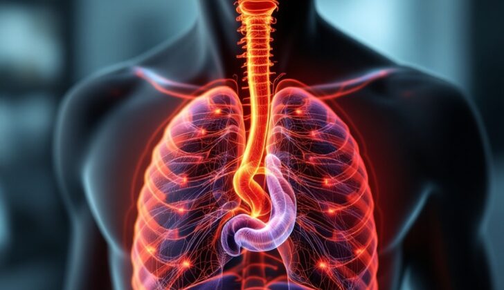
What is Esophageal Cyst?
Esophageal cysts are uncommon and normally formed at birth – a condition first discussed by Blasius in 1711. They were also described by Roth in 1881, who divided them into two types: simple cysts covered by a cellular layer or “foregut” cysts, which include a subgroup called ‘duplication cysts’. Esophageal duplication cysts have two layers of smooth muscle on their surface, are covered by squamous cells or intestinal-style cells, and are either joined to the esophagus or located within its wall. These cysts are usually discovered during childhood but can also appear in adults.
Sometimes, people with these cysts don’t show any symptoms, or they might experience difficulty breathing, difficulty swallowing (dysphagia), and/or chest pain. Esophageal cysts are often unexpectedly found during endoscopy (a test where a flexible tube with a light and camera attached to it is inserted into your digestive tract) or imaging procedures, such as a CT scan, an MRI, or a barium swallow test (a series of x-rays of the esophagus).
While the most common treatment for esophageal cysts is surgical removal through an operation known as thoracotomy, less invasive techniques, such as endoscopic (using an endoscope), laparoscopic (minimally invasive surgery), or thoracoscopic (surgery with the help of a tiny video camera) approaches, are becoming more widely used.
What Causes Esophageal Cyst?
We’re not entirely sure why esophageal cysts, or fluid-filled sacs that appear in the esophagus, develop. It’s believed that they may form when the primitive, or early-stage, esophagus doesn’t develop correctly during the fourth to eighth weeks of gestation (when the baby is still in the womb), although we don’t have a clear explanation for why these cysts occur in different places throughout the digestive tract.
These esophageal cysts are most common on the right side of the esophagus. This preference for the right side is thought to be linked to how the organs inside the chest cavity develop and rotate during fetal development.
Risk Factors and Frequency for Esophageal Cyst
Esophageal cysts are quite uncommon – they occur in about 1 in 8200 births and are twice as likely to occur in males. They make up about 10 percent of all tumors in the chest area (mediastinal) found in children. They are also estimated to account for 10 to 15 percent of all duplication cysts in the digestive tract that originate from the esophagus. The majority of these cysts, as much as 80 percent, are found in children.
Signs and Symptoms of Esophageal Cyst
Esophageal cysts, which are often diagnosed during early childhood, can also appear in adults. Sometimes, these esophageal duplication cysts can be linked to abnormalities in the backbone. The symptoms of these cysts come as a result of the pressure or displacement they put on surrounding structures in the middle part of the chest (mediastinum). These symptoms can include trouble breathing, vomiting, difficulty swallowing (dysphagia), chest pain, and poor growth. If the cyst involves abnormal gastric tissue (ectopic gastric epithelium), there might also be vomiting of blood (hematemesis). Some cases have noted neurological problems, such as nerve root compression along with limited neck motion. While it’s rare, these cysts can potentially lead to cancer.
When diagnosing esophageal duplication cysts, doctors also consider other possible causes of cysts in the mediastinum. These can include bronchogenic cysts, pericardial cysts, cystic degeneration of mediastinal tumors, or other types of cysts like a hydatid cyst or Mullerian cysts.
Testing for Esophageal Cyst
When checking for esophageal cysts, your doctor might take a history and perform a physical examination. Then, they use imaging techniques and look at the specific characteristics of the cells in the cyst for further understanding.
Imaging studies like chest X-rays and barium swallow studies can help reveal any kind of abnormal shape or area in the middle area (mediastinum) of your chest, like a mass pressing on your esophagus, and any change in position or narrowing of the windpipe.
A computed tomography (CT) scan, which is a specialized X-ray, may show a fluid-filled sac originating from the esophagus. Intravenously administered contrast agents, which help produce clearer images, cannot color this sac. An added advantage of a CT scan is that it can also rule out issues with the lungs and thoracic spine. It can also provide detailed structural information if surgery is needed.
Magnetic resonance imaging (MRI) can also provide in-depth details of this area. In general, endoscopy, which is an examination using a thin tube inserted through the throat, can show a lump caused by the cyst pressing on the inside of the esophagus. The interior of the esophagus generally appears normal.
A specific type of endoscopy called endoscopic ultrasound (EUS) is considered the best method to evaluate esophageal cysts. EUS lets doctors determine the details of the cyst, like whether it’s near the esophagus or surrounded by two layers of muscle, which helps distinguish it from other types of cysts like bronchogenic cysts. Plus, EUS can be used to take a sample (fine needle aspiration) of fluids or cells from the cyst for further examination.
EUS generally reveals that the cyst is dark, evenly filled, and has smooth edges in the submucosal wall, which is the innermost layer of the esophagus. In some cases, it may show up with different echo densities due to the presence of pus, blood, or thick contents in the cyst.
Three criteria are essential for classifying an esophageal cyst:
1. The cyst is part of or attached to the esophageal wall.
2. The cyst is covered by two layers of muscle.
3. The lining of the cyst is squamous, columnar, cuboidal, pseudostratified or lined with hair-like structures.
A correct diagnosis of an esophageal cyst is usually made by combining imaging and pathology results.
Treatment Options for Esophageal Cyst
There are no medical treatments for esophageal cysts, which are sac-like growths in the esophagus. If you’re experiencing symptoms due to these cysts, your doctor will likely recommend surgery, which is considered the best way to treat them. Simple cysts can usually be removed, while more complex cysts often have to be totally cut out.
In the past, doctors would perform a surgery known as a posterolateral thoracotomy to take out the cyst. But these days, there are less invasive surgical options that also provide better cosmetic results. These include methods like video-assisted thoracoscopic surgery (VATS) and robotic-assisted thoracoscopic surgery (RATS), where the surgeons use a tiny camera and special tools to perform the surgery with small cuts.
There’s also a procedure called endoscopic submucosal tunnel dissection (ESTD), which is another way to cut out the cyst less invasively. This method is still being studied to better understand long-term results and potential complications. For cysts that extend into the abdominal cavity, your doctor might suggest laparoscopic resection, another minimally invasive surgery technique, where small incisions are made in your abdomen instead of large cuts.
If you have a cyst but aren’t experiencing any symptoms, it’s currently unclear whether treatment is needed. Surgery could be considered to prevent potential complications down the line, like ulcers, perforations (holes in the esophagus), and a small chance of cancer.
What else can Esophageal Cyst be?
These are some types of cysts and similar conditions doctors may consider when diagnosing a patient:
- Bronchogenic cyst
- Cervical duplication cyst
- Thyroglossal duct cyst
- Neuroenteric cyst
- Lipoma
- Lymphangioma
- Hemangioma
- Anterior meningocele
- Pericardial cyst
What to expect with Esophageal Cyst
Patients generally do well both in the immediate aftermath and in the long run after treatment involving full surgical removal of the impacted area. It’s not common for the issue to return, but there have been reported cases of this happening.
Possible Complications When Diagnosed with Esophageal Cyst
Esophageal cysts can lead to several complications, including heartburn, reflux esophagitis (irritation of the esophagus), rupture, obstruction, bleeding, infections, and even cancer. When an intervention is carried out to treat these cysts, there may be additional risks such as injuries to the trachea and esophagus, development of pseudodiverticulum (a false pouch in the esophagus), and nerve damage which might result in paralysis.
Common Complications:
- Heartburn
- Reflux esophagitis
- Rupture
- Obstruction
- Bleeding
- Infections
- Potential for cancer
- Injuries to the trachea and esophagus from intervention
- Development of pseudodiverticulum from intervention
- Nerve damage or paralysis from intervention
Preventing Esophageal Cyst
It’s important that both patients and their family members understand how to spot the signs of esophageal cysts. This knowledge helps in seeking timely medical help if symptoms appear. For patients who are undergoing surgery, it’s crucial they are made aware of the possible complications that might arise from the procedure. Post-surgery, they should have appointments lined up to see their doctor for follow-up care and check-ups, to ensure everything is healing properly. Remember, thorough health education can help prevent complications and improve recovery.


