What is Gastroschisis?
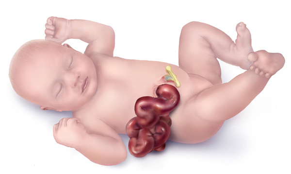
Gastroschisis is a relatively uncommon condition where a baby’s abdomen doesn’t completely close up, and parts of the bowel stick out through an opening near the belly button. Unlike some other health issues, this isn’t usually linked to genetic conditions. Because the bowel isn’t covered inside the womb, it can sometimes appear thickened, enlarged, and covered with a fibrous layer due to inflammation. Babies with this condition often don’t grow as well in the womb.
We usually discover gastroschisis during an ultrasound around the midpoint of pregnancy, seeing that parts of the bowel are floating free in the womb. A blood test can also indicate this condition by showing higher-than-normal amounts of a substance called alpha-fetoprotein (AFP). It’s important to note that compared to other conditions that lead to abdominal openings, like omphalocele, only about one in ten babies with gastroschisis have issues beyond the digestive system. Still, up to a quarter of them may have additional digestive problems.
We can categorize babies born with gastroschisis as “simple” or “complex”. We make this distinction based on whether the baby has other issues like a blocked or narrowed intestine, a hole in the bowel, tissue death, an improperly formed bowel, or a twisted bowel. This helps us predict outcomes and plan care.
It’s often best for the baby to be born in a hospital with specialist services such as high-risk pregnancy care, newborn care, advanced newborn care facilities, and a pediatric surgeon. In most cases, it’s safe for the baby to be born via normal delivery, which usually happens between the 37th and 38th week of pregnancy. After birth, we take care to cover and protect the exposed bowel. We also insert a feeding tube and intravenous lines, and make sure the baby’s breathing is stable.
We can treat Gastroschisis by either surgery or slowly pushing the bowel back into the abdomen using a spring-loaded bag. While a small percentage of babies have severe issues like bowel obstructions, loss of bowel, and long hospital stays, it’s encouraging to note that over 90% of babies with gastroschisis survive.
What Causes Gastroschisis?
Gastroschisis is a condition that occurs when the front wall of a baby’s body doesn’t form properly during embryonic development, causing some of the intestines to protrude outside the body. We don’t fully understand why this happens yet.
Several factors might contribute to the development of gastroschisis. These include smoking tobacco, exposure to certain environmental chemicals (like nitrosamines and atrazine), use of drugs that reduce inflammation (such as aspirin and ibuprofen), and certain cold medicines (like pseudoephedrine and phenylpropanolamine).
Risk Factors and Frequency for Gastroschisis
Gastroschisis, a birth defect of the belly, occurs in 1 out of every 4000 live births and is on the rise globally. It affects male and female infants equally. However, it’s more likely to occur in singleton pregnancies (i.e., pregnancies with one baby), in women below 20 years of age and those from Hispanic backgrounds.
Signs and Symptoms of Gastroschisis
Gastroschisis is a condition that can be seen in newborns, in which there is a hole in the abdominal wall next to the belly button. Through this hole, the infant’s intestines usually stick out. The size of the hole in the abdominal wall is typically about 4 cm. In contrast to a similar condition called omphalocele, there is no protective sac around the organs in gastroschisis. The intestines outside the belly can become thick and often stick together. This is influenced by the contact with the fluid in the womb. Because the intestines are matted together, some parts may become narrowed or even rupture while the baby is still in the womb. Other organs such as the stomach, liver, and bladder can also protrude through this hole. In rare situations, the intestines are visible on a small lump that extends through a tiny hole. This is known as a vanishing gastroschisis and can mean that there is a significant loss of the length of the intestine.
Testing for Gastroschisis
Gastroschisis, a type of birth defect where the baby’s stomach and intestines are found outside of the baby’s body, can often be spotted during a routine ultrasound around the midpoint of pregnancy (20 weeks). This condition causes a higher-than-normal level of a protein called alpha-fetoprotein (AFP) in the mother’s blood.
During an ultrasound, the doctors might see an area of damage to the right side of the baby’s belly button, with the baby’s bowel floating freely. The baby’s bowel, having been exposed to the fluid surrounding the baby inside the womb (the amniotic fluid), might look thickened and inflated.
If gastroschisis isn’t diagnosed until birth, it can affect how the newborn is cared for. A late diagnosis usually indicates that the baby’s bowel hasn’t changed much as a result of being in contact with the amniotic fluid. But a late diagnosis could also mean that some of the baby’s bowel had to be removed if the opening through which the bowel had slipped out of the baby’s belly was very small and damaged the bowel.
Babies with gastroschisis might not grow as expected inside the womb, a condition known as intrauterine growth restriction (IUGR). The mother’s pregnancy might also be at a higher risk for sudden loss of the pregnancy or an unexpected early delivery.
Approximately 10 percent of babies with gastroschisis might have other complications not related to the digestive system. Up to a quarter of these babies might have further digestive problems such as a blocked or narrowed intestine, twisted intestines, dead tissues in the bowel, or other serious issues. Rarely, this is because of abnormal chromosomes (the genetic material found in the cells of the body), seen in 1 percent of cases, usually in babies with other complications.
Treatment Options for Gastroschisis
During pregnancy, it is important to monitor infants with gastroschisis, a birth defect where an infant’s intestines are outside of the body, for proper growth. In some cases, up to 60% of these infants might not grow as expected. Regular ultrasounds (every 3 to 4 weeks starting at 24 weeks of pregnancy) track the baby’s growth and amniotic fluid volume, the fluid that surrounds the baby in the uterus. Too much or too little amniotic fluid could indicate potential problems – too little fluid can risk cord compression, while too much could suggest a blockage in the baby’s intestines.
While the risk of genetic anomalies in babies with gastroschisis generally aligns with normal population risks, any additional structural abnormalities may increase this risk. In such cases, procedures such as amniocentesis, a test in which a sample of the amniotic fluid, which surrounds the fetus in the womb, is collected and analyzed, can be useful for the parents and doctors to better understand and prepare for the baby’s needs after birth.
Delivery methods for babies with gastroschisis are determined by several factors including gestation age (how far along the pregnancy is), ultrasound findings, and other test results. While the gastroschisis condition itself doesn’t necessarily mean the baby can’t be born vaginally, the timing and approach to delivery are often discussed and decided upon by a team of specialists. Delivery typically occurs around the 36th week of pregnancy. If the baby’s liver is also outside of the body, then a cesarean-section (C-section) may be considered.
Once the baby with gastroschisis is born, immediate care is crucial. Due to the exposed intestines, newborns with gastroschisis can lose more body fluid than usual. However, providing too much fluid can lead to swelling and other complications. The exposed intestines are usually protected with specific equipment, and the baby is also given medicines through a vein. Additional care includes checking the baby’s airway and reducing the pressure on their abdomen from fluid build-up.
Surgical treatment options to return the exposed intestines to the baby’s abdomen include immediate repair soon after birth or a multi-step process involving a temporary protective pouch (silo) with gradual reinsertion of the intestines. Both options have been found to have similar outcomes in terms of hospital stay, time to start eating, and time on a breathing machine. In most cases, the belly button (umbilicus) is preserved during the operation for a better cosmetic result.
Post-surgery, the intestines are closely inspected for any obstruction or narrow points. Complete attachment of the intestines usually isn’t attempted during the initial surgery due to swelling. If required, a temporary opening (ostomy) can be made in the intestines to allow feeding while the swelling reduces. Care is taken to monitor bowel blood flow during and after surgery due to various factors that can restrict it. High pressure within the abdomen after surgery (over 10-15 mmHg) can affect kidney and intestinal blood flow, and pressures over 20 mmHg can cause significant organ dysfunction and complications.
What else can Gastroschisis be?
If a newborn baby has gastroschisis, a condition where the baby’s abdomen isn’t fully formed and organs protrude out, it can be confused with other conditions that have similar appearances. These alternative conditions are largely related to the abdominal wall and the umbilical cord.
- The first condition doctors might consider is Omphalocele. This condition is identified by a membrane protecting the bowel contents and an intact umbilical cord. This membrane can break before or during birth. The location of the liver and the place where the umbilical cord vessels join help distinguish between Omphalocele and gastroschisis.
- An Umbilical Hernia is another potential diagnosis. With this condition, the umbilical cord attaches to the hernia sac.
- Pentalogy of Cantrell is yet another possible condition. It is characterized by a lower breastbone defect, a defect in the front part of the diaphragm, a defect in the pericardium which is the thin sac that contains the heart, an omphalocele, and heart defects present at birth.
- Bladder Exstrophy could also be a diagnosis. It is indicated by a low umbilical cord attachment and an inability to visualize the bladder.
- Cloacal Exstrophy is a disorder that affects the intestines, bladder, and genitals and could be confused with gastroschisis. Here, the umbilical cord has a low insertion with exposed bowel and bladder and an omphalocele.
- The last diagnosis to consider is the Amniotic Band Sequence, also called limb body wall complex. This condition affects the limbs of the baby, causing them to have constriction rings.
Doctors use these and other clues to figure out the accurate diagnosis for a newborn baby showing signs of gastroschisis.
What to expect with Gastroschisis
Babies born with gastroschisis, an abdominal wall defect, generally have very good long-term prospects. In fact, stats show that 98 percent of babies with gastroschisis born in North America survive.
To further classify the condition, doctors may refer to it as “simple” or “complex” which depends if there is additional intestinal damage or issues. Up to 75 percent of gastroschisis cases are simple, with the remaining 25 percent being complex. Babies born with complex gastroschisis can experience more complications like issues with their digestive system, breathing, and infections while they’re newborns. Some serious conditions associated with complex gastroschisis include higher risk of in-hospital death, short bowel syndrome, bowel obstruction, necrotizing enterocolitis. They are also usually given parenteral nutrition and tube feedings on discharge. Infants with this complex condition are likely to stay in the hospital 2 months longer than usual.
Also, most infants with gastroschisis face a different placement of their intestines, which isn’t generally repaired. They have a higher chance of volvulus, an issue where the intestine twists. Furthermore, these babies may present intestinal adhesions, which can cause blockage in the intestine. 15% to 30% of gastroschisis cases have connections with Cryptorchidism, characterized by undescended testicles. Close monitoring is done for the first year before any operative measure is taken.
Long-term outcomes aren’t fully clear but are generally positive. Babies without an umbilicus, due to surgery, may face some social stress due to the cosmetic appearance of their abdomen. In these instances, new belly button (umbilicoplasty or umbilical reconstruction surgery) can be considered. As per early studies, it is suggested that these babies are usually within the normal range for brain development, learning, and overall health quality.
Possible Complications When Diagnosed with Gastroschisis
Complications in newborns who have a condition called gastroschisis can include premature birth – 28% of such infants are born prematurely compared to 6% among those without this condition. They may also encounter complications due to the need for total parenteral nutrition (TPN), an essential nutrient solution administered directly into the bloodstream, which can result in bloodstream infections. Various bowel complications may also occur, including necrotizing enterocolitis (NEC), a serious intestinal disease, and infections of the abdomen from surgery to close the gastroschisis.
Newborns with gastroschisis are categorized into either a ‘simple’ or ‘complex’ category based on whether they have intestinal complications or not. These complications may include bowel blockages, reduced blood flow to the bowel, perforation, or development of NEC. Regrettably, newborns with the ‘complex’ category have higher mortality rates, require multiple surgeries, and have longer hospital stays. Additionally, they have increased risks of serious infections, prolonged cholestasis (a condition where bile cannot flow from the liver to the duodenum), and may need intestinal transplantation because of intestinal failure.
It’s worth noting that up to 10% of newborns with gastroschisis also have associated bowel blockages, most commonly in the jejunum or ileum sections of the small intestine. These newborns generally experience more severe outcomes. The timing of surgery to treat these blockages depends on the condition of the bowel, as the bowel may not hold stitches well if there’s a lot of inflammation. A study found no significant difference in surgery outcomes, whether it was performed early or late, or in the possibility of early feeding. If known, the blocked part of the bowel can be reduced and reassessed every 4 to 6 weeks to allow for decreased adhesions and inflammation. A blocked bowel may also be diagnosed after several weeks of no bowel function with a confirming contrast study.
Lastly, ‘closing gastroschisis’ refers to when the gastroschisis defect size decreases before birth. As this hole gets smaller, it causes diminishing blood supply to the bowel, leading to bowel blockages and possible loss of bowel. When a large portion of the intestine is lost before birth, it typically results in a condition called short bowel syndrome.
Preventing Gastroschisis
When a baby is diagnosed with gastroschisis while still in the womb, families are offered a prenatal consultation. This is an opportunity to meet a team of medical professionals who will explain that gastroschisis is a condition where a baby’s abdominal wall doesn’t form correctly on the right side of the umbilical cord. This causes the baby’s intestines to stick out of the belly, and they can sometimes float in the amniotic fluid. Doctors can usually spot this on an ultrasound when they see the intestines outside of the baby’s body. In some cases, other organs can also be visible through the opening, such as the liver, stomach, and bladder.
When the baby is ready to be born, it’s important that they are delivered in a hospital that can provide immediate surgical care. There is no specific best time for delivery, but it usually happens near term. After birth, the baby will need a tube inserted into its stomach to drain fluids, and they may also need help breathing. The doctors will also give the baby fluids through a vein. The exposed intestines will be covered to keep them moist and protected.
The surgical team will then decide on the best approach for treatment. Sometimes, the intestines can be placed back into the belly gradually using a temporary bag (called a silo), or the opening in the belly can be closed up right away. The surgeons will talk with the family about the different methods for closing the opening and decide which is best. Since the intestines have been exposed to the amniotic fluid, it may be a while before the baby can digest food normally, so they will receive nutrients through their veins for a few weeks.
There are risks involved with this condition, such as possible organ damage when the intestines are put back in the belly or possible problems with the blood supply to the intestines. In the womb and after birth, some babies may face blood supply issues if the belly is too small or the intestines are outside the body. It’s also possible that up to one-third of babies with gastroschisis will get a serious infection, and 10%-15% of babies might have a condition where the intestines are not connected properly and will need more surgery.
Once the baby is home, they can usually eat a normal diet and do normal activities. Parents will be taught how to take care of the baby’s surgical wound. It’s important to call the surgical team if the baby starts to throw up food or if the surgical wound turns red.


