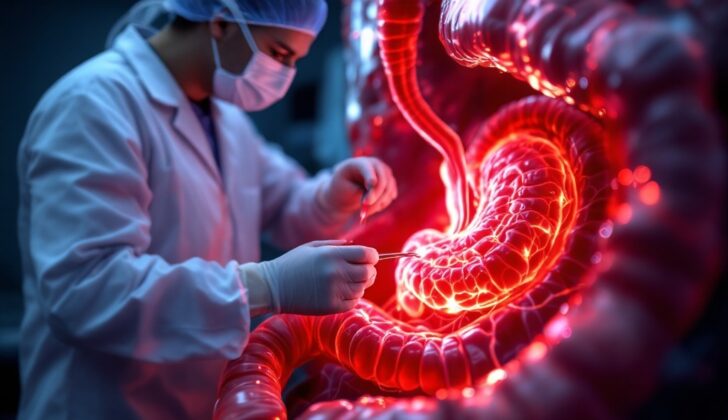
What is Hemosuccus Pancreaticus?
Gastrointestinal bleeding, or GI bleeding, is a common health condition that can be quite serious, often putting a lot of strain on healthcare facilities. There are many health problems that might cause GI bleeding. It can be sorted based on the location or the cause of the bleeding. Common causes of GI bleeding include peptic ulcers, hemorrhoids, esophagitis, tears in the esophagus, esophageal varices, inflammatory bowel disease, polyps, diverticulitis and colon cancer. Some rare conditions, including hemosuccus pancreaticus, hemobilia, aortoenteric fistula, may also cause it.
Hemosuccus pancreaticus is a very rare cause of GI bleeding. It’s when blood comes from a small opening in the digestive system known as the ampulla of Vater, moving through the key duct in the pancreas and into the beginning of the small intestine, known as the duodenum. There are instances when the blood travels through the auxiliary pancreatic duct into a small bit of tissue on the small intestine called the minor duodenal papillae. The first case of this was reported in 1931 with the term ‘hemosuccus pancreaticus’ being coined in 1970. This condition could result in anything from a mild to severe level of bleeding, leading to a significant decrease in blood volume which could result in shock and even death. It’s often tough to diagnose at early stages because it happens so rarely and it’s in a hard-to-reach area. Also, the bleeding can stop and start. Because most physicians aren’t familiar with it, it proves to be a challenging condition to identify. It’s important to consider this condition when a patient with a history of chronic pancreatitis presents with sudden GI bleeding.
What Causes Hemosuccus Pancreaticus?
Hemosuccus Pancreaticus, or HP, has many different potential causes:
– Inflammation of the Pancreas: Also known as pancreatitis, this has been found to be responsible for around 80% of HP cases. It can be due to acute, chronic or hereditary reasons. Inflammation of the pancreas can cause changes in the pancreas’ ducts, often leading to the walls of these blood vessels breaking. This can be also caused by gallstones in the duct, and the development of what are called pseudocysts (or fluid-filled sacs) in the pancreas. The pseudocyst can activate enzymes that destroy the blood vessels, resulting in bleeding.
– Arterial Aneurysm: An aneurysm, which is an overfilling of a blood vessel either inside or outside of the pancreas, is considered the second most common cause of HP, being responsible for about 6% to 17% of cases. The splenic artery is usually the one most likely to be affected, but others like the hepatic, gastroduodenal, and pancreatoduodenal arteries can also be affected. Aneurysms are typically caused by a number of reasons which include conditions that weaken the walls of the arteries, inflammation of blood vessels, and a deficiency of a protein involved in protecting the body from neutrophil elastase (which destroys elastin, a protein in connective tissue).
– Pancreatic Tumors: Tumors in the pancreas, whether they are harmless or cancerous, have been known to cause HP. Even secondary tumors, especially those resulting from kidney cancer, can trigger HP. The tumors can invade nearby organs and create a connection between the tumor and the duct, making it more susceptible to bleeding.
– Medical Causes: HP can sometimes occur as a side effect of certain medical procedures. These procedures include biopsies using fine needle aspiration, stent placement in the pancreatic duct, ultrasound of the pancreas, a procedure to examine the pancreatic and bile ducts (ERCP), pancreatic duct sphincterotomy (a type of surgery), drainage of a pseudocyst, or stomach-reducing surgeries (bariatric surgeries).
– Infectious Diseases: Some infections like brucellosis and syphilis have been known to trigger HP.
– Congenital anomalies: Conditions present from birth, like Heterotopic pancreas and Pancreatic Divisum, also can cause HP. They lead to recurring pancreatitis episodes and have been known to cause HP, particularly with bleeding into the accessory pancreatic duct.
– Other factors: These include trauma (like from an accident or injury) or the twisting of blood vessels (arteriovenous malformation). There has been a case of HP reported in a patient with Glanzmann thrombasthenia, a rare bleeding disorder.
Risk Factors and Frequency for Hemosuccus Pancreaticus
HP, a rare cause of acute bleeding in the digestive tract, is estimated to occur in about 1 out of every 1,500 cases of GI bleeding. It’s more commonly found in males, with a ratio of about 7 to 1. Since it’s a rare condition, it often goes unnoticed. HP typically begins to show symptoms in individuals who are between 50-60 years old. There’s a strong connection between HP and exposure to alcohol as well as other risk factors connected to the development of chronic pancreatitis. As of now, no research has shown that any race or ethnicity is more prone to this condition than others in relation to the rate of occurrence.
Signs and Symptoms of Hemosuccus Pancreaticus
Hemosuccus pancreaticus (HP) is a health condition that often presents with repeated episodes of acute pancreatitis. It’s characterized by abdominal pain which usually starts from the front and extends to the back. Melena, or dark, tarry stools – often considered the hallmark sign of this condition. The most prominent symptoms are those related to gastrointestinal (GI) bleeding like melena, bright red blood in stools (hematochezia), and less commonly vomiting of blood (hematemesis). It rarely leads to life-threatening complications like rupture into the abdominal cavity. Despite originating from an artery, the bleeding is typically not severe but can occasionally present as heavy hematemesis and low blood volume shock.
Patients usually complain of abdominal pain, mostly focused in the upper middle area which can spread towards the back. The pain is generally not severe enough to cause changes in vital signs. Characteristically, the pain intensity fluctuates over time due to the temporary closing of ducts in the pancreas. Some patients, specifically those with tumors at the head of the pancreas, may have yellowing of the skin and eyes (jaundice) due to obstruction. A significant number of patients might show signs of acute or chronic anemia. Heavy alcohol consumption is a strong risk factor for HP.
- Abdominal pain, often radiating to the backside
- Dark, tarry stools (melena)
- Bright red blood in stools (hematochezia)
- Vomiting of blood (hematemesis) – less common
- Upper middle abdominal pain that spreads to the back
- Fluctuating pain intensity over time
- Jaundice – in cases with pancreatic head tumors
- Signs of acute or chronic anaemia
- Heavy alcohol consumption
There are also other symptoms such as vomiting, additional jaundice caused by a blood clot blocking the pancreaticobiliary reflux, and unexpected weight loss. During a physical examination, a palpable pulsation with a russhing sound (thrill murmur) might be detected if there’s an aneurysm present.
Testing for Hemosuccus Pancreaticus
The diagnosis of Hemosuccus Pancreaticus (HP), a rare condition that causes bleeding in the digestive system, can be hard. This is because of its rarity, the tricky location it affects within the body, the changeable nature of its symptoms, and the many causes that can trigger it. Usually, blood test results will be normal for this condition, unless it’s caused by a backup of bile, which is a digestive fluid, into the pancreas (pancreaticobiliary reflux), which can make a protein called bilirubin levels go up. If Pancreatitis, inflammation of the pancreas, is the cause, then enzymes named amylase and lipase levels can go up.
The diagnosis is mostly done through imaging techniques because blood tests can’t conclusively diagnose HP. Several imaging techniques can help in spotting HP. These include a procedure where a tiny camera is passed down your throat into your stomach (Esophagogastroduodenoscopy), a contrast-enhanced CT scan (which uses X-rays to produce detailed images), angiography (a type of X-ray using a special dye), an ultrasound test done through an endoscope (endoscopic ultrasound or EUS), and a procedure using an endoscope to examine the pancreas ducts (ERCP).
Regarding endoscopy, it’s vital once there’s a sign of bleeding in the gastrointestinal (digestive) tract. This helps identify possible common causes of such bleeding like a peptic ulcer, inflamed stomach lining, and enlarged veins in the stomach. However, spotting bleeding from the area where the bile and pancreatic ducts open inside the duodenum (ampulla) is rather tricky since it happens sporadically and it’s harder to get a clear view of the ampulla with a forward-viewing endoscope. Nonetheless, an ERCP can help clearly visualize and pinpoint any bleeding’s source. Finding clotted blood blocking the pancreatic ducts is a clue that suggests a possible HP diagnosis.
Abdominal ultrasound, whether using doppler or dynamic techniques which visualize internal structures and blood flow, can help display the existence of an Aneurysm (a bulge in the wall of a blood vessel) or pancreatic pseudocyst (fluid-filled sac in the pancreas). The ultrasound gives positive results in about 38% of cases.
A Contrast-enhanced CT scan is excellent for exhibiting pancreatic conditions such as chronic pancreatitis, pseudocysts, and pseudoaneurysms (blood-filled sacs due to vessel wall leakage). Signs that indicate HP on a CT scan are usually detected as blood clots within the pancreatic duct. The CT scan might also show persistent visibility of the dye within the aneurysmal vessels, the existence of a pseudocyst, or/and a sustained presence of the contrast dye after the arterial phase. These findings indicate an urgent need for angiography, a specific type of x-ray to confirm the diagnosis and potentially treat the problem. The CT scan has a 90% success rate in these cases.
Angiography, which is considered the best diagnostic and therapeutic tool, works by focusing on the celiac trunk, superior mesenteric artery, and branches. Angiography can identify the problematic artery, allows for a detailed look at the arterial anatomy and provides a chance for a therapeutic intervention. Usually, it yields positive results in over 90% of the cases.
Treatment Options for Hemosuccus Pancreaticus
If you have gastrointestinal (GI) bleeding, doctors primarily need to gauge how severe the blood loss is, stabilize your condition, and constantly monitor you. The immediate treatment involves providing oxygen therapy, supplying fluids intravenously (through your veins), and preparing for a possible blood transfusion if required. After stabilizing you, the next objective is to locate the source of the bleeding using various imaging techniques, with the ultimate aim of stopping the bleeding.
For pancreatic bleeding (HP), the medical community has not yet pinpointed any highly effective medications. Solely depending on supportive therapy, such as providing oxygen and fluids, carries a high risk (90%) for life-threatening outcomes. So, doctors usually opt for either interventional radiology (using image-guided techniques to treat the condition) or open surgery. The choice of treatment depends on your specific health condition and symptoms at the time of diagnosis. In most cases, interventional radiology is the primary technique used.
Interventional radiology is often selected as the first line of treatment for patients with a stable condition and suspected HP. It is typically carried out in patients who have undergone angiography, a type of X-ray used to check blood vessels. The success rate of interventional radiology ranges from 67% to 100%. It includes techniques like angiography with embolization (blocking the blood vessel) using artificial materials, balloon tamponade (blocking arterial flow using a balloon), or stent placement (inserting a tube to keep the passageway open). The most commonly used method is coil embolization, which induces clotting within the bleeding vessels. However, this method can sometimes reduce blood supply within the vessel. Despite the high initial success rate, there’s a 30% chance of bleeding recurring after embolization. Balloon tamponades and stent placements act as a temporary solution before a scheduled surgery. Interference using interventional radiology has few major complications other than a superficial skin infection at the site of intervention in about 0.5% of cases.
If embolization fails, it might be due to the inability to identify or isolate the bleeding artery or due to spasm (sudden contraction) of the bleeding vessel.
Surgical interventions are typically considered if you have:
– Continuous uncontrolled bleeding and unstable body functions.
– Clear symptoms but negative angiographic results.
– Complications like pancreatic abscess (an enclosed area of infection), gastric outlet obstruction (blockage of the channel leading from stomach), obstructive jaundice (blockage of the bile duct), or severe pain.
– Hemorrhagic pseudoaneurysms (false blood vessel formed due to a leak in the arterial wall).
– Unsuccessful embolization or recurrent bleeding after the embolization procedure.
– Severe cases, with or without pancreaticoduodenectomy (a surgical procedure to remove a part of the pancreas).
During the surgery, if it’s difficult to remove the pseudocyst (a collection of fluid in the abdomen) due to its location, doctors might tie off the artery on both sides of the bleeding source. Alternatively, they may cut off the drainage to the pseudocyst that enters the GI tract. In these instances, ultrasound during the operation, and inspection of the pancreas might be required to locate the source of bleeding and decide the extent of removal. Surgically addressing this condition is successful in 70% to 80% of cases, but there’s a risk of life-threatening outcomes ranging from 10% to 50% and a chance of recurrent bleeding ranging between 0% and 5%.
New treatment methods such as endoscopic-guided ultrasound with or without creating a direct channel from the pseudocyst to the stomach, using fibrin glue and histoacryl adhesives, are revealing promising results. They are particularly beneficial for patients who either do not exhibit clear signs in their X-ray or angiographic images, are allergic to contrast material used in X-rays, or are not suitable candidates for surgery. However, the overall safety and benefits of these new treatments still need further investigation.
What else can Hemosuccus Pancreaticus be?
Differentiating diseases of the digestive system can be quite complex given the many possibilities. In the case of upper and lower gastrointestinal (GI) bleeding, which refers to bleeding in different parts of the digestive system, the potential causes can be divided into the following categories:
For upper GI bleeding which involves the esophagus, stomach, and part of the small intestine, potential causes include:
- Peptic ulcer disease (caused by H. pylori, excessive NSAID use, stress, or specific types of ulcers)
- Gastritis and duodenitis (inflammation of the stomach and duodenum)
- Esophageal varices (abnormally large veins in the esophagus)
- Mallory-Weiss tear (a tear at the junction of the stomach and esophagus)
- Esophagitis (inflammation in the esophagus)
- Upper GI tumors
- Dieulafoy’s lesion (an uncommon cause of bleeding in the stomach)
- Arteriovenous malformation (abnormal connection between arteries and veins)
- Portal hypertension gastropathy (a condition related to liver disease)
- Hemobilia (bleeding from the bile duct)
- Aortoenteric fistula (an abnormal connection between the aorta and digestive system)
- Bleeding disorder
- Gastric antral vulvular ectasia (a rare condition causing stomach bleeding)
As for lower GI bleeding, which covers the second part of the small intestine and the large intestine, potential causes can be:
- Angiodysplasia (abnormal blood vessels in the colon)
- Diverticulosis (small pouches on the colon walls)
- Meckel’s diverticulum (a pouch present from birth on the wall of the lower part of the small intestine)
- Benign or malignant tumors
- Inflammatory bowel disease
- Infectious colitis (infection in the colon)
- Ischemic colitis (inflammation of the colon caused by reduced blood flow)
- Anorectal etiologies (problems originating in the anus or rectum)
- Damage to the colon wall caused by radiation therapy
- Aortoenteric fistula (an abnormal connection between the aorta and intestines).
What to expect with Hemosuccus Pancreaticus
In general, HP – a medical condition known as Haptoglobin – has a death rate of about 9.6%. This rate tends to rise when HP is complicated by a malignant or cancerous growth. If the patient receives only basic or supportive care, the death rate can soar to as high as 90%.
Possible Complications When Diagnosed with Hemosuccus Pancreaticus
The primary problems that can arise with HP – Hairy Palms condition include:
- Severe bleeding in the upper or lower digestive tract
- Long-term anemia (low red blood cell count)
- Breaking or tear in an organ, leading to a pooled area of blood outside the blood vessels in the back of the abdomen
- Shock due to severe loss of blood
- Failure of multiple organs
- Potential death
- Bleeding or pooled blood at the scanned site
- Bleeding happening again in 30% of cases
- Internal bleeding due to damage to the arterial wall during the scan
- Allergic reactions to the local anesthetic, the dye used for the scan, or calm-inducing medications
- Immediate damage to the kidneys
- Bleeding happening again in about 0% to 5% of cases
- Internal bleeding in the abdomen and/or a surgical cut that separates/breaks open
- Inflammation in the abdomen, abscess (pus) formation, and widespread infection in the blood
- Blockage in the intestines
- Blood clots in deep veins and lung embolism, which is a sudden blockage in a lung artery due to a blood clot
- Potential death in about 20% to 25% of cases
If an angiographic intervention is performed (a type of X-ray to see how the blood flows through the arteries), some potential side effects might happen:
For abdominal surgical interventions, there are associated risks involved:
Preventing Hemosuccus Pancreaticus
Rectal bleeding can be seriously worrisome and challenging to deal with, as it could pose a high risk of causing further health problems or even death. It can sometimes be tough to pinpoint the exact cause of the bleeding.
Medical staff may have to carry out several thorough physical exams and ask for the patient’s medical history many times. They might also need to conduct extensive blood tests to check things like blood cell count, blood type, your body’s ability to stop bleeding (bleeding diathesis), your body’s response to irritation, injury, or infection (inflammatory markers), and signs that suggest possible cancer (cancer blood screening). Since there can be a wide range of potential causes, it’s crucial to carry out the right tests to precisely identify the source of the bleeding and deliver the appropriate treatments as quickly as possible.
For patients who have rectal bleeding, one possible cause might be a condition called hemosuccus pancreaticus (HP), especially in patients who have long-term inflammation of the pancreas (chronic pancreatitis) and those who consume alcohol heavily. If HP is suspected, the best way to manage the condition will depend on how the patient is doing and any additional risk factors they might have. Even though procedures like angiography (an X-ray that uses a special dye and a camera to take pictures of the blood flow in a blood vessel) and surgery may carry a risk of bleeding again and health complications, they still offer the best chances of finding out what’s causing the bleed and effectively treating it.
Patients with chronic pancreatitis are advised to avoid alcohol and stop smoking to reduce the likelihood of their condition worsening to cancer, which carries a 4% to 5% risk. They’re also recommended to eat a low-fat diet that’s high in protein and carbohydrates. Strictly taking in fat-soluble vitamins and pancreatic enzyme supplements as prescribed is also important. In addition, they should have their blood sugar levels checked every year for diabetes and a DEXA scan (a test that measures bone density) for osteoporosis, a condition that makes your bones weak and more likely to break.


