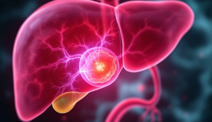
What is Hepatic Cystadenoma?
It used to be thought that liver cysts, or fluid-filled sacs in the liver, were relatively rare. However, with the progression and increased availability of imaging techniques, these are being identified more often. It’s estimated that 5 to 10% of people worldwide have cysts in their liver. There are many possible causes for liver disease, such as infections, inflammation, cancer, hereditary conditions, and injuries.
A specific type of liver cyst is the mucinous cystic neoplasm, which includes biliary cystadenomas (BCA) and cystadenocarcinomas (BCAC). These terms might sound complex but they’re just types of liver cysts. Combined, BCA and BCAC make up less than 5% of all liver cyst diseases. They often cause vague abdominal symptoms and are usually discovered unexpectedly through imaging tests.
The appearance of these cysts on imaging tests can suggest their type, but there could be overlap with other cysts, leading to some uncertainty. BCAs are thought to have the potential to turn into cancer. Even though only a few cases of this happening have been reported, the current agreement is that the best treatment is to surgically remove them completely.
What Causes Hepatic Cystadenoma?
The definite cause of hepatic cystadenomas, which are a type of cyst in the liver, is still unknown. One idea is that they might come from the early cells that grow into the lining of bile duct in the liver. However, there’s another theory which suggests these growths might be due to cell implantation. This theory is supported by the fact that they show similar characteristics to ovarian tissue, respond to female hormones like estrogen and progesterone, and are often found in a specific part of the liver called segment 4.
Risk Factors and Frequency for Hepatic Cystadenoma
Biliary cystadenomas and cystadenocarcinomas (BCACs and BCAs) are rare types of liver cysts, making up only a small portion of the global cases. The chance of having a BCA inside the liver is estimated to be between 1 in 20,000 to 1 in 100,000. These cysts are most commonly found in women aged 40 to 50 years old. In fact, about 85% of them originate from the part of the liver that produces bile. They grow quite slowly but can eventually reach up to 30 cm in size.
- BCACs and BCAs are rare liver cysts.
- Their occurrence rate ranges from 1 in 20,000 to 1 in 100,000.
- They are most common in women aged 40 to 50 years old.
- About 85% of these cysts come from the part of the liver that produces bile.
- Despite growing slowly, they can grow up to 30 cm in size.
Signs and Symptoms of Hepatic Cystadenoma
Hepatic cystadenomas or liver cysts can have a wide range of symptoms, but often, they don’t cause any noticeable problems. This means many people only discover they have a cyst when they’re being tested for something else. When symptoms do occur, they typically include abdominal pain, bloating, feeling sick, and vomiting. In rare cases, the cyst can lead to more serious issues like jaundice, inflammation of the bile duct, bleeding inside the cyst, or even rupture of the cyst. It’s important to note that there are currently no physical signs that would suggest if a cyst could turn into cancer.
- Many people have no symptoms
- Common symptoms include abdominal pain, bloating, nausea, and vomiting
- More serious symptoms can include jaundice, bile duct inflammation, internal bleeding in the cyst, or rupture of the cyst
- There are no known physical signs of cancerous potential
Testing for Hepatic Cystadenoma
Hepatic cystadenomas, a type of tumor in the liver, are usually diagnosed using a combination of ultrasound, CT (Computed Tomography) scans, MRI (Magnetic Resonance Imaging) scans, and information from clinical observations and pathology tests. These tumors are usually found within the liver, tending to be single, slow-growing, multi-chambered cystic tumors filled with a clear, mucus-like fluid. They are more commonly found in women and are often located within the fourth segment of the liver.
On an ultrasound, these tumors appear as a clear, well-defined spot surrounded by a bright, echo-making capsule with multiple septations or internal walls. In a CT scan, the tumors appear as a fluid-filled cystic mass with a rim that resembles soft tissue, along with internal walls, and possibly calcifications on the capsule and lumpy areas on the wall.
On an MRI scan, these tumors typically display characteristics common to fluid-filled cystic lesions, including low signals on T1 (a type of MRI image) and high signals on T2 (another type of MRI image). However, these signals can vary based on the protein content of the cyst and the presence of hemorrhage, or solid portions. Importantly, these tumors do not have any connections with the bile duct system. These tumors can show mild enhancement of the capsule and internal walls after the injection of contrast agents in both CT and MRI scans.
The existence of internal debris, widening of the bile duct and lumps that enhance after contrast injection raises concerns for a rare type of liver tumor known as Biliary Cystadenocarcinoma.
Usually, the laboratory tests of patients with hepatic cystadenomas are within the normal ranges, although some patients may have elevated liver enzyme values, in particular, bilirubin levels. Elevated levels of specific markers in the blood, namely carbohydrate antigen 19-9 and carcinoembryonic antigens, may be indicative of the tumor turning malignant, but this is neither a highly sensitive nor a specific sign. The practice of using a fine-needle to extract fluid from the cyst (Fine-Needle Aspiration) is no longer recommended due to the risk of spreading the tumor cells and leading to a condition known as peritoneal carcinomatosis, where the cancer spreads within the abdomen.
Treatment Options for Hepatic Cystadenoma
Hepatic cystadenoma, or a particular form of liver cyst, can potentially turn into cancer over time. The signs of this condition on medical imaging tests like ultrasound or CT scans may hint at its presence, but these signs often overlap with those of other conditions and aren’t always clear.
Due to the small number of recorded instances of hepatic cystadenoma, there are no established guidelines recommending the best way to treat it. Certain treatments, such as percutaneous ablation (which uses heat to destroy the cyst) and unroofing techniques (which involve cutting off the top of the cyst), don’t always work effectively. They may even come with an 80% chance of the cyst returning.
Therefore, the preferred approach is to remove the cyst entirely through surgery, because of the possible risk of the cyst turning malignant (into cancer) and recurring. However, if complete surgical removal isn’t possible, a technique called enucleation, which involves carefully removing the cyst without damaging the surrounding tissue, can be used.
What else can Hepatic Cystadenoma be?
Diagnosing Biliary Cystadenoma (BCA) from Biliary Cystadenocarcinoma (BCAC) before surgery can be difficult. However, since the treatment for these conditions is the same, the main goal is to rule out other less harmful cystic conditions using imaging techniques. The most common liver cyst is the simple hepatic cyst. Health professionals can tell the difference between simple cysts and BCAs by the absence of internal dividers and protrusions in the former.
Distinguishing a cyst filled with blood can be tricky due to its complex appearance in ultrasound imaging. However, in a CT scan, these cysts look smooth and consistent without internal complications unlike BCAs. Also, blood clots in these cysts usually appear brighter in MR images.
There are also other diagnoses to consider, such as:
- Pyogenic hepatic abscesses
- Hydatid disease
- Mesenchymal hamartomas
- Undifferentiated embryonal sarcoma
These conditions usually present with fever and are not more common in females like BCAs. Moreover, related imaging findings such as surrounding inflammation, segmental differences in blood supply, and internal gas are more indicative of an infectious cause. Mesenchymal hamartomas and undifferentiated embryonal sarcoma also do not have a higher prevalence in females and are more commonly seen in children and younger adults.
What to expect with Hepatic Cystadenoma
The data regarding the prognosis or the likely course and outcome of hepatic cystadenoma, after undergoing a resection, a medical term for surgical removal, is quite limited due to the rare nature of this condition. However, from the few reported cases, it’s been observed that patients who have the entire growth removed do well – with only a 5% to 10% chance of the growth recurring again.
Possible Complications When Diagnosed with Hepatic Cystadenoma
Complications associated with hepatic cystadenomas, or cysts on the liver, can include blockage of the bile duct causing jaundice, inflammation of the bile duct, rupture of the cyst within the abdomen, or bleeding inside the cyst. These issues can often be the first signs of the condition. One of the major risks is that a benign cystadenoma can transform into a malignant cystadenocarcinoma. Documents report that this malignant transformation can happen in as many as 20% of cases.
Here are some complications:
- Obstructive jaundice or yellowing of the skin caused by a blockage in the bile duct
- Inflammation of the bile duct
- Rupture of the cyst within the abdomen
- Bleeding inside the cyst
- A benign cyst transforming into a malignant cystadenocarcinoma. (This can occur in as many as 20% of cases)
Preventing Hepatic Cystadenoma
It’s important that patients understand that BCAs, or Bile duct cystic adenomas, are quite rare and health professionals have yet to pinpoint specific risk factors that could lead to them. Additionally, patients should be aware that if the BCA is not entirely removed, there is a chance it could come back or potentially turn cancerous. This information is based on current medical research and documents.


